Review Article
Key words
NADPH oxidase; NADPH oxidase inhibitors; reactive oxygen species; reactive nitrogen species; oxidation-reduction; cardiovascular diseases.
Redox Unbalance: NADPH Oxidase as Therapeutic Target in Blood
Pressure Control
Luiza A. Rabêlo
1,2, Valéria Nunes de Souza
1, Lucas José Sá da Fonseca
1, Walkyria O. Sampaio
2,3Universidade Federal de Alagoas1, Maceió, AL; Universidade Federal de Minas Gerais2, Belo Horizonte, MG; Centro Universitário de Belo
Horizonte - Uni-BH3, Belo Horizonte, MG - Brazil
Mailing address: Luiza A. Rabêlo •
Praça Afrânio Jorge SN - Instituto de Ciências Biológicas e da Saúde - Prado - 5701-002 - Maceió, AL - Brazil
E-mail: [email protected]
Manuscript received February 28, 2009; revised manuscript received May 22, 2009; accepted June 8, 2009.
Multiple enzyme systems produce RONS and their derivatives in the vascular system, including cyclo-oxigenase, lipoxigenase, P450 cytochrome, xanthine oxidase (XO), myeloperoxidase (MPO), nitric oxide synthase (NOS) and NADPH oxidase - this latter one of the most important sources of this substance, both in endothelial cells as in smooth muscle cells1-22.
Since the findings of Baehner et al23 40 years ago - which
allowed the discovery of NADPH oxidase24 - several studies
addressed the relation between said enzyme complex and the redox unbalance (oxidative stress). Increase in production of superoxide anion (•O
2-) and other RONS is implied in
arteriosclerosis, arterial high blood pressure, cell proliferation and hypertrophy. However, the role of NADPH oxidase in such processes remain unknown, which is mainly attributable to occurrence of multiple isoforms of Nox (subunits which form the NADPH oxidase) and their vehicles, as well as to the lack of specific inhibitors25,26. The enzyme complex act as electron
donor for reduction of O2 into •O
2-, following the reaction 2O2
+ NAD(P)H è 2O2- + NAD(P) + H+10,16,22,25,27.
•
O
2-
: production by NADPH oxidase and its
relation with the
•NO
Groundbreaking experiments of Furchgott and Zawadzki28
first demonstrated the existence of endothelium-derived relaxing factorwhich was subsequently identified as •NO29,
produced based on L-arginine by the action of endothelial nitric oxide synthase (eNOS)29-32 in the presence of
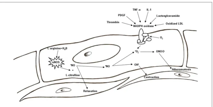
co-factors, mainly the tetrahydrobiopterin (H4B; Figure 1). The
•NO diffuses into vascular smooth muscle cells and activates
the guanylate cyclase, promoting cyclic GMP mediated vasodilation29-33. In normal conditions, the •NO performs key
role in maintenance of vascular wall in quiescent status by inhibition of inflammation, cell proliferation and thrombus, reducing vascular tone, activation of platelet and leukocytes, proliferation of smooth muscle cells, extracellular matrix deposition and death of endothelial cells34-36. •NO is also a
free radical and, when produced jointly with •O
2-, react in an
extremely swift way to form a highly reactive proinflammatory species: the peroxynitrite anion (•ONOO-; vasodilatator less
powerful than •NO). Thus, most of the cytotoxicity attributed
to •NO comes from the •ONOO-5,37. This last substance is
an important lipid peroxidation mediator, including LDL oxidation, event of capital importance to atherogenesis5.
Physiologically, a certain amount of intracellular •O 2- is
required for normal signaling performed by •NO. Nevertheless,
pathologically, the extracellular increase of the first species
Abstract
Several studies refer to reactive oxygen and nitrogen species (RONS) as important agents in the pathogenesis of a number of heart diseases, including high blood pressure, arteriosclerosis and heart failure. Such species are highly bioactive molecules and a short life due chiefly to reduction of molecular oxygen. The enzyme complex of NADPH oxidase is the main source of these reactive species in vascular system. Under physiological conditions, formation and elimination of these substances seem balanced in vascular wall. During redox Unbalance, nonetheless, there is increase in NADPH oxidase activity and predominance of pro-oxidizing agents, surpassing the anti-oxidant capacity of the organism self-defense. Besides this, such enzyme hyperactivity reduces the bioavailability of nitric oxide, capital for vasodilation and maintenance of normal vascular function. In spite of NADPH oxidase being directly connected to the endothelial dysfunction, it was firstly described as for its expression in phagocytes, where its activity determines efficiency of organism defense mechanisms against pathogens. Slight differences between structural units of NADPH oxidases, depending on the type of cell which expresses it, may create therapeutic implications, allowing to selectively inhibiting redox unbalance triggered by NADPH oxidase, without compromising, however, its participation in physiological cellular signaling which make sure protection against micro-organisms.
Introduction
Figure 1 -Mechanisms of activation of NADPH oxidase and its relation with the •NO metabolism.
decreases the bioavailability of •NO, reducing its diffusion
into the vascular smooth muscle38,39. In certain circumstances,
chronic production of RONS (increase in the NADPH oxidase activity, for instance) exceeds the capacity of cell enzymatic antioxidants (such as the glutathione peroxidase-1 - main oxidant enzyme in cytosol and in mitochondria35,36 - and
the forms of superoxide dismutase linked to the membrane, responsible for dismuting the superoxide anion into H2O2) and cell non-enzymatic antioxidants, contributing to vascular disease due to sustained endothelial activation29,40.
Large amounts of •O
2- formed capture most or all the •NO,
promoting the formation of •ONOO-34,41. The •O
2- generated
may also be transformed into hydroxyl radical, which diffuses into the vascular smooth muscle and induces the production of vasoconstrictive endoperoxides, such as prostaglandin H2 and prostanoids41, derived from the peroxidation of the
arachidonic acid catalyzed by free radical, considered as indicators of systemic increase of redox unbalance in vivo42.
The excess of •O
2- production, with subsequent decrease
of •NO’s bioavailability, may be also caused by transition
metals, such as iron, copper, mercury and lead. Some of these environment pollutants are capable of increasing the production of free radicals by means of activation of enzyme complex NADPH oxidase43,44, and this mechanism may be
seen as probable cause of high blood pressure induced by said chemical species.
Studies by Wiggers et al45 showed that the exposure of
rats to low concentrations of mercury induces endothelial dysfunction both in conductance and resistance vessels. Authors imply this event is likely arisen out of reduction of
•NO’s bioavailability due to the increase of vascular production
of •O
2-. Thus, it is possible that treatment with mercury affect
the protein expression or, more specifically, the activity of NADPH oxidase.
Such unbalance is related to RONS due to the fact that the O2 is germane to cell respiration, and there are several enzyme systems which use this substrate as an electron acceptor1,20. Family of such species includes highly bioactive
molecules with short life due chiefly to O2 reduction. Physiologically, the formation and elimination of RONS are balanced in the vascular wall46, whose key role is played by
the endothelium in maintenance of vascular tone and in blood pressure by releasing vasoactive substances, such as the •NO47. The increase of RONS due to redox unbalance
was found in elder rats’ and mice’s aorta, carotid, mesenteric and coronaries, implying that this unbalance is associated to ageing, with the increase of NADPH oxidase activity48.
Experimental evidences indicate that the oxidative damage induced by reactive species derives from the increase of •O
2
-production and its metabolism and/or reduction of •NO25,49
bioavailability. However, in spite of the excess of RONS being toxic, physiological concentrations of these species may function as mediator signs of several responses, including cell migration and growth20,22,50, once, under normal conditions
and in most arteries, the production of •NOis predominant,
in such a way that this radical takes small amounts of formed
•O
2-40,51-53, making sure the maintenance of balance in organic
oxidative status.
Non-selective inhibition of NADPH oxidase
compromises physiological cell signaling
Review Article
Rabêlo et al
Role of NADPH oxidase in redox unbalance
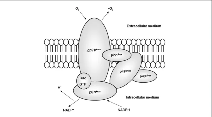
oxidase”)7, and a smaller sub-unit, the p22phox. Besides
these, the enzyme comprises three cytoplasmatic subunits (p47phox, p67phox e p40phox) and a small regulatory protein G (Rac2). Activation of NADPH oxidase begins with serine phosphorylation of the cytoplasmatic subunit p47phox, triggering its migration to the membrane, where, jointly with Rac, gets associated with the cytochrome b558, beginning the enzyme’s catalytic activity1,5,17,37,53-58 (Figure 2).
NADPH oxidase expressed in vascular cells differs from those found in phagocytes, both for its biochemical structure and for its functions5. It was initially found in neutrophils,
which are well-known as essential defenders against micro-organisms released by a combination of several mechanisms, including the generation of •O
2- produced by endothelial
subunits p40phox, p47phox and gp91phox of the NADPH oxidase54. Intriguingly, the catalytic groups gp91phox,
p22phox, p47phox and p67phox were also found in adventitial fibroblasts7,13,22,55.
Identification of subunits homologous to gp91phox resulted in the formation of Nox family (from “nonphagocytic NADPH oxidase”), currently consisting of seven members (Nox1, Nox2 [formally known as gp91phox], Nox3,Nox4, Nox5, Duox1 and Duox2 [dual oxidase])10,56,57. The main
components of enzyme complex NADPH oxidase, Nox1 and Nox4, are highly expressed in vascular cells and increased during the vascular remodeling process, such as in high blood pressure and atherosclerosis7. These slight differences
in structural expression facilitate development of specific inhibitors of the vascular NADPH oxidase which do not compromise the physiological signaling and the phagocytic functions mediated by RONS5.
Several characteristics of the NADPH oxidases are striking. First, the vascular enzyme produces •O
2- in low levels and for
long periods, mostly intracellularly, where it plays the role of cell signalling1,5,14,16,46,50,59 (Figure 3). Second, gp91phox is
usually substituted for homologous Nox1 or Nox4, particularly in smooth muscles. Third, while the homologous Nox and the subunit p22phox connected to the membrane are capital to maintaining a stable unit which supports the electron transfer for generation of •O
2-, the role of cytosolic components
in NADPH oxidase remain blurred, whose last aspect has implications on activation and specificities of the inhibitors of said enzyme1,5,14,16,46. Besides this, the expression of subunits
seems to vary in smooth muscle cells of different vessels. Human vascular smooth muscle cells (HVSMC) seem to express a subunit gp91phox similar to neutrophillic60 subunit.
The homologous of gp91phox, the Nox1, is expressed in rat vascular smooth muscle cells (RVSMC) and the ones of mice61 and aortic smooth muscle cells (ASMC), and do not
appear in human vascular smooth muscle cells60. Nox1 is 56%
homologous to human gp91phox and, jointly with p22phox, is the probable functional component of cytochrome b558 in ASMCs and RVSMC62. Nox4, another homologous of
gp91phox, is expressed in RVSMC, ASMC and HVSMC. Endothelial cells are mainly expressed in subunits Nox2 and Nox4. Expression of Nox1 is less prominent in endothelium and may be increased during leukocyte adhesion process and shearing stress. Nox2 and Nox4 seem to be the most important functional subunits in endothelial cells, contributing similarly in production of reactive species63.
There is also evidence that the NADPH oxidase is found in the cell nucleus, acting in the gene expression. Several groups
Figure 2 -Structure of phagocytic NADPH oxidase. The gp91phox is the NADPH binder with electron transport function in active NADPH oxidase. The extracellular production of •O
identified subunits of NADPH oxidase in internal membranes. However, consequences of the NADPH oxidase activation in between are not clear as of yet20.
Enzyme activation: central role of subunits
p47phox and gp91phox
The most relevant beginning event for the activation of NADPH oxidase involves the phosphorylation of p47phox and the consequent interaction with gp91phox1,16. Bond
between homologous domains of two structures Src produce self-inhibition of bond with p22phox. Such self-inhibiting interaction is lost in phosphorylation, allowing bond between the subunits p47phox and p22phox1, and the activation of
NADPH oxidase is made. Confirming this, a gene deletion of subunit gp91phox provides protection against ischemic stroke in mice, as it helps to prevent hematoencephalic barrier dysfunction64.
NADPH oxidases are activated and regulated by several factors, as mechanical forces, hormones and cytokines, considering the thrombin, the Platelet-Derived Growth Factor (PDGF), the α Tumor Necrosis Factor (TNF-α), the lactosylceramide, the Interleukin-1 and the oxidized LDL stand out among these factors (Figure 1). The last one increases the production of RONS and the expression of Nox4 in human endothelium1,5. Cultivated macrophages accumulate large
amounts of cholesterol when exposed to modified LDL (receptors for LDL are abundant in these cells, indicating that such structures would be involved in the formation of artherosclerotic foam cells)58,65.
In vascular smooth muscle cells, the angiotensin II (Ang II) is a potent stimulator of NADPH oxidase activity. This angiotensinergic peptide is a known aspect in pathogenesis of most cardiovascular disorders, and it may induce formation of
Figure 3 -NADPH oxidase inhibition mechanism by apocynin. The inhibition is only effective in presence of apocynin dimers, a condition which is possible when myeloperoxidase (MPO) is added to vascular cells without such enzyme. Adapted from Touyz, 2008.
•O
2-, partly due to vascular NADPH oxidases5. The activation
mediated by Ang II was first demonstrated by Griendling et al66, underscoring the importance of Ang II concentrations
in the increase of NADPH oxidase’s activities in vascular smooth muscle cells1,15,16. Despite the signaling by Ang II
stimulating NADPH oxidase is not thoroughly studied, PLD, PKC, PLA2, PI3K and thiol oxireductases pathways seem to be involved67-69. In vascular smooth muscle, Src tyrosine
kinase regulates the NADPH oxidase activity stimulated by Ang II, inducing phosphorylation and translocation of subunit p47phox15. Besides this, Src is capital for the effects of Ang II in
the NADPH’s subunits synthesis, stimulating the increase in the expression of gp91phox, p22phox, p47phox and p67phox60. In
addition to this, phosphorylation of Src precedes the activation of ERK1/2 and JNK cascades in vascular smooth muscle, leading to migration, cell growth, apoptosis and deposition of collagen70, activating focal adhesion complexes, and has
important participation in vascular inflammatory response71.
These proliferative and inflammatory pathways provide positive feedback to the activation of NADPH oxidase in the vascular system. In vascular smooth muscle, the EGF receptor transactivation seems to be involved, leading to activation of PI3 kinase and small RacG protein minutes after the receptor AT1 of angiotensin is activated5,11,14,16,20,46,72,73. Ang II acts by
increasing the NADPH oxidase subunits’ expression in hours or days5,11,14,15,46. In animal models proposed by Rajagopalan
et al11, the production of vascular •O
2-, the NADPH oxidase
activity and the level of expression of p22phox, gp91phox, p67phox and Nox1 have increased (there was 100% for the NADPH oxidase).
In the event of activation by Ang II, the H2O2, generated from the •O
2- produced by NADPH oxidase, is capital for the
hypertrophy of vascular smooth muscle cells54, also acting as
Review Article
Rabêlo et al
Role of NADPH oxidase in redox unbalance
beds, both in human and mice, endogenous H2O2, in low concentrations, acts as an endothelium-dependent relaxing factor, promoting peripheral, coronary hyperpolarization and vasodilation, as well as in cerebral arteries74,75. In cultivation
of smooth muscle cells, Ang II induces hypertrophy mediated by H2O2, by means of activation of the expression of proto-oncogenes, MAP kinase and Akt/PKB54. In mice with deficiency
in gene which expresses the gp91phox, the Ang II fails to induce cardiac hypertrophy17. Besides this, experimental evidences
show that the •NO is a chemoattraction factor for angiogenesis,
and inhibiting its production blocks vascular neoformation49.
In addition to this, Ang II also promotes mechanisms involved in inflammatory response, and it may simultaneously stimulate the production of •NO and •O
2- by endothelial cells, favorable
situation to formation of •ONOO-1,13,46,76. Intriguingly, the
activation of NADPH oxidase by Ang II is lessened by Ang-(1-7)12, and biologically active component of renin-angiotensin
system17,25,77. Ang-(1-7) seems to inhibit phosphorylation of
Src induced by Ang II12, promoting endothelium-dependent
vasodilation due to induction of •NO36 release. As for the
humoral activation mechanisms, the aldosterone regulates the MAP kinase and the generation of •O
2- by NADPH oxidase
through c-Src-dependent mechanisms72. Some therapeutic
actions of hypotensor drugs (angiotensin receptor blockers and angiotensin converter enzyme inhibitors) are attributed
to inhibitors of NADPH oxidase, with consequent reduction in production of RONs25. However, there is no record that these
therapeutic strategies which have as target such species may bring direct benefit to control of high blood pressure patients18.
Cardiovascular and neurodegenerative
diseases are related to increased activity of
NADPH oxidase
Clinical studies show that RONS play a significant role in high blood pressure. The production of these species undergoes increase in patients suffering from essential high blood pressure, renovascular hypertension, malignant hypertension and preeclampsia hypertension25. Besides this,
increased production of •O
2- by NADPH oxidase in vessels
is related to risk factors for arteriosclerosis (Fig. 1) and harm to endothelial function in coronary heart disease patients5.
Production of •O
2- appears increased in atheroschlerotic
blood vessels2,40,42, contributing to the beginning of
pro-inflammatory events, with gene transcription regulation of vascular cell adhesion molecules and chemoattractive proteins for monocytes78. Patients suffering from arteriosclerosis present
both endothelial dysfunction and redox unbalance. However, the accurate association mechanism for these two events in
human is unknown as of yet42.
The endothelial dysfunction is related to the increase in the NADPH oxidase activity (by increase of expression of its catalytic subunits14,53,79), ever since atherosclerotic lesions
in human coronaries show strong expression of gp91phox in the vulnerable area of the plaque. Besides this, Nox4 increases during the stage of formation of atheroma, whose appearance happens in a reduced form in advanced lesions. Great saphenous veins and internal mammary arteries of patients with diabetes mellitus present an increase in the NADPH oxidase activity and in decoupled eNOS (in this status, the enzyme acts as producer of •O
2- instead of •NO)
when compared to control group, evincing the relationship between NADPH oxidase and the atherosclerotic lesions14.
Vitamin supplementing may reduce vascular redox unbalance and improve total oxidative status. For instance, some animal models in which antioxidant properties of vitamins C and E are associated to decrease in activation of NADPH oxidase42,80 and increase in SOD activity (the largest
system of cellular defense against •O
2- in vascular system46). It
may be advocated that hydrophobic vitamin E could inhibit the formation of enzyme complex of the NADPH oxidase subunits49. Despite these observations, clinical tests with
antioxidant vitamins have not shown therapeutic benefits as for mortality in cardiovascular events25,46. Besides this, depending
mainly on the posology, such substances may present pro-oxidant properties with harmful organic interactions. As the data from major prospective clinical studies has failed to demonstrate beneficial effects of antioxidants25, a balanced diet
without vitamin supplementing is commonly recommended, ever since potential harms of supplementing are still undefined.
In high blood pressure, the product of the activation of NADPH oxidase, the •O
2-, produces the oxidation of
H4B, leading to an increase in the eNOS decoupling, and the levels of this type of oxygen increase, causing the bioavailability of •NO14 to drop. Thus, H
4B is believed to be
deficient in condition associated with altered endothelial function, such as hypercholesterolemia, diabetes, high blood pressure and tobacco addiction. Stroes et al81 showed that
the treatment with H4B increase vasodilation in human with hypercholesterolemia, insofar the removal of this biopterine implies in eNOS40 decoupling. In support to these findings,
the eNOS superexpression prevents hypoxia-induced pulmonary vascular remodeling52, causing structural changes
and alterations in cellular processes that result in increase of the median layer: vascular lumen ratio82,83.
Another factor implied in the redox unbalance is the endotoxemia. During its progress, release of lipopolysaccharides promotes increase of expression and activity of xanthine oxidase and of NADPH oxidase46. The increase of NADPH
oxidase-dependent RONS in endosomes is important to pro-inflammatory immune response20. Activated neutrophils
produce •O
2-, hydroxyl radical (•OH) and hypochlorous acid
(HOCl), in addition to microbicide peptides and proteases84
responsible for the efficient immune response. Neutrophillic activation in phagocytosis of pneumococci induces the production of RONS by NADPH oxidase. In addition to this, the block of NADPH oxidase activation during acute bacterial meningitis in experimental animals provokes deficient defense
against Streptococcus pneumoniae85.
As for the production of •NO, clinical and experimental
studies have shown that administering L-arginine recovers the synthesis of this radical and the vascular function in several cardiovascular diseases, implying that impairment of precursor amino acids availability is present in these disorders. Reduced levels of L-arginine also promote eNOS decoupling, resulting in increase of RONs34.
•NO-donor drugs (nitrodilators) have been used to treat
coronary heart diseases, high blood pressure and heart failure, having anti-inflammatory actions on vascular wall as suppressors of lipoprotein oxidation, inhibitors of migration and proliferation of vascular smooth muscle cells and platelet aggregation inhibitors. They act as suppressors of the expression of adhesion molecules and chemiokines after the stimulation with proinflammatory factors. Such aspects prevent the inflammatory cell infiltration into the vascular wall, where there is also opposition to pro-inflammatory actions of RONS. In addition to this, it was recently found that •NO
seems to have anti-inflammatory effect of artery wall due to its suppression of RONS generator enzymes, particularly the NADPH oxidase5.
Nox proteins are also related to other countless organic disorders, including cancer, bone reabsorption and Alzheimer Disease, as they are expressed in several tissues1,80,86. Evidences
demonstrate the role of Nox2 in neurodegenerative processes, such as Parkinson’s disease, stroke, cerebral trauma and meningitis, mainly due to neurotoxicity of RONS57,87. Thus, the
Nox inhibitors present significant clinical potential, particularly if they do not inhibit the activity of phagocytes1.
Inhibitors of NADPH oxidase: potential
therapeutic target
Pharmacological inhibitors of NADPH oxidase which block directly the activity of this enzyme have been described as consisting of peptide and non-peptide structures. The DIP-diphenyleneiodonium5,10 and
4’-hydroxy-3-methoxyacetophenone (the apocynin, isolated from the medicinal plant Picrorhiza kurroa)88 has been broadly used to
block the activity of the NADPH oxidase in vitro5. Currently,
the DPI-diphenyleneiodonium is known to be non-selective and act as non-specific oxidant6. In its turn, the apocynin
has multiple biological actions in addition to antioxidant effects5, being characterized as an inhibitor of NADPH
oxidase since the 80’s6. Studies by Godbole et al89 have
demonstrated that the apocynin recovers the production of nitrite and the vasodilation in ordinary femoral arteries of swines89. As the DIP do not stand for a specific inhibitor of
the NADPH oxidase, the apocynin became very popular6,
with 692 records of publications on the PubMed database until January 15, 2009, 571 out of which are specific for the key “apocynin NADPH oxidase inhibition”, demonstrating strong correlation between the terms.
Review Article
Rabêlo et al
Role of NADPH oxidase in redox unbalance
HEK293 cells with no MPO, the formation of apocynin dimers does not occur even with superexpression of Nox isoforms6.
When human MPO is added, dimers may be identified in cells with superexpression of Nox isoforms or in cells co-incubated with H2O2. In absence of H2O2 or Nox, dimer production by HEK293 cells is inhibited. In stimulated leukocytes, dimers are promptly detectable, insofar as in stimulated vascular cells, there is not such production, even in presence of MPO. Thus, the authors suggest that the apocynin would act as anti-oxidant and not as inhibitor of NADPH oxidase in non-phagocytic cells6,88, and the inhibiting action for the NADPH oxidase
would be restricted to leukocytes which express MPO6 (Fig.
3). However, peroxidases other than the MPO may influence the apocynin activity, and it is possible that vascular cells have peroxidative enzymes capable of activating this inhibitor. Such fact is confirmed by findings on endothelial cells, in which apocynin dimers were identified, inhibiting, depending on concentration, the NADPH oxidase activity, the formation of RONS and the cell proliferation88.
Pagano et al90 developed the chimeric peptide gp91ds-Tat,
comprised of nine amino acids of HIV protein coat and nine amino acids of gp91phox1,10. This substance presents reaction
with p47phox and interferes in bond of this subunit with the gp91phox. The “Tat” portion allows the gp91ds-Tat to penetrate into the cell, and, thus, it is effective inhibitor of oxidases which contain gp91phox. However, despite the peptides as the one referred to are useful in experimental interventions, biotransformation processes render its oral administration difficult, limiting its potential in clinical therapeutics1.
Peptidic inhibitors of NADPH oxidase are also described, such as the antibiotic PR-39, endogenously secreted by intestine cells and neutrophils of humans and swines. They act both as immunity cells and as inhibitors to the non-phagocytic NADPH oxidase activity. Initially, the PR-39 was seen as a specific inhibitor of the NADPH oxidase. However, recent studies have shown that the antibiotic presents several effects, partly because it binds to SH3 domains of other proteins and also because it interacts with membrane lipides5.
Conclusion
This study identified that the NADPH oxidase emerged as one of the main structures involved in vascular redox unbalance. As the •NO is also capable of suppressing the
NADPH oxidase activity, this event is believed to present direct implications not only in the development of vascular disease, but also acute recovery of ischemia and reperfusion lesion, in which NADPH-dependent unbalance is of major contribution1,5.
The development of oxidase specific inhibitors, based on Nox units, may experimentally provide tools to enlighten on the role of these enzymes. It may also be useful in the treatment of several diseases1, mainly when it comes to
selective enzyme inhibition aiming to reduce the vascular damage arising out of excessive production of RONS without, nonetheless, compromising the cell signaling mechanism which depend upon said species.
Acknowglements
The authors would like to thank the CNPq, the FAPEAL and the PPSUS-2006/Ministry of Health-FAPEAL for the financial support for this project.
Potential Conflict of Interest
No potential conflict of interest relevant to this article was reported.
Sources of Funding
This study was partially funded by CNPq, PROCAD-NF/ CAPES and FAPEAL.
Study Association
This study is not associated with any post-graduation program.
References
1. Cai H, Griendling KK, Harrison DG. The vascular NAD(P)H oxidases as therapeutic targets in cardiovascular diseases. Trends Pharmacol Sci. 2003; 24: 471-8.
2. Cangemi R, Angelico F, Loffredo L, Ben MD, Pignatelli P, Martini A, et al. Oxidative stress-mediated arterial dysfunction in patients with metabolic syndrome: effect of ascorbic acid. Free Radic Biol Med. 2007; 43: 853-9. 3. Cosentino F, Barker J, Brand MP, Heales SJ, Werner ER, Tippins JR, et al.
Reactive oxygen species mediate endothelium-dependent relaxations in tetrahydrobiopterin-deficient mice. Arterioscler Thromb Vasc Biol. 2001; 21: 496-502.
4. Custodis F, Baumhäkel M, Schlimmer N, List F, Gensch C, Böhm M, et al. Heart rate reduction by ivabradine reduces oxidative stress, improves endothelial function, and prevents atherosclerosis in apolopoprotein e deficient mice. Circulation. 2008; 117: 2377-87.
5. Dusting GJ, Selemidis S, Jiang F. Mechanisms for suppressing NADPH oxidase in the vascular wall. Mem Inst Oswaldo Cruz. 2004; 100 (Suppl.I): 97-103. 6. Heumüller S, Wild S, Barbosa-Sicard E, Schmidt HHHW, Busse R, Schröder K,
et al. Apocynin is not an inhibitor of vascular reduced nicotinamide-adenine dinucleotide phosphate oxidases but an antioxidant. Hypertension. 2008; 51: 1-7.
7. Lassègue B, Clempus RE. Vascular NAD(P)H oxidase: specific features, expression, and regulation. Am J Physiol Regul Comp Physiol. 2003; 285: R277-R297.
8. Moens AL, Takimoto E, Tocchetti CG, Chakir K, Bedja D, Cormaci G, et al. Reversal of cardiac hypertrophy and fibrosis from pressure overload by tetrahydrobiopterin: efficacy of recoupling nitric oxide synthase as a therapeutic strategy. Circulation. 2008; 117: 2626-36.
9. Molnar J, Yu S, Mzhavia N, Pau C, Chereshnev I, Dansky HM. Diabetes induces endothelial dysfunction but does not increase neointimal formation in high-fat diet fed C57BL/6J mice. Circ Res. 2005; 96; 1178-84. 10. Paravicini TM, Touyz RM. NADPH oxidase, reactive oxygen species, and
hypertension: clinical implications and therapeutic possibilities. Diabetes Care. 2008; 31: S170-S180.
Angiotensin II-mediated hypertension in the rat increases vascular superoxide production via membrane NADH/NADPH oxidase activation: contribution to alterations of vasomotor tone. J Clin Invest. 1999; 97: 1916-23.
12. Sampaio WO, Castro CH, Santos RAS, Schiffrin EL, Touyz RM. Angiotensin-(1-7) counterregulates angiotensin II signaling in human endothelial cells. Hypertension. 2007; 50: 1093-8.
13. Schiffrin EL, Touyz RM. Inflammation and vascular hypertrophy induced by angiotensin II: role of NADPH oxidase-derived reactive oxygen species independently of blood pressure elevation? Arterioscler Thromb Vasc Biol. 2003; 23: 707-9.
14. Taniyama Y, Griendling KK. Reactive oxygen species in the vasculature: molecular and cellular mechanisms. Hypertension. 2003; 42: 1075-81. 15. Touyz RM, Yao G, Schiffrin EL. c-Src Induces phosphorylation and translocation
of p47phox: role of superoxide generation by angiotensin II in human vascular smooth muscle cells. Arterioscler Thromb Vasc Biol. 2003; 23: 1-9. 16. Touyz RM, Chen X, Tabet F, Yao G, He G, Quinn MT, et al. Expression of a
functionally active gp91phox-containing neutrophil-type NAD(P)H oxidase in smooth muscle cells from human resistance arteries: regulation by angiotensin II. Circ Res. 2002; 90: 1205-13.
17. Touyz RM, Mercure C, He Y, Javeshghani D, Yao G, Callera GE, et al. Angiotensin II-dependent chronic hypertension and cardiac hypertrophy are unaffected by gp91phox-containing NADPH oxidase. Hypertension. 2005; 45: 530-7.
18. Touyz RM, Schiffrin EL. Reactive oxygen species and hypertension: a complex association. Antioxid Redox Signal. 2008; 10: 1041-4.
19. Ülker S, McMaster D, McKeown PP, Bayraktutan U. Antioxidante vitamins C and E ameliorate hyperglycaemia-induced oxidative stress in coronary endothelial cells. Diabetes, Obes Metab. 2004; 6: 442-51.
20. Ushio-Fukai M. Localizing NADPH oxidase-derived ROS. Sci STKE. 2006; 349: re8.
21. Yokoyama M, Inoue N. How vascular NAD(P)H oxidase activity and nox isoform expression are regulated. Arterioscler Thromb Vasc Biol. 2004; 24: 1540-1.
22. Paravicini TM, Gulluyan LM, Dusting GJ, Drummond GR. Increased NADPH oxidase activity, gp91phox expression, and endothelium-dependent vasorelaxation during neointima formation in rabbits. Circ Res. 2002; 91: 54-61.
23. Baehner RL, Gilman N, Karnovsky ML. Respiration and glucose oxidation in human and guinea pig leukocytes comparative studies. J Clin Invest. 1970; 49: 692-700.
24. Soberman RJ. The expanding network of redox signaling: new observations, complexities, and perspectives. J Clin Invest. 2003; 111: 571-4.
25. Touyz RM. Reactive oxygen species, vascular oxidative stress, and redox signaling in hypertension: what is the clinical significance? Hypertension. 2004; 44: 248-52.
26. Zhang Q, Malik P, Pandey D, Gupta S, Jagnandan D, Chantemele EB, et al. Paradoxical activation of endothelial nitric oxide synthase by NADPH oxidase. Arterioscler Thromb Vasc Biol. 2008; 28: 1627-33.
27. Zhang L, Sheppard OR, Shah AM, Brewer AC. Positive regulation of the NADPH oxidase NOX4 promoter in vascular smooth muscles cells by E2F. Free Radic Biol Med. 2008; 45 (5): 679-85.
28. Furchgott RF, Zawadzki JV. The obligatory role of endothelial cells in the relaxation of arterial smooth muscle by acetylcholine. Nature. 1980; 288: 373-6.
29. Deanfield JE, Halcox JP, Rabelink TJ. Endothelial function and disfunction: testing and clinical relevance. Circulation. 2007; 115: 1285-95.
30. Evora PRB, Pearson PJ, Rodrigues AJ, Viaro F, Schaff HV. Relaxamento dependente do endotélio causado pela poli-L-arginina: implicações sobre a hiperpolarização como mecanismo de vasodilatação. Arq Bras Cardiol. 2003; 80: 621-5.
31. Pereira LMM, Mandarim-de-Lacerda CA. Estudo quantitativo da microcirculação miocárdica na hipertensão arterial por progressiva inibição da síntese do óxido nítrico. Arq Bras Cardiol. 1999; 73: 407-12.
32. Santhanam L, Lim HK, Lim HK, Miriel V, Brown T, Patel M, et al. Inducible NO synthase dependent S-nitrosylation and activation of arginase1 contribute to age-related endothelial dysfunction. Circ Res. 2007; 101: 692-702.
33. Fitzgerald SM, Kemp-Harper BK, Parkington HC, Head GA, Evans RG. Endothelial dysfunction and arterial pressure regulation during early diabetes in mice: roles for nitric oxide and endothelium-derived hyperpolarization factor. Am J Physiol Regul Integr Comp Physiol. 2007; 293: R707-R713. 34. Durante W, Johnson FK, Johnson RA. Arginase: a critical regulator of nitric
oxide synthesis and vascular function. Clin Exp Pharmacol Physiol. 2007; 34: 906-11.
35. Blankenberg S, Rupprecht HJ, Bickel C, Torzewski M, Hasfner G, Tiret L, et al. Glutathione peroxidase 1 activity and cardiovascular events in patients with coronary artery disease. N Engl J Med. 2003; 349: 1605-13. 36. de Haan JB, Witting PK, Stefanovic N, Pete J, Daskalakis M, Kola I, et al. Lack
of the antioxidant glutathione peroxidase-1 does not increase atherosclerosis in C57BL/J6 mice fed a high-fat diet. J Lipid Res. 2006; 47: 1157-67.
37. Touyz RM, Schiffrin EL. Reactive oxygen species in vascular biology: implications in hypertension. Histochem Cell Biol. 2004; 122: 339-52.
38. Adachi T, Weisbrod RM, Pimentel DR, Ying J, Sharov VS, Schöneich C, et al. S-glutathiolation by peroxinitrite activates SERCA during arterial relaxation by nitric oxide. Nat Med. 2004; 10: 1200-7.
39. Nakata S, Tsutsui M, Shimokawa H, Suda O, Morishita T, Shibata K, et al. Spontaneous myocardial infarction in mice lacking all nitric oxide synthase isoforms. Circulation. 2008; 117: 2211-23.
40. Laursen JB, Somers M, Kurz S, McCann L, Warnholtz A, Freeman BA, et al. Endothelial regulation of vasomotion in ApoE-deficient mice: implications for interactions between peroxynitrite and tetrahydrobiopterin. Circulation. 2001; 103: 1282-8.
41. Vanhoutte PM. Endothelium-derived free radicals: for worse and for better. J Clin Invest. 2001; 107: 23-4.
42. Lavi S, Yang EH, Prasad A, Mathew V, Barsness GW, Rihal CS, et al. The interaction between coronary endothelial dysfunction, local oxidative stress, and endogenous nitric oxide in humans. Hypertension. 2007; 51: 127-33.
43. Yu BP. Cellular defenses against damage from reactive oxygen species. Physiol Rev. 1994; 74: 139-62.
44. Yu Z, Zhang J, Wang X, Chen J. Excessive copper induces the production of reactive oxygen species, which is mediated by phospholipase D, nicotinamide adenine dinucleotide phosphate oxidase and antioxidant system. J Integr Plant Biol. 2008; 50 (2): 157-67.
45. Wiggers GA, Peçanha FM, Briones AM, Perez-Girón JV, Miguel M, Vassallo DV, et al. Low-mercury concentrations cause oxidative stress and endothelial dysfunction in conductance and resistance arteries. Am J Physiol Heart Circ Physiol. 2008; 295: H1033-H1043.
46. Wassmann S, Wassman K, Nickenig G. Modulation of oxidant and antioxidant enzyme expression and function in vascular cells. Hypertension. 2004; 44: 381-6.
47. Ülker S, McKeown PP, Bayraktutan U. Vitamins reverse endothelial dysfunction through regulation of eNOS And NADPH oxidase activities. Hypertension. 2003; 41: 534-9.
48. Pacher P, Beckman JS, Liaudet L. Nitric oxide and peroxynitrite in health and disease. Physiol Rev. 2007; 87: 315-424.
49. Chen X, Touyz RM, Park JB, Schiffrin EL. Antioxidant Effects of Vitamins C and E are associated with altered activation of vascular NADPH oxidase and superoxide dismutase in stroke-prone SHR. Hypertension. 2001; 38 [part2]: 606-11.
50. Görlach A, Brandes RP, Nguyen K, Amidi M, Dehghani F, Busse R. A gp91phox containing NADPH oxidase selectively expressed in endothelial cells is a major source of oxygen radical generation in the arterial wall. Circ Res. 2000; 87: 26-32.
51. Hildeman DA, Mitchell T, Kappler T, Marrack P. T cell apoptosis and reactive oxygen species. J Clin Invest. 2003; 111: 575-81.
Review Article
Rabêlo et al
Role of NADPH oxidase in redox unbalance
of pulmonary arteries induced by chronic hypoxia. Br J Pharmacol. 2006; 148: 714-23.
53. Xu P, Costa-Gonçalves AC, Todiras M, Rabelo LA, Sampaio WO, Moura MM, et al. Endothelial dysfunction and elevated blood pressure in mas gene-deleted mice. Hypertension. 2008; 51 [part2]: 574-80.
54. Jankowski A, Grinstein S. A noinvasive fluorimetric procedure for measurement of membrane potential: quantification of the NADPH oxidase-induced despolarization in activated neutrophils. J Biol Chem. 1999; 274: 26098-104.
55. Cifuentes ME, Pagano PJ. c-Src and smooth muscle NAD(P)H oxidase: assembling a path to hypertrophy. Arterioscler Thromb Vasc Biol. 2003; 23: 919-21.
56. Chen K, Kirber MT, Xiao H, Yang Y, Keaney JF Jr. Regulation of ROS signal transduction by NADPH oxidase 4 localization. J Cell Biol. 2008; 181: 1129-39. 57. Schäppi MG, Jaquet V, Belli DC, Krause K. Hyperinflammation in chronic
granulomatous disease and anti-inflammatory role of the phagocyte NADPH oxidase. Semin Immunopathol. 2008; 30: 255-71.
58. Rueckschloss U, Galle J, Holtz J, Zerkowski H, Morawietz H. Induction of NAD(P)H Oxidase by oxidized low-density lipoprotein in human hendothelial cells: antioxidative potencial of hydroxymethylglutaryl coenzyme a reductase inhibitor therapy. Circulation. 2001;104:1767-72. 59. Nathan C. Specificity of a third kind: reactive oxygen and nitrogen
intermediates in cell signaling. J Clin Invest. 2003; 111: 769-78.
60. Touyz RM, Chen X, Tabet F, Yao G, He H, Quinn MT, et al. Expression of a functionally active gp91phox-containing neutrophil-type NAD(P)H oxidase in smooth muscle cells from human resistance arteries: regulation by angiotensin II. Circ Res. 2002; 90: 1205-13.
61. Ambasta RK, Schreiber JG, Janiszewski M, Busse R, Brandes RP. Noxa1 is a central component of the smooth muscle NADPH oxidase in mice. Free Radic Biol Med. 2006; 41 (2): 193-201.
62. Lassègue B, Sorescu D, Szöes K, Yin Q, Akers M, Zhang Y, et al. Novel gp91phox homologues in vascular smooth muscle cells: nox 1 mediates angiotensin-II-induced superoxide formation and redox-sensitive signaling pathways. Cir Res. 2001; 88: 888-94.
63. Petry A, Djordjevic T, Weitnauer M, Kietzmann T, Hess J, Gorlach A. Nox2 and Nox4 mediate proliferative response in endothelial cells. Antioxid Redox Signal. 2006; 8 (9-10): 1473-84.
64. Kahles T, Luedike P, Endres M, Galla HJ, Steinmetz H, Busse R, et al. NADPH oxidase plays a central role in blood-brain barrier damage in experimental stroke. Stroke. 2007; 38: 3000-6.
65. Steinbretcher UP. Oxidation of human low density lipoprotein results in derivatization of lysine residues of apolipoprotein B by lipid peroxide decomposition products. J Biol Chem. 1987; 262: 3603-8.
66. Griendling KK, Minuri CA, Ollerenshaw TD, Alexander RW. Angiotensin II stimulates NADH and NADPH oxidase activity in cultured vascular smooth cells. Circ Res. 1994; 74: 1141-8.
67. Touyz RM, Schiffrin EL. Signal transduction mechanisms mediating the physiological and pathophysiological actions of angiotensin II in vascular smooth muscle cells. Pharmacol Rev. 2000; 52 (4): 639-72.
68. Zafari AM, Ushio-Fukai M, Minieri CA, Akers M, Lassegue B, Griendling KK. Arachidonic acid metabolites mediate angiotensin II-induced NADH/ NADPH oxidase activity and hypertrophy in vascular smooth muscle cells. Antioxid Redox Signal. 1999; 1 (2): 167-79.
69. Janiszewiski M, Lopes LR, Carmo AO, Pedro MA, Brandes RP, Santos CX, et al. Regulation of NAD(P)H oxidase by associated protein disulfide isomerase in vascular smooth muscle cells. J Biol Chem. 2005; 280 (49): 40813-9. 70. Kyam M, Yoshizumi M, Tsuchiya K, Kagami S, Izawa Y, Fujita Y, et al. Src and
Cas are essentially but differentially involved in angiotensin II-stimulated migration of vascular smooth muscle cells via extracellular signa-regulated kinase 1/2 and c-jun NH2-terminal kinase activation. Mol Pharmacol. 2004; 65: 832-41.
71. Gual JL. Role of focal adhesion kinase in integrin signaling. Int J Bioch Cell Biol. 1997; 29: 1085-96.
72. Callera GE, Touyz RM, Tostes RC, Yogi A, He Y, Malkinson S, et al. Aldosterone activates vascular p38MAP kinase and NADPH oxidase via c-Src. Hypertension. 2005; 45: 773-9.
73. Kawahara T, Quinn MT, Lamberth JD. Molecular evolution of the reactive oxygen-generating NADPH oxidase (Nox/Doux) family of enzymes. BMC Evol Biol. 2007; 7: 109.
74. Rabêlo LA, Cortes SF, Alvarez-Leite J, Lemos VS. Endothelium dysfunction in LDL receptor knockout mice: a role for H2O2. Br J Pharmacol. 2003; 138: 1215-20.
75. Thengchaisri N, Shipley R, Ren Y, Parker J, Kuo L. Exercise training restores coronary arteriolar dilation to NOS activation distal to coronary artery occlusion: role of hydrogen peroxide. Arterioscler Thromb Vasc Biol. 2007; 27: 791-8.
76. Marnet LJ, Riggins JN, West JD. Endogenous generation of reactive oxidants and eletrophiles and their reaction with DNA and protein. J Clin Invest. 2003; 111: 583-93.
77. Kucharewicz I, Pawlak R, Matys T, Pawlak D, Buczko W. Antithrombotic effect of captopril and losartan is mediated by angiotensin-(1-7). Hypertension. 2002; 40: 774-9.
78. Tarpey MM, White CR, Suarez E, Richardson G, Radi R, Freeman BA. Chemiluminescent detection of oxidants in vascular tissue: lucigenin but not coeleterazine enhances superoxide formation. Circ Res. 1999; 84: 1203-11. 79. Stocker R, Huang A, Jeranian E, Hou JY, Wu TT, Thomas SR, et al.
Hypochlorous acid impairs endothelium-derived nitric oxide bioactivity through a superoxide-dependent mechanism. Arterioscler Thromb Vasc Biol. 2004; 24: 2028-33.
80. Klein JA, Ackerman SL. Oxidative stress, cell cycle and neurodegeneration. J Clin Invest. 2003; 111: 785-93.
81. Stroes E, Kastelein J, Cosentino F, Erkelens W, Wever R, Koomans H, et al. Tetrahydrobiopterin restores endothelial function in hypercholesterolemia. J Clin Invest. 1997; 99: 41-6.
82. Bahia L, Aguiar LGK, Villela NR, Bottino D, Bouskela E. O endotélio na síndrome metabólica. Arq Bras Endocrinol Metab. 2006; 50 (2): 291-303.
83. Touyz RM. Vascular remodeling, retinal arteries, and hypertension. Hypertension. 2007; 50: 603-4.
84. El-Benna J, Dang PM, Gougerot-Pocidalo M. Priming of neutrophil NADPH oxidase activation: role of p47phox phosphorylation and NOX2 mobilization to the plasma membrane. Semin Immunopathol. 2008; 30: 279-89. 85. Paul R, Obermaier B, Van Ziffle J, Angele B, Pfister HW, Lowell CA, et
al. Myeloid Src kinases regulate phagocytosis and oxidative burst in pneumococcal meningites by activating NADPH oxidase. J Leukoc Biol. 2008; 84 (4): 1141-50.
86. Delwing D, Delwing D, Bavaresco CS, Wyse ATS. Protective effect of nitric oxide synthase inhibition or antioxidants on brain oxidative damage caused by intracerebroventricular arginine administration. Brain Res. 2008; 1193: 120-7.
87. Li B, Guo Y, Sun M, Dong H, Wu S, Wu D, et al. The NADPH oxidase is involved in lipopolysaccharide-mediated motor neuron injury. Brain Res. 2008; 1226: 199-208.
88. Touyz RM. Apocynin, NADPH oxidase and vascular cells: a complex matter. Hypertension. 2008; 51: 172-4.
89. Godbole AS, Lu X, Guo X, Kassab GS. NADPH oxidase has a directional response to shear stress. Am J Physiol Heart Circ Physiol. 2009; 296: H152-H158.