Original Paper
Cell Physiol Biochem 2011;27:305-312 Accepted: March 03, 2011
Cellular Physiology
Cellular Physiology
Cellular Physiology
Cellular Physiology
Cellular Physiology
and Biochemistr
and Biochemistr
and Biochemistr
and Biochemistr
and Biochemistry
yy
yy
Tobacco Smoke Induces Ventricular Remodeling
Associated with an Increase in NADPH Oxidase
Activity
Bruna P.M. Rafacho
1, Paula S. Azevedo
1, Bertha F. Polegato
1, Ana
A.H. Fernandes
2, Maria A. Bertoline
3, Denise C. Fernandes
3,
Fernanda Chiuso-Minicucci
4, Meliza G. Roscani
1, Priscila P. dos
Santos
1, Luiz S. Matsubara
1, Beatriz B. Matsubara
1, Francisco R.M.
Laurindo
3, Sergio A.R. Paiva
1, Leonardo A.M. Zornoff
1and Marcos
F. Minicucci
11Internal Medicine Department, Botucatu Medical School, Univ Estadual Paulista (UNESP), Botucatu, 2Chemistry and Biochemistry Department, Institute of Biological Sciences, Univ Estadual Paulista
(UNESP), Botucatu, 3Vascular Biology Laboratory, Heart Institute (InCor), University of São Paulo School
of Medicine, São Paulo, 4Department of Microbiology and Immunology, Institute of Biosciences, São
Paulo State University, Botucatu
Key Words
Oxidative stress • Tobacco smoke exposure • Cardiac remodeling
Abstract
Background: Recent studies have assessed the direct effects of smoking on cardiac remodeling and function. However, the mechanisms of these alterations remain unknown. The aim of this study was to investigate de role of cardiac NADPH oxidase and antioxidant enzyme system on ventricular remodeling induced by tobacco smoke. Methods: Male Wistar rats that weighed 200-230 g were divided into a control group (C) and an experimental group that was exposed to tobacco smoke for a period of two months (ETS). After the two-month exposure period, morphological, biochemical and functional analyses were performed. Results: The myocyte cross-sectional area and left ventricle end-diastolic
dimension was increased 16.2% and 33.7%, respectively, in the ETS group. The interstitial collagen volume fraction was also higher in ETS group compared to the controls. In addition to these morphological changes, the ejection fraction and fractional shortening were decreased in the ETS group. Importantly, these alterations were related to augmented heart oxidative stress, which was characterized by an increase in NADPH oxidase activity, increased levels of lipid hydroperoxide and depletion of antioxidant enzymes (e.g., catalase, superoxide dismutase and glutathione peroxidase). In addition, cardiac levels of IFN-γ, TNF-α and IL-10 were not different between the groups. Conclusion: Cardiac alterations that are induced by smoking are associated with increased NADPH oxidase activity, suggesting that this pathway plays a role in the ventricular remodeling induced by exposure to tobacco smoke.
Introduction
Exposure to tobacco smoke is a major risk factor for coronary heart disease (CHD), which is currently the leading cause of preventable death. Smoking increases the risk of death from CHD at least two-fold, and increases the risk of stroke by approximately 50% [1, 2]. It has been estimated that 440,000 deaths occur annually in the United States due to smoking [3].
More than 4,720 chemical compounds, in addition
to an estimated 1015-1017 free radicals, are present in
cigarette smoke[4, 5]. These compounds can cause
vascular and tissue inflammation by stimulating inflammatory cells to produce reactive oxygen species (ROS). Initially, ROS production was thought to be an uncontrollable process that was associated with a pathological disease or a process that occurs at high levels (micromolar scale) in phagocytes to kill bacteria. Recently, ROS generation on a smaller scale (nanomolar scale) has been characterized in many cell types as a controlled, agonist-triggered and enzyme-dependent physiological process [6]. Isoforms of the nicotinamide adenine dinucleotide phosphate (NADPH) oxidase complex (Nox family NADPH oxidases) are the main cellular and vascular source of ROS with purposes of compartment-specific redox signaling [5]. NADPH oxidases are highly regulated, transmembrane enzymes present in a variety of phagocytic and nonphagocytic cells in several tissues, such as the vascular endothelium and the heart. NADPH oxidases catalyze the transfer of electrons from NADPH to molecular oxygen, leading to superoxide
anion (O2-) and other derived products [6]. There is
increasing evidence that NADPH oxidase activity is the main source of ROS involved in noxious effects of cigarette smoke [7-9].
In addition to the well-known pathological effects of cigarette smoke on vascular systems, recent studies have also assessed the direct effects of smoking in cardiac remodeling and function. Such studies have shown that tobacco smoke induces left atrium and ventricle enlargement, myocyte hypertrophy and systolic dysfunction [10-14]. The potential mechanisms for these alterations include hemodynamic and neurohormonal changes [15], metalloproteinases [16] and mitogen-activated protein kinases activation [17]. These and other effects may be associated with oxidative stress [18, 19].
It is well known that oxidative stress affects the extracellular matrix, myocyte contractile proteins and
myocyte hypertrophy; this oxidative stress can ultimately cause myocyte death via necrosis or apoptosis. Animals that have been exposed to tobacco smoke undergo cardiac structural changes that can be observed with electron microscopy; interestingly, these changes can be alleviated with beta-carotene supplementation [18]. Rats exposed to cigarette smoke undergoing experimental myocardial infarction show decreased reduced/oxidized glutathione ratios in heart and liver tissue [19]. However, no studies have addressed whether NADPH oxidase participates in the cardiac remodeling induced by smoking.
The aim of this study is to investigate the role of cardiac NADPH oxidases and antioxidant enzyme systems on ventricular remodeling induced by tobacco smoke.
Materials and Methods
All experiments and procedures were performed in concordance with the National Institute of Health’s Guide for the Care and Use of Laboratory Animals and were approved by the Animal Ethics Committee of our institution.
Male Wistar rats that weighed 200-230 g were divided into 2 experimental groups: a control group that was not exposed to cigarette smoke (group C; n=10), and a group that was exposed to cigarette smoke for 2 months (group ETS n=10). Food and water were supplied ad libitum. The rats were observed for a period of 60 days, during which morphological, biochemical and functional analyses were performed. The ETS rats were exposed to cigarette smoke in a chamber (dimensions 95x80x65 cm) that was connected to a smoking device based on a model published by Wang et al. [20] and adapted by Paiva et al. [21]. The smoke was drawn out of filtered commercial cigarettes (composition per unit: 1.1 mg nicotine, 14 mg tar and 15 mg carbon monoxide) with a vacuum pump and exhausted into the smoking chamber. During the first week, the number of cigarettes was gradually increased from 5 to 10 cigarettes over a 30 min period, administered twice each afternoon. From this point on, 10 cigarettes were used for each exposure, and the rats were exposed four times/day, twice in the morning and twice in the afternoon.
Echocardiographic analysis
left lateral position. Targeted 2-D M-mode echocardiograms were obtained from short-axis views of the left ventricle (LV) at or just below the tip of the mitral-valve leaflets, as well as at the level of aortic valve and left atrium. M-mode images of the LV, left atrium and aorta were recorded on a black-and-white thermal printer (Sony Up-890MD) at a sweep speed of 100 mm/s.All of the tracings were manually measured with a caliper by the sameobserver using the leading-edge method recommended by the AmericanSociety of Echocardiography [22]. The data represent the mean of measurements from at least five consecutive cardiac cycles.The LV end-diastolic dimension (LVDD) and posterior wall thickness(LVWT) were measured at maximal diastolic dimension, whereas the end-systolicdimension (LVSD) was measured at maximal anterior motion of the posteriorwall. The left atrium was measured at its maximal diameter, and the aortawas measured at the end of diastole. The LV systolic function was assessed by calculatingthe fractional shortening{(LVDD – LVSD)/LVDD x 100} and the ejection fraction{(LVDD3 – LVSD3)/LVDD3}.
The transmitral diastolic flow (Eand A) velocities were obtained from the apical four-chamber view.The E/A ratio was used as an index of LV diastolic function.
Morphometric analysis
At the completion of the functional analyses, the right and left ventricles (including the interventricular septum) were dissected, separated and weighed. Transverse sections of the LV were fixedin 10% buffered formalin and embedded in paraffin. Five-micron-thick sections were stained with hematoxylin and eosin (HE) or the collagen-specificstain picrosirius red (Sirius red F3BA in aqueous saturated picricacid). The myocyte cross-sectional area was determined for a minimumof 100 myocytes per HE-stained cross section. The measurements were obtained from digital images (400x magnification)that were collected with a video camera that was attached to a Leica microscope; the images were analyzed with the Image-Pro Plus 3.0 software(Media Cybernetics; Silver Spring, MD). The myocyte cross-sectional area was measured with a digital pad, and the selected cells were transversely cut so that the nucleus was in thecenter of the myocyte [21]. The interstitial collagen volume fraction was determined for the entire cardiac section that was stained with picrosirius red by analyzing digital images that were captured under polarized light (200xmagnification). The cardiac tissue components were identifiedaccording to the following staining patterns: red for collagen fibers, yellowfor myocytes and white for interstitial space. The collagen volumefraction was calculated as the sum of all the connective tissue areasdivided by the sum of all the connective tissue and myocyte areas.On average, 35 microscopic fields were analyzed per heartwith a 20x lens. Perivascular collagen was excluded from thisanalysis [21].
Cardiac lipid hydroperoxide and antioxidant enzyme analysis
Left ventricle samples (200 mg) were homogenized in 5 mL of chilled phosphate buffer (0.1 M, pH 7.4) in a motor-driven Teflon glass Potter Elvehjem. The homogenate
was centrifuged at 12,520 x g for 15 min at 0ºC, and supernatant was used for measure total protein as described previously [23], lipid hydroperoxide (LH) through hydroperoxide-mediated oxidation of Fe2+ as previously
published [23], and antioxidants enzyme activities. Glutathione peroxidase (GSHPx, E.C.1.11.1.9), superoxide dismutase (SOD, E.C.1.15.1.1) and catalase (CAT, E.C.1.11.1.6) activity was assessed as previously specified [23-25]. The enzyme activity assays were performed at 25°C with a micro-plate reader (lQuant-MQX 200 with Kcjunior software connected to computer system control, Bio-Tec Instruments, Winooski, Vermont, USA). The absorbance was measured using a Pharmacia Biotech spectrophotometer (UV/visible Ultrospec 5000 with Swift II Applications software connected to computer system control, 974213, Cambridge, England, UK) at 560 nm. All of the reagents were from Sigma (St. Louis, Missouri, USA) [23].
NADPH oxidase activity in cardiac tissue
NADPH oxidase activity was assessed by HPLC analysis of dihydroethidium (DHE) oxidation products. Left ventricle samples (50 mg) were homogenized in lysis buffer (50 mM Tris, pH 7.4, 0.1 mM EDTA, 0.1 mM EGTA, 10 g/ml aprotinin, 10 g/ml leupeptin and 1 mM phenylmethylsulfonyl fluoride); samples were sonicated (3 cycles of 10 s each at 8 W) and centrifuged (18,000 g for 15 min) to separate the mitochondria and nuclei. The supernatants were further centrifuged at 100,000 g for 1 h to obtain a membrane-enriched fraction. HPLC analysis was subsequently used to measure the NADPH oxidase activity of such membrane fraction [6, 26-28].
monitored by fluorescence detection with an excitation wavelength of 510 nm and emission wavelength of 595 nm [6, 28].
NADPH oxidase activity was quantified by comparing the EOH integrated peak areas between the experimental and standard solutions under identical chromatographic conditions [27].
Evaluation of cardiac cytokine production
Cardiac cytokine production was performed as previously described [29-30]. Cytokine levels of IFN-γ, TNF-α and IL-10 in cardiac homogenate were evaluated by ELISA according to manufacturer instructions (R & D Systems, Minneapolis, MN, USA).
Statistical analysis
Statistical comparisons between the groups were performed with a Student’s t-test for parameters with normal distribution. Otherwise, comparisons between groups were made with the Mann-Whitney U test. The data are expressed as the mean ± SD or the median (including the lower quartile and upper quartile). Data analysis was carried out with the SigmaStat software for Windows v2.03 (SPSS Inc, Chicago, IL). P-values less than 0.05 were considered to be statistically significant.
Results
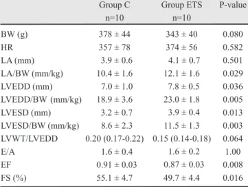
The echocardiographic data are listed in Table 1. The ETS group had enlargement of the left chamber compared to the controls. The body weight (BW)-corrected left atrium (LA) and left ventricle end-diastolic dimension (LVDD) was increased 16.3% and 33.7%, respectively, in the ETS group. In addition to these morphological changes, the ejection fraction (EF) and fractional shortening (FS) were decreased in the ETS group. Rats that were exposed to tobacco smoke had approximately a 10% reduction in FS compared to the control rats (C = 55.1 ± 4.7% and ETS = 49.7 ± 4.4 %; p = 0.016).
The morphological data are listed in Table 2. The BW corrected left ventricular weight (LVW) was elevated in the ETS group, although this difference was not statistically significant. In contrast, the myocyte cross-sectional area (CSA), which is an index of ventricular hypertrophy, was increased 16.2%
in the ETS group (C = 303 ± 28 µm2 and ETS = 352 ± 44
µm2; p = 0.008). The interstitial collagen volume
fraction was also higher in ETS group compared to the controls (C = 2.2 %, range 1.9-2.8% and ETS = 3.1%, range 2.6-3.6%; p = 0.009).
The data describing the cardiac lipid hydroperoxide and antioxidant enzyme activities are listed in Table 3.
Lipid hydroperoxide was increased almost 2-fold in the ETS group compared to the controls (C = 329.8 nmol/g, range 320.8-366.8 nmol/g and ETS = 629.3 nmol/g, range 675.5-741.3 nmol/g; p < 0.001). In addition, cardiac antioxidant enzyme activity (e.g., catalase, superoxide dismutase and glutathione peroxidase) were lower in the ETS group compared to the controls.
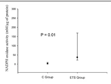
Figure 1 shows the cardiac NADPH oxidase activity, which was increased in the ETS group (n=6) compared to the controls (n=4) (C = 2.42 mM/µg
Group C n=10
Group ETS n=10
P-value
BW (g) 378 ± 44 343 ± 40 0.080
HR 357 ± 78 374 ± 56 0.582
LA (mm) 3.9 ± 0.6 4.1 ± 0.7 0.501 LA/BW (mm/kg) 10.4 ± 1.6 12.1 ± 1.6 0.029 LVEDD (mm) 7.0 ± 1.0 7.8 ± 0.5 0.036 LVEDD/BW (mm/kg) 18.9 ± 3.6 23.0 ± 1.8 0.005 LVESD (mm) 3.2 ± 0.7 3.9 ± 0.4 0.013 LVESD/BW (mm/kg) 8.6 ± 2.3 11.5 ± 1.3 0.003 LVWT/LVEDD 0.20 (0.17-0.22) 0.15 (0.14-0.18) 0.064
E/A 1.6 ± 0.4 1.6 ± 0.2 1.00
EF 0.91 ± 0.03 0.87 ± 0.03 0.008 FS (%) 55.1 ± 4.7 49.7 ± 4.4 0.016
Table 1. Echocardiographic data. C: control animals; ETS: animals exposed to tobacco smoke; BW: body weight; HR: heart rate; LA: left atrium; LV: left ventricle; LVEDD: LV end-diastolic dimension; LVESD: LV end-systolic dimension; LVWT: LV posterior wall thickness; EF: ejection fraction; FS: fractional shortening; E: peak velocity of early ventricular filling; A: peak velocity of transmitral flow during atrial contraction. The data are expressed as the mean ± SD or the median (including the lower quartile and upper quartile).
Table 2. Morphological data. C: control animals; ETS: animals exposed to tobacco smoke; BW: body weight; LVW: left ventricular weight; RVW: right ventricular weight; CSA: cross-sectional area; IC: interstitial collagen volume fraction. The data are expressed as the mean ± SD or the median (including the lower quartile and upper quartile).
n=10 n=10
P-value
BW (g) 378 ± 44 343 ± 40 0.079
LVW/BW (mg/g) 1.94±0.11 2.07±0.16 0.048 RVW/BW (mg/g) 0.52 (0.48-0.55) 0.58 (0.51-0.64) 0.251 CSA (mm2) 303 ± 28 352 ± 44 0.008 IC (%) 2.2 (1.9-2.8) 3.1 (2.6-3.6) 0.009
of protein, range 1.78-7.52 mM/µg and ETS = 37.02 mM/µg of protein, range 24.59-145.45 mM/µg; p = 0.01) In addition, there were no differences of cardiac
levels of IFN-γ (C = 6.89 (5.83-12.16) pg/ml and
ETS = 7.70 (2.99-12.29) pg/ml; p = 0.914), TNF-α
(C = 12.27 (11.44-35.63) pg/ml, ETS = 14.96 (12.46-20.98) pg/ml; p = 0.762) and IL-10 (C = 11.50 (11.09-19.61) pg/ml and ETS = 13.39 (8.90-19.19) pg/ml; p = 0.914 ) between the groups.
Discussion
Our study shows that tobacco smoke exposure induces morphological alterations and systolic dysfunction in rats. Importantly, these alterations were related to augmented heart oxidative stress, which was characterized by increased NADPH oxidase activity, increased levels of lipid hydroperoxide and depletion of antioxidant enzymes.
In accordance with previous reports, our data indicate that tobacco smoke exposure increases the size of the left cardiac chambers, induces myocardial hypertrophy and fibrosis and disrupts systolic function. Changes in the ventricular mass, volume and geometry are characteristic features of ventricular remodeling [31-33]. Although ventricular remodeling initially can be a compensatory process, ventricular remodeling eventually leads to progressive ventricular dysfunction, heart failure and sudden death [31-33].
The mechanisms involved in ventricular remodeling induced by tobacco smoke exposure remain poorly understood. However, a recent study has shown that smoking increases ROS production and oxidative stress, which may lead to vascular remodeling [34]. Overall cellular oxidative burden is regulated by a balance between the rates of ROS generation and a variety of
antioxidant enzymes/pathways, including catalase, superoxide dismutase, glutathione peroxidase, thioredoxin and small molecules such as vitamins. ROS are generally believed to be harmful because they cause oxidative damage to DNA, protein, lipids and other macromolecules [35]. At nanomolar concentrations, these free radicals may play an important role in physiological processes, for example, by functioning as a second messenger in signal transduction pathways. Thus, another, and perhaps the most significant, way excessive ROS can be deleterious is through disruption of signaling networks [6, 36].
In the vascular endothelium, it is well established that cigarette smoke induces ROS production via NADPH oxidase activation [7-9]. Previous studies also suggest that tobacco smoke may induce ventricular remodeling via changes in oxidative stress [18, 19]. However, the role that NADPH oxidase plays in cardiac remodeling that is induced by smoking is unknown.
Fig. 1. The cardiac NADPH oxidase activity, which was increased in the EST group compared to the controls. Table 3. Total protein, lipid hydroperoxide concentration and antioxidant enzyme activity in the cardiac tissue. C: control animals; ETS: animals exposed to tobacco smoke; LH: lipid hydroperoxide; CAT: catalase; GSH-Px: glutathione peroxidase; SOD: superoxide dismutase; TP: total protein. The data are expressed as the mean ± SD or the median (including the lower quartile and upper quartile).
N=6 N=8 P-value
LH (nmol/g tissue) 329.8 (320.8-366.8) 692.3 (675.5-741.3) <0.001
CAT (µmol/mg protein) 1.08 ± 0.18 0.56 ± 0.14 <0.001
GSH-Px (nmol/mg tissue) 37.84 ± 4.64 29.95 ± 4.59 0.008
SOD (nmol/mg of protein) 3.96 (3.83-4.11) 2.30 (1.96-2.95) <0.001
TP (mg/100 mg tissue) 55.68 ± 2.16 46.82 ± 2.71 <0.001
In this study, we found that cigarette smoke exposure induces NADPH oxidase activation in the cardiac tissue. In addition, the cardiac antioxidant enzymes were depleted, and lipid hydroperoxide was increased in rats that were exposed to cigarette smoke. Our data reinforce the role of oxidative stress in cardiac damage induced by smoking; furthermore, the data suggest, for the first time, that NADPH oxidase plays a key role in the ventricular remodeling induced by tobacco smoke exposure.
NADPH oxidase activity was also increased in other models of ventricular remodeling and heart failure [37-39]. Two main isoforms are expressed in the heart - NADPH oxidase 2 and NADPH oxidase 4 [39], and experimental studies suggest that both isoforms may be involved in left ventricular hypertrophy. Additionally, NADPH oxidase 4 appears to be involved in pressure overload hypertrophy [37]. NADPH oxidase activity in left ventricular hypertrophy is modulated by mitogen-activated protein kinase (MAPK) activation. Interestingly, the MAPK pathway is also activated by tobacco exposure [17]. Thus, the myocyte hypertrophy that was present in the rats that were exposed to tobacco smoke may be associated with MAPK activation and increased NADPH oxidase activity.
In addition to the cardiomyocyte, other cell types present in cardiac tissue may have contributed to increased NADPH oxidase activity and ROS generation, such as endothelial and smooth muscle cells from microvessels, adventitial and interstitial fibroblasts and, particularly, infiltrating inflammatory cells. However, in this study there are no alterations in cardiac cytokine profile induced by cigarette smoke exposure, suggesting that inflammatory cells may not contribute to the increased NADPH oxidase activity seen in this model. Whether any of these cell types contributed
to increased ROS should be determined in future studies. Cardiac fibrosis is a hallmark of deleterious cardiac remodeling. In the present study, tobacco smoke exposure increased the interstitial collagen volume fraction. Collagen accumulation is associated with conduction abnormalities and progressive cardiac dysfunction. Although the collagen increase in ETS group was not associated with increased mortality, it may have contributed to left ventricular dysfunction. There is strong evidence that NADPH oxidase is involved in the development of interstitial fibrosis [39, 40]. Indeed, in a pressure overload model, interstitial fibrosis was inhibitedin NADPH oxidase 2-deficient mice, even though the extent of hypertrophy was similar to the controls [40]. Thus, this data suggest that different pathways modulate by NADPH oxidase activity are involved in fibrosis and hypertrophy.
Because NADPH oxidase plays a direct role in pathophysiological cardiac remodeling, tobacco-induced changes in NADPH oxidase activity may influence adverse ventricular remodeling via direct oxidative damage or by activating signal transduction pathways that cause myocyte hypertrophy, cardiac fibrosis and systolic dysfunction. Further studies are needed to elucidate the specific mechanism involved in NADPH oxidase mediated cardiac remodeling in this model.
In conclusion, cardiac alterations caused by smoking are associated with increased NADPH oxidase activity, suggesting that this enzymatic pathway plays a role in the ventricular remodeling induced by tobacco smoke exposure.
Acknowledgements
This study was funded by CAPES (“Coordenação de Aperfeiçoamento de Pessoal de Nível Superior”).
Reference
1 Ockene IS, Miller NH: Cigarette smoking,
cardiovascular disease, and stroke: a statement for healthcare professionals from the American Heart Association. American Heart Association Task Force on Risk Reduction. Circulation 1997;96:3243-3247.
2 Shinton R, Beevers G: Meta-analysis of
relation between cigarette smoking and stroke. BMJ 1989;298:789-794.
3 Andrews JO, Tingen MS: The effect of
smoking, smoking cessation, and passive smoke exposure on common laboratory values in clinical settings: A review of the evidence. Crit Care Nurs Clin N Am 2006;18:63-69.
4 Green CR, Rogman A: The tobacco
5 Yao H, Edirisinghe I, Yang S, Rajendrasozhan S, Kode A, Caito S, Adenuga D, Rahman I: Genetic ablation of NADPH oxidase enhances susceptibility to cigarette smoke-induced lung inflammation and emphysema in mice. Am J Pathol 2008;172:1222-1237.
6 Fernandes DC, Wosniak J Jr, Pescatore
LA: Analysis of DHE-derived oxidation products by HPLC in the assessment of superoxide production and NADPH oxidase activity in vascular systems. Am J Physiol Cell Physiol 2007;292:C413-C422.
7 Jaimes EA, DeMaster EG, Tian RX, Raij
L: Stable compounds of cigarette smoke induce endothelial superoxide anion production via NADPH oxidase activation. Arterioscler Thromb Vasc Biol 2004;24:1031-1036.
8 Orosz Z, Csiszar A, Labinskyy N, Smith
K, Kaminski PM, Ferdinandy P, Wolin MS, Ungvari Z: Cigarette smoke-induced proinflammatory alterations in the endothelial phenotype: role of NAD(P)H oxidase activation. Am J Physiol Heart Circ Physiol 2007;292:H130-H139.
9 Stokes KY: NAD(P)H oxidase: where
there’s smoke, there’s fire. Am J Physiol Heart Circ Physiol 2007;292:H119-H120.
1 0 Brooks WW, Bing OH, Huber GL,
Abermann WH: Contractile performance of rat myocardium after chronic tobacco smoke inhalation. Arch Environ Heath 1982;37:93-97.
1 1 Paiva SAR, Zornoff LAM, Okoshi MP,
Okoshi K, Cicogna AC, Campana AO: Behavior of cardiac variables in animals exposed to cigarette smoke. Arq Bras Cardiol 2003;18:225-228.
1 2 Castardeli E, Paiva SA, Matsubara BB,
Matsubara LS, Minicucci MF, Azevedo PS, Campana AO, Zornoff LA: Chronic cigarette smoke exposure results in cardiac remodeling and impaired ventricular function in rats. Arq Bras Cardiol 2005;84:320-324.
1 3 Castardeli E, Duarte DR, Minicucci MF,
Azevedo PS, Matsubara BB, Matsubara LS, Campana AO, Paiva SA, Zornoff LA: Exposure time and ventricular remodeling induced by tobacco smoke exposure in rats. Med Sci Monit 2008;14:BR62-66.
1 4 Zornoff LA, Matsubara BB, Matsubara
LS, Minicucci MF, Azevedo PS, Campana AO, Paiva SA: Cigarette smoke exposure intensifies ventricular remodeling process following myocardial infarction. Arq Bras Cardiol 2006;86:276-282.
1 5 Tanus-Santos JE, Sampaio RC, Hyslop S,
Franchini KG, Moreno H Jr: Endothelin ET(A) receptor antagonism attenuates the pressor effects of nicotine in rats. Eur J Pharmacol 2000;396:33-37.
1 6 Castardeli E, Duarte DR, Minicucci MF,
Azevedo PS, Matsubara BB, Matsubara LS, Campana AO, Paiva SA, Zornoff LA: Tobacco smoke-induced left ventricular remodelling is not associated with metalloproteinase-2 or -9 activation. Eur J Heart Fail 2007;9:1081-1085.
1 7 Gu L, Pandey V, Geenen DL, Chowdhury
SAK, Piano MR: Cigarette smoke-induced left ventricular remodelling is associated with activation of mitogen-activated protein kinases. Eur J Heart Fail 2008;10:1057-1064.
1 8 Zornoff LA, Matsubara LS, Matsubara
BB, Okoshi MP, Okoshi K, Dal Pai-Silva M, Carvalho RF, Cicogna AC, Padovani CR, Novelli EL, Novo R, Campana AO, Paiva SA: Beta-carotene supplementation attenuates cardiac remodeling induced by one-month tobacco-smoke exposure in rats. Toxicol Sci 2006;90:259-266.
1 9 Duarte DR, Minicucci MF, Azevedo PS,
Matsubara BB, Matsubara LS, Novelli EL, Paiva SA, Zornoff LA: The role of oxidative stress and lipid peroxidation in ventricular remodeling induced by tobacco smoke exposure after myocardial infarction. Clinics 2009;64:691-697.
2 0 Wang X-D, Liu C, Bronson RT, Smith
DE, Krinsky NI, Russel RM: Retinoid signaling and activator protein-1 expression in ferrets given b-carotene supplements and exposure to tobacco smoke. J Natl Cancer Inst 1999;91:60-66.
2 1 Paiva SAR, Zornoff LAM, Okoshi MP,
Okoshi K, Matsubara LS, Matsubara BB, Cicogna AC, Campana AO: Ventricular remodeling induced by retinoic acid supplementation in adult rats. Am J Physiol 2003;284:H2242-H2246.
2 2 Lang RM, Bierig M, Devereaux RB,
Flachskampf FA, Foster E, Pellikka PA, Picard MH, Roman MJ, Seward J, Shanewise JS, Solomon SD, Spencer KT, Sutton MS, Stewart WJ, Chamber Quantification Writing Group: Recommendations for chamber quantification: a report from the American Society of Echocardiography’s Guidelines and Standards Committee and the Chamber Quantification Writing Group, developed in conjunction with the European Association of Echocardiography, a branch of the European Society of Cardiology. J Am Soc Echocardiogr 2005;18:1440-1463.
2 3 Burneiko RCM, Diniz YS, Galhardi CM,
Okoshi K, Matsubara LS, Matsubara BB, Cicogna AC, Campana AO: Interaction of hypercaloric diet and physical exercise on lipid profile, oxidative stress and antioxidant defenses. Food Chem Toxicol 2006;44:1167-1172.
2 4 Nakamura W, Hosoda S, Hayashi K:
Purification and properties of rat liver glutathione peroxidase. Biochem Biophys Acta 1974;358:251-261.
2 5 Ewing JF, Janero DR: Microplate
superoxide dismutase assay employing a nonenzimatic superoxide generator. Anal Biochem 1995;232:243-248.
2 6 Zhao H, Kalivendi S, Zhang H, Joseph J,
Nithipatikom K, Vasquez-Vivar J, Kalyanaraman B: Superoxide reacts with hydroethidine but forms a fluorescent product that is distinctly different from ethidium: potential implications in intracellular fluorescence detection of superoxide. Free Radic Biol Med 2003;34:1359-1368.
2 7 Zielonka J, Zhao H, Xu Y, Kalyanaraman
B: Mechanistic similarities between oxidation of hydroethidine by Fremy’s salt and superoxide: stopped-flow optical and EPR studies. Free Radic Biol Med 2005;39:853-863.
2 8 Laurindo FR, Fernandes DC, Santos CX:
Assessment of superoxide production and NADPH oxidase activity by HPLC analysis of dihydroethidium oxidation products. Methods Enzymol 2008;441:237-260.
2 9 Azevedo PS, Minicucci MF,
Chiuso-Minicucci F, Justulin LA Jr, Matsubara LS, Matsubara BB, Novelli EL, Seiva L, Ebaid G, Campana AO, Zornoff LAM, Paiva SAR: Ventricular remodelling induced by tissue vitamin A deficiency in rats. Cell Physiol Biochem 2010;26:395-402.
3 0 Minicucci MF, Azevedo PS, Oliveira SA
Jr, Martinez PF, Chiuso-Minicucci F, Polegato BF, Justulin LA Jr, Matsubara LS, Matsubara BB, Paiva SA, Zornoff LA: Tissue vitamin A insufficiency results in adverse ventricular remodeling after experimental myocardial infarction. Cell Physiol Biochem 2010;26:523-530.
3 1 Pfeffer MA, Braunwald E: Ventricular
remodeling after myocardial infarction: experimental observations and clinical implications. Circulation 1990;81:1161-1172.
3 2 Cohn JN, Ferrari R, Sharpe N: Cardiac
remodeling- concepts and clinical implications: a consensus paper from an international forum on cardiac remodeling. J Am Coll Cardiol 2000;35:569-582.
3 3 Zornoff LAM, Paiva SAR, Duarte DR,
3 4 Minicucci MF, Azevedo PS, Paiva SA, Zornoff LA: Cardiovascular remodeling induced by passive smoking. Inflamm Allergy Drug Targets 2009;8:334-339.
3 5 Giordano FJ: Oxygen, oxidative stress,
hypoxia, and heart failure. J Clin Invest 2005;115:500-508.
3 6 Droge W: Free radicals in the
physiological control of cell function. Physiol Rev 2002;82:47-95.
3 7 Nakagami H, Takemoto M, Liao JK:
NADPH oxidase derived superoxide anion mediates angiotensin II-induced cardiac hypertrophy. J Mol Cell Cardiol 2003;35:851-859.
3 8 Li JM, Gall NP, Grieve DJ, Chen M, Shah
AM: Activation of NADPH oxidase during progression of cardiac hypertrophy to failure. Hypertension 2002;40:477-484.
3 9 Nabeebaccus A, Zhang M, Shah AM:
NADPH oxidases and cardiac remodeling. Heart Fail Rev 2011;16:5-12.
4 0 Johar S, Cave AC, Narayanapanicker A,