1 8 Arq Bras Oftalmol. 2013;76(1):18-20
Artigo Original |
Original articlePanretinal photocoagulation versus intravitreal injection retreatment pain in
high-risk proliferative diabetic retinopathy
Dor em panfotocogulação retiniana versus injeção intravítrea em pacientes com
retinopatia diabética proliferativa de alto risco
Célia Regina FaRiasde aRaújo luCena1,josé aFonso Ramos Filho1,andRé máRCio VieiRa messias1,josé apaReCidoda silVa2,Felipe piaCentini paesde almeida1, ingRid uRsula sCott3, jeFFeRson augusto santana RibeiRo1,4, RodRigo joRge1
Submitted for publication: July 18, 2012 Accepted for publication: November 21, 2012
Study carried out at School of Medicine of Ribeirão Preto, Universidade de São Paulo.
1 Physician, Department of Ophthalmology, Ribeirão Preto School of Medicine, Universidade de São
Paulo - USP - Ribeirão Preto, SP, Brazil;
2 Physician, Department of Psychology, Faculty of Philosophy, Sciences and Literature, Universidade
de São Paulo - USP - São Paulo, SP, Brazil;
3 Physician, Departments of Ophthalmology and Public Health Sciences, Penn State College of
Medicine, Hershey, PA, USA;
4 Physician, School of Health Sciences, Universidade Estadual do Amazonas - UEA - Manaus, AM,
Brazil.
Funding: Supported by CNPq: Grant number: 306692/2008-2.
Disclosure of potential conflicts of interest: C.R.F.A.Lucena, None; J.A.Ramos Filho, None; A.M.V.Messias, None; J.A.Silva, None; F.P.P.Almeida, None; I.U.Scott, None; J.A.S.Ribeiro, None; R.Jorge, None.
Corresponding author: Rodrigo Jorge. Divisão de Oftalmologia, Faculdade de Medicina de Ribeirão Preto. Avenida Bandeirantes, 3900 - Ribeirão Preto (SP) - 14049-900 - Brazil
E-mail: [email protected]
The present study was part of a master degree thesis from C. R. Lucena.
Review Board approval: 11976/2008.
Clinical Trial register: NCT01009021.
ABSTRACT
Purpose: To compare pain related to intravitreal injection and panretinal photo-coagulation in the management of patients with high-risk proliferative diabetic retinopathy.
Methods: Prospective study including patients with high-risk proliferative diabetic retinopathy and no prior laser treatment randomly assigned to receive panretinal photocoagulation (PRP group) or panretinal photocoagulation plus intravitreal ranibizumab (PRPplus group). In all patients, panretinal photocoagulation was administered in two sessions (weeks 0 and 2), and intravitreal ranibizumab was administered at the end of the first laser session in the PRPplus group. Retreatment was performed at weeks 16 and 32 if active new vessels were detected at fluores-cein angiography. Patients in the PRPplus group received intravitreal ranibizumab and patients in the PRP group received 500-μm additional spots per quadrant of
active new vessels. After the end of retreatment, a 100-degree Visual Analog Scale
was used for pain score estimation. The patient was asked about the intensity of pain during the whole procedure (retinal photocoagulation session or intravitreal ranibizumab injection). Statistics for pain score comparison were performed using a non-parametric test (Wilcoxon rank sums).
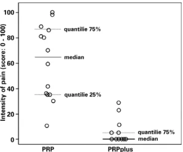
Results: Seventeen patients from PRPplus and 14 from PRP group were evaluated for pain scores. There were no significant differences between both groups re-garding gender, glycosylated hemoglobin and disease duration. Mean intravitreal injection pain (±SEM) was 4.7 ± 2.1 and was significantly lower (p<0.0001) than mean panretinal photocoagulation pain (60.8 ± 7.8). Twelve out of 17 patients
from the PRPplusgroup referred intensity pain score of zero, while the minimal
score found in PRP group was found in one patient with 10.5.
Conclusion: In patients with highrisk proliferative diabetic retinopathy who nee -ded retreatment for persistent new vessels, there was more comfort for the patient when retreatment was performed with an intravitreal injection in comparison with retinal photocoagulation. Further larger studies are necessary to confirm our preliminary findings.
Keywords: Pain; Intravitreal injections; Diabetic retinopathy; Light coagulation; Vascular endothelial growth factor A
RESUMO
Objetivo: Comparar a dor relacionada à injeção intravítrea e panfotocoagulação no tratamento de pacientes com retinopatia diabética proliferativa de alto risco. Métodos: Estudo prospectivo incluindo pacientes com retinopatia diabética prolife-rativa de alto risco e nenhum tratamento a laser prévio aleatoriamente designados para receber panfotocoagulação retiniana (grupo PRP) ou panfotocoagulação e ra nibizumabe intravítreo (grupo PRPplus). Em todos os pacientes, a panfotocoagula-ção foi administrada em duas sessões (semanas 0 e 2), e ranibizumabe intravítreo foi administrado no final da primeira sessão de laser no grupo PRPplus. Retratamento foi realizado nas semanas 16 e 32 se neovasos ativos fossem detectados na angiofluores-ceinografia, utilizando ranibizumabe intravítreo no grupo PRPplus e laser adicional grupo PRP. Após o fim do retratamento, uma Escala Analógica Visual de 100-unidades foi utilizada para a estimativa da pontuação da dor. O paciente foi questionado sobre a intensidade da dor durante todo o procedimento (sessão de fotocoagulação de retina ou injeção intravítrea de ranibizumabe). A comparação dos índices de dor foi realizada utilizando um teste não-paramétrico (Wilcoxon rank sums).
Resultados: Dezessete pacientes do grupo PRPplus e 14 do grupo PRP foram avaliados para os índices de dor. Não houve diferenças significativas entre os dois grupos quanto ao sexo, hemoglobina glicosilada e duração da doença. A média de dor da injeção intravítrea (±SEM) foi 4,7 ± 2,1, significativamente menor (p<0,0001) do que a dor média da panfotocoagulação (60,8 ± 7,8). Doze dos 17 pacientes do grupo PRPplus referiram pontuação de intensidade da dor zero, enquanto que o índice mínimo no grupo PRP foi encontrado em um paciente com 10,5.
Conclusão: Em pacientes com retinopatia diabética proliferativa de alto risco que necessitaram de retratamento por neovasos persistentes, houve mais conforto para o paciente quando o retratamento foi realizado com uma injeção intravítrea em com -paração com fotocoagulação da retina. Estudos posteriores são necessários para confirmar nossos achados preliminares.
Almeida PP, et al.
1 9
Arq Bras Oftalmol. 2013;76(1):18-20
INTRODUCTION
Retinal new vessels (NV) represent an important risk factor for se vere vision loss in patients with diabetes mellitus(1). About 60% of patients with proliferative diabetic retinopathy (PDR) respond to panretinal photocoagulation (PRP) with regression of
neovasculari-zation within 3 months(2). However, many patients require additional
laser treatment, and 4.5% ultimately require pars plana vitrectomy
despite PRP(3).
Besides additional laser photocoagulation, intravitreal injection of anti-vascular endothelial growth factor (VEGF) agents have beco-me an interesting alternative for new vessels regression(4,5). However, both procedures (PRP and injection) may induce discomfort and pain. Pain regarding PRP treatment is a well-recognized concern and
may even prevent its completion(6), while pain regarding intravitreal
injection may disturb patient compliance and peribulbar block may be necessary for uncooperative patients(7). Since intravitreal injection has become a trendy alternative therapeutic strategy and PRP is the standard of care for high-risk PDR, we decided to compare pain related to both procedures in the management of patients with PDR.
METHODS
The study protocol adhered to the tenets of the Declaration of Hel sinki and was approved by the local Institutional Review Board, and all patients gave written informed consent before entering the study.
This is a prospective, open label, randomized study in which two
groups of diabetic patients were followed(5). Between February 2009
and December 2009, all patients evaluated at the Retina and Vitreous Section of the Department of Ophthalmology, School of Medicine of Ribeirão Preto, who presented with high-risk PDR and had not received any prior retinal laser treatment were invited to participate in the study.
Patients were included if they had high-risk PDR, according to
Early Treatment Diabetic Retinopathy Study (ETDRS) guidelines(1,8),
as follows: 1) presence of NV at the disc (NVD) greater than ETDRS standard photograph 10A, or; 2) presence of NVD associated with vitreous or pre-retinal hemorrhage, or; 3) NV elsewhere (NVE) cove-ring more than a half disc area associated with vitreous or pre-retinal hemorrhage. Exclusion criteria included: 1) history of prior laser treat ment or vitrectomy in the study eye; 2) history of thromboem-bolic event (including myocardial infarction or cerebral vascular ac cident); 3) major surgery within the prior 6 months or planned within the next 28 days; 4) uncontrolled hypertension (according to guidelines of the seventh report of the joint National Committee on Preven tion, Detection, Evaluation, and Treatment of High Blood
Pressure [JNC-7])(9); 5) known coagulation abnormalities or current
use of anticoagulation medication other than aspirin; or 6) any condition affecting documentation or follow-up.
During the study enrollment period, high-risk PDR was identiied in one eye of 5 patients and in both eyes of 35 patients based on clinical examination and conirmed on luorescein angiography. At baseline, each patient received a detailed ophthalmologic examina-tion including measurement of the logarithm of the minimum angle of resolution (logMAR) ETDRS and best corrected visual acuity (BCVA) according to a standardized refraction protocol (using modiied ETDRS charts 1, 2, and R), as well as applanation tonometry, dilated slit lamp biomicroscopic examination (including grading of lenticular opacities using the Lens Opacities Classification System III)(10), and binocular indirect fundoscopic examination. Digital red free fundus photography and luorescein angiography were performed using two fundus camera systems (HRA-OCT, Heidelberg, Germany/TRC-50IA/ IMAGEnet; Topcon, Tokyo, Japan).
All patients received PRP, which was performed in two sessions (at
week 0 and week 2) according to ETDRS guidelines(11). Six hundred to
eight hundred 500µm spots were performed per session, at the dis-cretion of the treating investigator. If patients had clinically signiicant
macular edema(12) macular focal/grid laser was performed during
the irst PRP session. Patients could be retreated with focal/grid laser at the 16 and 32-week study visits. If both eyes were eligible for the study, the eye with best visual acuity was included.
Patients were enrolled in groups of two. The technician was asked to pick up one of two identical opaque envelopes, one contai-ning the designation for PRP, whereas the other contained the de-signation for PRP/intravitreal ranibizumab injection (IVR) treatment. The second patient was automatically assigned with the second en velope. For the 20 eyes selected to receive the combined treat-ment (PRP plus intravitreal ranibizumab), one intravitreal injection of 0.5 mg (0.05 ml) of ranibizumab was performed approximately 60 minutes after the completion of the first PRP session (week 0) by a single retinal specialist. Ranibizumab was injected into the vitreous cavity via a 29 gauge needle inserted through the inferotempo-ral pars plana 3.0 - 3.5 mm posterior to the limbus using topical propa racaine drops under sterile conditions (eyelid speculum and
povidone-iodine)(13). Patients were instructed to instill one drop of
0.3% ciprofloxacin into the injected eye four times daily for 1 week after the procedure.
Retreatment was performed at weeks 16 and 32 if active new vessels were detected at luorescein angiography. Patients in the PRPplus group received IVR and patients in the PRP group received 500-μm additional spots per quadrant of active new vessels.
Fifteen minutes after the end of retreatment (PRP session or
re-treatment IVR injection),a masked examiner used a 100-degree Visual
Analog Scale (VAS) for pain score estimation(7). The numbers of the
scale were visible only on the examiner’s side, so that patients could not choose the same number to guide pain scores. Prior to rating level of pain, each patient was asked to slide the marker along the entire scale, with the aid of the examiner. At point 0, the examiner clariied to the patient that this point of the scale represented “no pain at all”; at point 100, the examiner clariied to the patient that this point of the scale represented “the most intense pain one could ever feel”. The patient was asked about the intensity of pain during the whole procedure (PRP session or IVR injection). Statistics for pain score comparison were performed using a non-parametric test (Wil-coxon rank sums).
RESULTS
Seventeen patients from the PRP plus patients and 16 patients from the PRP groups completed the 16-week visit, but 17 patients from PRPplus and 14 from PRP were evaluated for pain scores. Pa-tients’ demographics are summarized in table 1. Mean age of PRP patients was signiicantly higher than PRPplus patients; there were no signiicant diferences between both groups regarding gender, glycosylated hemoglobin (HbA1c) and disease duration. Mean ± SD age (years) was 63.5 ± 8.9 and 51.1 ± 11.3 (p=0.0018); mean ± SD HbA1c (%) was 9.3 ± 1.1 and 9.1 ± 0.8 (p=0.5391); and mean ± SD di -sease duration (years) was 12.9 ± 8.8 and 14.7 ± 6.9 (p=0.5326) for PRP and PRPplus respectively. Mean IVR pain (±SEM) was 4.7 ± 2.1 and was signiicantly lower (p<0.0001) than mean PRP pain (60.8 ± 7.8). Twelve
out of 17 patients from the PRPplusgroup referred intensity pain
score of zero, meaning that they had no pain at all during intravitreal injection, while the minimal score found in PRP group was found in one patient with 10.5, but no other patient in this group had score under 30. Figure 1 shows the distribution of intensity pain scores in both groups; there was a statistically signiicant diference between groups (P<0.0001; Wilcoxon).
DISCUSSION
Panretinal photocoagulation versus intravitreal injection retreatment pain in high-risk proliferative diabetic retinopathy
2 0 Arq Bras Oftalmol. 2013;76(1):18-20
CONCLUSION
In addition to the beneicial efects regarding BCVA and new vessels regression of PRPplus ranibizumab in comparison to PRP alone for high-risk PDR treatment reported elsewhere, there is also more comfort for the patient when retreatment is performed with an intravitreal injection. Further larger studies are necessary to conirm our preliminary indings.
REFERENCES
1. Fundus photographic risk factors for progression of diabetic retinopathy. ETDRS report number 12. Early Treatment Diabetic Retinopathy Study Research Group. Oph thalmology. 1991;98(5 Suppl):823-33.
2. Vander JF, Duker JS, Benson WE, Brown GC, McNamara JA, Rosenstein RB. Long-term stability and visual outcome after favorable initial response of proliferative diabetic retinopathy to panretinal photocoagulation. Ophthalmology. 1991;98(10):1575-9. 3. Flynn HW Jr, Chew EY, Simons BD, Barton FB, Remaley NA, Ferris FL 3rd. Pars plana
vitrectomy in the Early Treatment Diabetic Retinopathy Study. ETDRS report number 17. The Early Treatment Diabetic Retinopathy Study Research Group. Ophthalmology. 1992;99(9):1351-7.
4. Jorge R, Oliveira RS, Messias A, Almeida FP, Strambe ML, Costa RA, et al. Ranibizu-mab for retinal neovascularization. Ophthalmology [Internet]. 2011[cited 2012 Jan 21];118(5):1004. Available from: http://www.sciencedirect.com/science/article/pii/ S0161642010013783
5. Ramos-Filho JA, Messias A, Almeida FP, Ribeiro JA, Costa RA, Scott IU, et al. Panretinal photocoagulation (PRP) versus PRP Plus intravitreal ranibizumab for high-risk proli-ferative diabetic retinopathy. Acta Ophthalmol Scand [Internet]. 2011[cited 2012 Jun 21];89(7):e567-72. Available from: http://onlinelibrary.wiley.com/doi/10.1111/j.1755-3768.2011.02184.x/full
6. De Faria Rodrigues A, Messias AM, Da Silva JA, Ribeiro JA, Jorge R, Scott IU. Diclofenac for panretinal photocoagulation pain. Ophthalmology [Internet]. 2010[cited 2012 Jun 21];117(12):2441.e1-3. Available from: http://www.sciencedirect.com/science/article/ pii/S0161642010006093
7. Cintra LP, Lucena LR, Da Silva JA, Costa RA, Scott IU, Jorge R. Comparative study of analgesic efectiveness using three diferent anesthetic techniques for intravitreal injection of bevacizumab. Ophthalmic Surg Lasers Imaging. 2009;40(1):13-8. 8. Four risk factors for severe visual loss in diabetic retinopathy. The third report from
the diabetic retinopathy study. The Diabetic Retinopathy Study Research Group. Arch Ophthalmol [Internet]. 1979[cited 2010 Jun 12];97(4):654-5. Available from: http:// archopht.jamanetwork.com/article.aspx?articleid=632929
9. Chobanian AV, Bakris GL, Black HR, Cushman WC, Green LA, Izzo JL Jr, Jones DW, Ma-terson BJ, Oparil S, Wright JT Jr, Roccella EJ; National Heart, Lung, and Blood Institute Joint National Committee on Prevention, Detection, Evaluation, and Treat ment of High Blood Pressure; National High Blood Pressure Education Program Coordinating Com-mittee. The Seventh Report of the Joint National Committee on Prevention, Detection, Evaluation, and Treatment of High Blood Pressure: the JNC 7 report. JAMA [Internet]. 2003 [cited 2010 Jun 21];289(19):2560-72. Erratum in: JAMA. 2003;290(2):197. Comment in: JAMA. 2003;290(10):1313; author reply 1314-5; JAMA. 2003;289(19):2573-5; JAMA. 2003;290(10):1312; author reply 1314-5; JAMA. 2003; 290(10):1313; author reply 1314-5. Available from: http://jama.jamanetwork.com/article.aspx?articleid=196589 10. Chylack LT Jr, Wolfe JK, Singer DM, Leske MC, Bullimore MA, Bailey IL, et al. The lens
opacities classiication system III. The Longitudinal Study of Cataract Study Group. Arch Ophthalmol [Internet]. 1993[cited 2010 Oct 12];111(6):831-6. http://archopht. jamanetwork.com/article.aspx?articleid=640261
11. Techniques for scatter and local photocoagulation treatment of diabetic retinopathy: Early Treatment Diabetic Retinopathy Study Report n. 3. The Early Treatment Diabetic Retinopathy Study Research Group. Int Ophthalmol Clin. 1987;27(4):254-64. 12. Photocoagulation for diabetic macular edema. Early Treatment Diabetic Retinopathy
Study report number 1. Early Treatment Diabetic Retinopathy Study Research Group. Arch Ophthalmol [Internet]. 1985[cited 2010 Apr 23];103(12):1796-806.Available from: http://archopht.jamanetwork.com/article.aspx?articleid=635820
13. Ribeiro JA, Messias AM, Scott IU, Jorge R. Alternative technique for reducing compound waste during intravitreal injections. Arq Bras Oftalmol [Internet]. 2009 [cited 2010 Dec 21];72(5):641-4. Available from: http://www.scielo.br/pdf/abo/v72n5/08.pdf 14. Catananti C, Gambassi G. Pain assessment in the elderly. Surg Oncol [Internet].
2010[cited 2011 Sep 21];19(3):140-8.Available from: http://www.sciencedirect.com/ science/article/pii/S0960740409001261
is less painful than a PRP session for diabetic patients with high-risk PDR. In fact, pain score associated with intravitreal injections of an-ti-VEGF agents in patients with diabetic retinopathy and neovascular
age-related macular degeneration (ARMD) ranges from 0 to 22(7),
while pain level associated with a PRP session ranges from 10 to 90(6). This diference may be explained by the longer pain stimulus during a PRP session. Another hypothesis is that there may be direct stimula-tion of the long ciliary nerves during a PRP session (especially if laser applied in the retina periphery at 3 and 9 o’clock meridians) usually merely under topical anesthesia for PRP contact lens positioning, while during intravitreal injections there would be needle conjunc-tival and uveal nociceptors stimulation, usually partially blocked by topical anesthesia(7).
Table 1. Patient demographics
PRP PRPplus P
Age (mean ± SD; in years) 63.5 ± 8.9 51.1 ± 11.3 0.0018 (t-Test)
Gender (Male/Female) 9/5 10/7 0.7557
(Likelihood Ratio)
Race (Black/Hispanic/Caucasian) 2/7/5 2/8/7 0.9461 (Likelihood Ratio)
Duration of diabetes (mean ± SD; in years)
12.9 ± 8.8 14.7 ± 6.9 0.5326 (t-Test)
HbA1c (mean ± SD) 09.3 ± 1.1 09.1 ± 0.8 0.5391 (t-Test)
HbA1c= glycosylated hemoglobin; PRP= panretinal photocoagulation; PRPplus= panretinal photocoagulation and intravitreal injection of ranibizumab; SD= stan-dard deviation.