IntroductIon
Lithium has been used therapeutically for almost 150 years. In the nineteenth century, Garrod and Hammond advocated the use of lithium salts for the treatment of gout and uric acid nephrolithiasis. Almost a century later, Cade, after observing the calming effect of lithium on guinea pigs, reported highly successful results in 10 manic patients who received the drug in 19491. However, in that same year, the
Food and Drug Administration (FDA) withdrew the drug from the U.S. market following the death of several patients due to lithium intoxication. Those patients, with heart failure or hypertension, received lithium chloride salts as a substitute for table salt. Therefore, studies on the effects of lithium as a mood stabilizer progressed slowly and only in 1970 the FDA approved its use in the treatment of mania1.
Lithium is now the drug of choice for the treatment of bipolar disorder, preventing recurrence and suicide attempts. Its use has successfully caused a dramatic decrease in manic and depressive symptoms in 70 to 80% of patients2.
However, despite such great success, lithium has a narrow therapeutic index, with therapeutic serum levels between 0.6 and 1.5 mEq/L3,4. In the initial phase of treatment, excessive
thirst, nausea, diarrhea, epigastric pain, muscle weakness and fatigue are symptoms that cause great discomfort to patients, often leading to low adherence to medication3.
The most common neurologic symptom is tremor, affecting
25-50% of users. Lithium can also often cause flattening or inversion of T waves on electrocardiogram (ECG), but clinically important cardiovascular effects are rare, except in cases of intoxication1. Symptoms of toxicity generally
correlate with serum lithium concentration in acute intoxi-cation, but may vary in patients on chronic lithium therapy5,
leading to the need for hemodialysis at severe levels1. Risk
factors for the development of lithium intoxication include advanced age, overdose, renal failure, drugs that affect renal function (nonsteroidal anti-inflammatory drugs [NSAIDs], angiotensin-converting enzyme [ACE] inhibitors, thiazide diuretics), decreased circulating blood volume (cirrhosis, congestive heart failure, nephrotic syndrome), decreased dietary sodium intake, diabetes mellitus, and diabetes insi-pidus induced by chronic lithium therapy4.
Lithium-induced nephrotoxicity is a complication known since lithium was introduced in the treatment of mood disorders6-8. Renal complications include impairment of
tubular function (particularly the development of nephro-genic diabetes insipidus and renal tubular acidosis) and progressive chronic kidney disease (secondary to lithium-induced chronic tubulointerstitial nephritis)7.
Hyperpara-thyroidism and hypercalcemia are also adverse effects of chronic lithium therapy. This review aimed to discuss clinical and laboratory alterations, pathophysiology and treatment of lithium-induced nephrotoxicity.
*Correspondence:
Rua Vicente Linhares, 1198
Fortaleza - CE,Brazil CEP: 60135-270 Phone: (85) 3224-9725 [email protected]
Lithium has been widely used in the treatment of bipolar disorder. Its renal toxicity includes impaired urinary concentrating ability and natriuresis, renal tubular acidosis, tubulointerstitial nephritis progres-sing to chronic kidney disease and hypercalcemia. The most common adverse effect is nephrogenic diabetes insipidus, which affects 20-40% of patients within weeks of lithium initiation. Chronic nephro-pathy correlates with duration of lithium therapy. Early detection of renal dysfunction should be achieved by rigorous monitoring of patients and close collaboration between psychiatrists and nephrologists. Recent experimental and clinical studies begin to clarify the mechanisms by which lithium induces changes in renal function. The aim of this study was to review the pathogenesis, clinical presentation, histopathological aspects and treatment of lithium-induced nephrotoxicity.
KeywOrds:Lithium. Toxicity. Kidney failure. Nephritis, Interstitial.
lithium
nephrotoxicity
Jobson lopesde oliveira1, Geraldo bezerrada silva Júnior2, Krasnalhia lívia soaresde abreu1, natáliade albuquerque rocha1, luiz Fernando leonavicius G. Franco1, sônia maria holanda almeida araúJo3, elizabethde Francesco daher4*
Study conducted at the Department of Clinical Medicine, School of Medicine, Universidade Federal do Ceará, Fortaleza, Ceará, Brazil
1. Graduação em Medicina - Alunos da Universidade Federal do Ceará, Fortaleza, CE
2. Graduação em Medicina - Especialista em nefrologia. Mestrando em Ciências Médicas pela Universidade Federal do Ceará, Fortaleza, CE 3. Mestrado em Clínica Médica - Doutoranda em Ciências Médicas pela Universidade Federal do Ceará, Fortaleza, CE
Pharmacological aspects of lithium
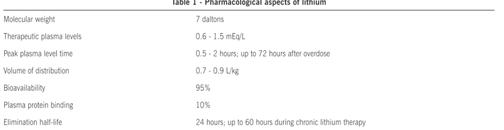
Lithium is a monovalent cation, with molecular weight of 7 daltons, of the family of alkali metals, the same of sodium and potassium. Typically, its concentration in body fluids is not significant (< 0.2 mEq/L). When administered orally, either as lithium citrate (liquid) or carbonate (capsule), lithium is completely absorbed in the upper gastrointestinal tract in about eight hours. Peak plasma levels occur one to two hours after oral administration of standard preparations and about four hours after administration of sustained-release preparations, in which these levels can continue to rise for three to four days in case of acute poisoning4,5.
Binding of lithium to serum proteins is less than 10%, being distributed in total body water with a volume of distribution of 0.7 to 0.9 L/kg. Tissue distribution of lithium occurs with preferential uptake in certain tissues. For example, significant delays are observed in reaching steady state concentrations in the brain compared to plasma concentrations.
Half-life of lithium within plasma varies considerably from patient to patient. In individuals with a normal glomerular filtration rate (GFR), lithium has an elimination half-life of 12 to 27 hours after a single dose. In elderly individuals and patients on chronic lithium therapy, its elimination half-life can increase considerably to approximately 60 hours9.
Lithium is excreted almost entirely by the kidneys and is freely filtered by the glomeruli. Fractional excretion of lithium is 20%, and 60% of the filtered lithium is reab-sorbed in the proximal tubule and 20% in the loop of Henle and the collecting duct9. In the proximal tubule, lithium is
reabsorbed similarly to sodium. Thus, states in which the need for sodium is increased (such as polyuria, diarrhea, congestive heart failure, cirrhosis, and use of NSAIDs) lead to an increase in the fraction of lithium reabsorbed, resulting in elevated serum levels of this cation4. Even at therapeutic
levels, lithium itself may facilitate retention of the drug by inducing polyuria and volume depletion5.
Conversely, any insult that reduces proximal tubule sodium reabsorption, such as carbonic anhydrase inhibi-tors, aminophylline, and osmotic diuretics, will stimulate lithium renal excretion, leading to decreased plasma lithium concentration4.
Several proteins that normally transport sodium in the nephron can also transport lithium, such as the sodium-hydrogen cotransporter in the proximal tubule (NHE3), the Na-K-2Cl cotransporter in the thick ascending limb of the loop of Henle, and the epithelial sodium channel (ENaC) in the cortical collecting tubule. ENaC’s permeability to lithium is 1.5-2-fold higher than its permeability to sodium7.
In contrast, lithium affinity in the basolateral Na/K-ATPase pump is at least an order of magnitude less than that for sodium or potassium4. Table 1 summarizes the main
phar-macological aspects of lithium.
Lithium-induced tubular dysfunction
The most prevalent form of lithium-induced renal injury is impaired urinary concentrating ability10, which can be
detected about eight weeks after lithium initiation11. In a
study involving 1,172 patients on chronic lithium therapy, Boton et al.11 found that impairment of urinary concentrating
ability was present in at least 54% of 1,105 unselected individuals. Initially reversible after cessation of lithium, tubular dysfunction translates into overt and irreversible polyuria and polydipsia with continued treatment, affecting 20 to 40% of patients12. This is the typical clinical picture
of nephrogenic diabetes insipidus, which is resistant to the action of arginine vasopressin (AVP)10.
Physiology of urinary concentrating mechanism
Two key processes are the basis of urinary concentrating ability in mammals. The first factor is the generation of a hypertonic medullary interstitium and the second is the insertion of water channels, the aquaporins, into the apical (AQP2) and basolateral (AQP3 and AQP4) membranes of the collecting duct, allowing tubular water reabsorption through a concentration gradient between the collecting duct lumen and the hyperosmotic medullary interstitium10,13,14. In
response to increased serum osmolality, AVP is released from the posterior pituitary, binds to V2 receptors in the baso-lateral membrane of collecting duct cells and, through the activation of adenylyl cyclase, triggers a series of intracellular events that culminate in the insertion of AQP2 molecules in the apical membrane, which is facing the tubular lumen.
Table 1 - Pharmacological aspects of lithium
Molecular weight 7 daltons
Therapeutic plasma levels 0.6 - 1.5 mEq/L
Peak plasma level time 0.5 - 2 hours; up to 72 hours after overdose
Volume of distribution 0.7 - 0.9 L/kg
Bioavailability 95%
Plasma protein binding 10%
Elimination half-life 24 hours; up to 60 hours during chronic lithium therapy
This process results in increased water permeability in this segment of the nephron, providing an antidiuretic effect13.
In addition to regulating the final concentration of urine, the collecting duct is also important in regulating fluid and electrolyte balance. About 5% of filtered sodium is reab-sorbed in this nephron segment. In collecting duct principal cells, sodium passively crosses the apical membrane through the amiloride-sensitive epithelial sodium channel (ENaC) along an electrochemical gradient maintained by the Na/K-ATPase pump of the basolateral membrane. Moreover, these cells also secrete potassium through a K channel of the apical membrane. The whole process is controlled by aldosterone, a mineralocorticoid hormone that mediates gene transcrip-tion of ENaC, basolateral Na/K-ATPase pump and apical K channel14 (Figure 1).
Pathogenesis of lithium-induced nephrogenic diabetes insipidus
The molecular mechanisms underlying lithium-induced water diuresis and natriuresis are far from well established7.
The postulated mechanisms include inhibition of adenylyl cyclase, decreased density of AVP receptors, and reduced
expression of AQP26. However, Li et al.15 showed that the
development of lithium-induced nephrogenic diabetes insi-pidus is dissociated from adenylyl cyclase activity. A study of healthy volunteers who received lithium carbonate for four weeks showed a significant reduction in urinary AQP2 excretion and in desmopressin (dDAVP)-stimulated urinary concentrating ability16. In the rat kidney medulla,
adminis-tration of lithium for 25 days induced a decrease in AQP2 expression17.
Filtered lithium enters collecting duct cells via ENaC, which has permeability to lithium 1.5-2-fold higher than to sodium. However, unlike sodium, lithium is not exported from the cell by the Na/K-ATPase, thus accumulating intracellu-larly. Lithium promotes the inhibition of glycogen synthase kinase 3 (GSK-3), an enzyme that controls the transport of water and sodium via AQP2 and ENaC, respectively. As a result of the effects of lithium, the cell becomes less respon-sive to the effects of aldosterone and AVP7 (Figure 1).
Another hypothesis to explain the development of nephro-genic diabetes insipidus arose from the observation that lithium can induce cyclooxygenase 2 (COX-2) expression in the kidney medulla via GSK-3 inhibition. Both COX-1 and COX-2 are expressed by the kidneys, and prostaglandins
a
Apical
Basolateral
b
Apical
Basolateral
Vesopressina
Vesopressina
H2O
H2O
H2O
H2O
AQP 2
AQP 2
AQP 3
AQP 4
AQP 3
AQP 4
K
+K
+K
+K
+2 K
+2 K
+Na
+K
+ATPase
Na
+K
+ATPase
ENaC
ENaC
ROMK
ROMK
Na
+GSK-38
3 Na
+Li
+Li
+3 Na
+Aldosterona
Aldosterona
Figure 1 – Transport of water, sodium and potassium in the principal cell of the collecting duct under physiological conditions (a) and in the presence of lithium (b). For further explanation, see text. Source: Grünfeld JP, Rossier BC. Lithium nephrotoxicity revisited. Nat Rev Nephrol
synthesized by them have been suggested to play an impor-tant role in lithium-induced polyuria18.
Treatment of lithium-induced nephrogenic diabetes insipidus
Amiloride is the treatment of choice for lithium-induced nephrogenic diabetes insipidus. This drug not only blocks sodium entry via ENaC, but also inhibits lithium entry into collecting duct principal cells. Amiloride partially restores the urinary concentrating ability, and this is associated with increased urinary AQP2 excretion10.
Tubular acidosis resulting from the use of lithium
Another tubular dysfunction resulting from long-term lithium treatment is hyperchloremic metabolic acidosis. This condition is believed to result from decreased proton secre-tion in the collecting duct and/or excessive backdiffusion of acid equivalents7.
Lithium-induced nephropathy
Lithium-induced tubulointerstitial nephritis is charac-terized by the presence of cortical and medullary inters-titial fibrosis and tubular atrophy19. In 1977, Hestbech
et al.20 demonstrated for the first time these changes
on renal biopsy specimens from a small group of patients treated with lithium for 2-15 years. In the same study, cortical and medullary tubular cysts occurred in 62.5% of specimens, and tubular dilatation in 33%19
(Figure 2). Studies using magnetic resonance (MRI) imaging, although not required for diagnosis, can also reveal the presence of renal microcysts in many patients21(Figure 3).
Data in the literature concerning glomerular involvement in lithium-induced nephrotoxicity are still scarce19.
Protei-nuria is not a common finding, being generally of low severity (< 1 g/day)22.The occurrence of nephrotic syndrome is
rare, and only 26 cases have been reported to date19,22.
Markowitz et al.19, in a study examining renal biopsies from
24 patients on long-term lithium treatment, found a surpri-singly high prevalence of focal segmental glomerulosclerosis (50%) and global glomerulosclerosis (100%), sometimes of equivalent severity to the chronic tubulointerstitial damage. The mechanism of glomerular injury may be secondary to direct cellular toxicity of lithium, resulting in minimal change disease or secondary focal segmental glomerulosclerosis6. In
minimal change disease, some patients recover after lithium withdrawal, suggesting a strong etiologic relationship23.
Risk factors for lithium-induced nephropathy
The main risk factor for the progression of lithium-induced nephropathy is the duration of drug administration24.
Once-daily regimens seem to be less deleterious than multiple daily dosing regimens, perhaps because of the possibility of renal tubular regeneration in those receiving a single daily dose. On the other hand, patients who switched from one regimen to another did not show significant changes in tubular function, except when the switch occurred before five
years of treatment25. Other risk factors for the progression
of renal injury include current lithium therapy, concomi-tant use of nephrotoxic drugs, advanced age, episodes of lithium intoxication, and presence of comorbidities such as hypertension, diabetes mellitus, hyperparathyroidism, and hyperuricemia8,26-28.
Figure 2 – Left: Severe tubulointerstitial nephropathy with the
additional inding of focal tubular cysts arising in a background of severe interstitial ibrosis and tubular atrophy. PAS stain, x40. Right:
High-power view (x100) of tubular cysts (c) lined by simple cuboidal epithelium, along with adjacent tubules with dilatation (d). Source: Markowitz GS, et al. Lithium nephrotoxicity: a progressive combined
glomerular and tubulointerstitial nephropathy. J Am Soc Nephrol 2000; 11: 1439-1448
C
C
d
d
Pathogenesis of lithium-induced nephropathy
The pathogenic mechanisms by which lithium causes tubulointerstitial nephritis and glomerular injury are not well understood7. Lithium plays a role as a modulator of
the pathway for inositol monophosphate, which results in decreased inositol levels and inhibition of cell cycle29. Thus,
accumulation of lithium in cells of the distal nephron via ENaC could account for chronic tubulointerstitial damage7.
Tam et al.23 proposed that the modulation of the
phosphoi-nositol pathway may also play a role in the pathogenesis of minimal change disease after lithium treatment.
Lithium-induced nephropathy and chronic kidney di-sease (CKD)
The relationship between lithium and the onset of progressive chronic kidney disease (CKD) was a controversial topic in the past. Johnson26. suggests that lithium-induced
chronic renal failure is very unusual. A study comparing 107 patients on lithium therapy with psychiatric control patients showed no relationship between creatinine clearance and duration of lithium treatment and lithium dosages30.
The risk of developing CKD in patients on long-term lithium treatment has now been well established by several clinical, histopathological and epidemiological studies7,19,24.
A French study of 54 patients with lithium-induced nephro-pathy demonstrated a mean annual loss of creatinine clea-rance of 2.29 mL/min24. In that country, the prevalence
of lithium-related chronic end-stage renal disease (ESRD)
is estimated as 2 per 1000 dialysis patients, accounting for 0.22% of all ESRD cases24. The progression of
lithium-induced CKD is slow, requiring a period of 10 to 20 years to progress to end-stage renal failure7,24.This may explain why
the association between CKD and lithium use was not well accepted in the past.
Treatment of lithium-induced nephropathy
Close monitoring of renal function of patients on lithium treatment is crucial for early detection of lithium-induced nephropathy6. Serum creatinine levels should be measured
every two months during the first six months of lithium therapy and every year thereafter27.
Cases in which impaired renal function is detected raise a dilemma7.Continuous lithium treatment is known to reduce
the risk of suicide among patients with bipolar disorder31.
Moreover, the risk of early recurrence of bipolar disorder appears to increase following discontinuation of lithium32.
Some patients, whose illness has been well controlled with lithium, refuse to switch or discontinue therapy. Thus, the decision to substitute lithium with another mood stabi-lizer should involve the patient, the psychiatrist and the nephrologist7,24.
The probability of recovery of renal function after lithium withdrawal increases when estimated creatinine clearance is greater than 40 mL/min24 and when serum creatinine levels
are less than 220 µmol/L19. A point at which the
discontinu-ation of lithium no longer alters the course of renal disease probably exists, that is, the process of renal fibrosis continues to progress despite removal of the triggering agent24.
In addition to renal function, other predictors of poor prognosis are the extent of interstitial fibrosis on renal biopsy and the presence of proteinuria7,24. This condition should
be adequately managed to reduce the progression of CKD. Whether amiloride can prevent the long-term effects of lithium nephropathy is not yet known. If such preventive action is identified, this finding might strongly suggest that lithium exerts a primary cytotoxic effect that is limited to the principal cells of the collecting duct and mediated by the entry of this cation through ENaCs7.
Lithium-induced hypercalcemia and hyperparathyroi-dism
An underrecognized side effect of lithium therapy is hyperparathyroidism with associated hypercalcemia and hypocalciuria33. The prevalence of hyperparathyroidism
among chronic lithium users (> 10 years) is approximately 10-15%34. The prevalence of lithium-induced hypercalcemia
is higher in patients with renal failure, in whom urinary excretion of calcium is reduced7. Nephrolithiasis and
nephro-calcinosis may result from hypercalcemia in these patients7.
Pathogenesis of lithium-induced hyperparathyroidism and hypercalcemia
One mechanism proposed to explain lithium-induced hyperparathyroidism is based on changes in calcium-sensing
Figure 3 – Magnetic resonance image in a 68-year-old patient shows
abundant microcysts in the cortical and medullary regions of both kidneys. Source: Farres MT, et al. Chronic lithium nephropathy: MR
receptor sensitivity in the parathyroid gland, causing a decrease in the set point for secretion of parathyroid hormone (PTH)33,35. If not corrected, persistent hyperparathyroidism
and resultant hypercalcemia may exacerbate psychiatric manifestations and lead to deleterious effects on bone mineral metabolism, urinary excretion and blood vessels33.
Treatment of lithium-induced hyperparathyroidism and hypercalcemia
The treatment of hypercalcemia in chronic lithium users may be clinical or surgical. Ablation of a single parathyroid adenoma usually leads to normocalcemia, even in patients who continue to receive lithium34. Some authors do not
recommend parathyroidectomy for the treatment of hyper-calcemia in these cases35, due to risks and technical
diffi-culties in cases of multiglandular disease (50% of cases)7,33.
Recently, cinacalcet, a calcimimetic drug, has been used for clinical treatment of hyperparathyroidism secondary to lithium. This drug enhances calcium-sensitive receptor sensitivity in the principal cells of the parathyroid gland, reducing the threshold for activation of these receptors to extracellular calcium. As a result, PTH secretion decreases, with consequent reduction in serum calcium levels33,35.
Serum calcium concentration should be measured every one to two years to detect hypercalcemia7.
c
onclusionFor over half a century, lithium has been the most widely used drug in the treatment of bipolar disorder. Lithium nephrotoxicity may present in different ways, from early alterations, such as nephrogenic diabetes insipidus and metabolic acidosis, to lesions secondary to chronic lithium therapy, such as hypercalcemia, tubulointerstitial nephro-pathy, and CKD. Blockade of the ENaC, such as by using amiloride, in the treatment of nephrogenic diabetes insipidus, is based on the observation of the key role played by these channels in the pathogenesis of this disease. Monitoring of serum creatinine and calcium levels in patients on lithium treatment is mandatory for early detection of CKD and hyper-calcemia. The decision to discontinue lithium therapy raises a dilemma, and should therefore be made jointly by the psychiatrist, the nephrologist and the patient. Paradoxically, lithium therapy is so effective in treating bipolar disorder that some patients are lost to follow-up, regular biochemical monitoring is not performed and, therefore, the discovery of renal failure is delayed.
Financial Support: CNPq
Conflict of interest: No conflicts of interest declared concerning the publication of this article.
r
eFerences1. Price LH, Heninger GR. Lithium in the treatment of mood disorders. N Engl J Med. 1994;331:591-8.
2. Muller-Oerlinghausen B, Berghofer A, Bauer M. Bipolar disorder. Lancet. 2002;359:241-7.
3. Ferrier N, Ferrie LJ, Macritchie KA. Lithium therapy. Adv Psychiatr Treat. 2006;12:256-64.
4. Timmer RT, Sands JM. Lithium Intoxication. J Am Soc Nephrol. 1999;10:666-74.
5. Borkan SC. Extracorporeal therapies for acute intoxications. Crit Care Clin. 2002;18:393-420.
6. Alexander MP, Farag YMK, Mittal BV, Rennke HG, Singh AK. Lithium toxicity: a double-edged sword. Kidney Int. 2008;73:233-7.
7. Grünfeld JP, Rossier BC. Lithium nephrotoxicity revisited. Nat Rev Nephrol. 2009;5:270-6.
8. Gitlin M. Lithium and kidney. Drug Saf. 1999;20:231-43.
9. Okusa MD, Crystal LJT. Clinical manifestations and managementof acute lithium intoxication. Am J Med. 1994;97:383-9.
10. Bedford JJ, Weggery S, Ellis G, McDonald FJ, Joyce PR, Leader JP,et al. Lithium-induced nephrogenic diabetes insipidus:renal effects of amiloride. Clin J Am Soc Nephrol. 2008;3:1324-31.
11. Boton R, Gaviria M, Batlle DC. Prevalence, pathogenesis,and treatment of renal dysfunction associated with chronic lithium therapy. Am J Kidney Dis. 1987;10:329-45.
12. Stone KA. Lithium-induced nephrogenic diabetes insipidus. J Am Board Fam Pract. 1999;12:43-7.
13. Singer G, Brenner B. Distúrbios hidreletrolíticos. In: Kasper DL, Fauci AS, Longo DL, Braunwald E, HauserSL, Jameson JL. Harrison medicina interna. 16ª ed. Rio de Janeiro: McGraw Hill; 2006.
14. George Jr. A, Neilson E. Biologia cellular e molecular do rim. In: Kasper DL, Fauci AS, Longo DL, Braunwald E, Hauser SL, Jameson JL. Harrison medicina interna. 17ª ed. Rio de Janeiro: McGraw Hill; 2008, p. 1741-8. 15. Li Y, Shaw S, Kamsteeg EJ, Vandewalle A, Deen PM. Development of lithium-induced nephrogenic diabetes insipidus is dissociated from adenyl cyclase activity.J Am Soc Nephrol. 2006;17:1063-72. 16. Walker RJ, Weggery S, Bedford JJ, McDonald FJ, EllisG, Leader JP.
Lithium-induced reduction in urinary concentrating ability and urinary aquaporin 2 (AQP2) excretion in healthy volunteers. Kidney Int. 2005;67:291-4.
17. Marples D, Christensen S, Christensen EI, Ottosen PD, Nielsen S. Lithium-induced down regulation of aquaporin-2water channel expression in rat kidney medulla. J Clin Invest. 1995;95:1838-45.
18. Rao R, Zhang MZ, Zhao M, Cai H, Harris RC, Breyer MD, et al. Lithium treatment inhibits renalGsK-3 activity and promotes cyclooxygenase 2-dependent polyuria. Am J Physiol Renal Physiol. 2005;288:F642-F9. 19. Markowitz GS, Radhakrishnan J, Kambham N, Valeri AM, Hines WH, D’Agati VD. Lithium nephrotoxicity: a progressive combined glomerular and tubulo interstitial nephropathy. J Am Soc Nephrol. 2000;11:1439-48. 20. Hestbech J, Hansen HE, Amdisen A, Olsen S. Chronic renal lesions following long term treatment with lithium. Kidney Int.1977;12:205-13. 21. Farres MT, Ronco P, Saadoun D, Remy P, Vincent F, Khalil A, et al. Chronic lithium nephropathy: MR imaging for diagnosis. Radiology. 2003;229:570-4.
22. Santella RN, Rimmer JM, Mac Pherson BR. Focal segmental glomeruloscle-rosis in patients receiving lithium carbonate. Am J Med. 1988;84:951-4. 23. Tamm VKK, Green J, Schwieger J, Cohen AH. Nephrotic syndrome and renal insufficiency associated with lithium therapy. Am J Kidney Dis. 1996;27:715-20.
25. Muir A, Davidson R, Silverstone T, Daway A, Forsling ML. Two regi-mens of lithium prophylaxis and renal function. Acta Psychiatr Scand. 1989;80:579-83.
26. Johnson, G. Lithium: early development, toxicity, and renal function. Neuropsychopharmacology. 1998;19:200-5.
27. Freeman MP, Freeman SA. Lithium: clinical considerations in Internal Medicine. Am J Med. 2006;119:478-81.
28. Bendz H, Sjodin I, Aurell M. Renal function on and off lithium in patients treated with lithium for 15years or more. A controlled, prospective lithium-withdrawal study. Nephrol Dial Transplant. 2996;11:457-60. 29. Quiroz JA, Gould TD, Manji HK. Molecular effects of lithium. Mol Interv.
2004;4:259-72.
30. Coskunol H, Vahip S, Mees ED, Başçi A, Bayindir O, Tuğlula I. Renal
side-effects of long-term lithium treatment. J Affect Disord. 1997;43:5-10. 31. Kessing LV, Søndergård L, Kvist K, Andersen PK. Suicide risk in patients
treated with lithium. Arch Gen Psychiatry. 2005;62:860-6.
32. Suppes T, Baldessarini RJ, Faedda GL, Tohen M. Risk of recurrence following discontinuation of lithium treatment in bipolar disorder. Arch Gen Psychiatry. 1991;48:1082-8.
33. Sloand JA, Shelly MA. Normalization of lithium-induced hypercalcemia and hyperparathyroidism with cinacalcet hydrochloride. Am J Kidney Dis. 2006;48:832-7.
34. Hundley JC, Woodrum DT, Saunders BD, Doherty GM, Gauger PG. Revis-iting lithium-associated hyperparathyroidism in the era of intraoperative parathyroid hormone monitoring. Surgery. 2005;138:1027-31. 35. Gregoor PS, Jong GM. Lithium hypercalcemia, hyperparathyroidism, and
cinacalcet. Kidney Int 2007;71:470.