5 artigo 339
OrigiNAl ArticlE
The authors declare that there was no conflict of interest in conducting this work
1 –Head of the Orthopedics and Traumatology Service, São José University Hospital, Minas Gerais School of Medical Sciences, Belo Horizonte (HUSJ-FCMMG-BH) and Titular Professor and Chair of Orthopedics and Traumatology, Minas Gerais School of Medical Sciences; Former President of SBOT.
2 – Member of the Knee Surgery Service, São José University Hospital, Minas Gerais School of Medical Sciences, Belo Horizonte (HUSJ-FCMMG-BH) and Adjunct Professor of Orthopedics and Traumatology, Minas Gerais School of Medical Science.
3 – Member of the Knee Surgery Service, São José University Hospital, Minas Gerais School of Medical Sciences, Belo Horizonte (HUSJ-FCMMG-BH) and Head of the Medical Department of Clube Atlético Mineiro; Physician of the Brazilian National Soccer Team.
4 – Orthopedist (R4) in the Knee Surgery Service, São José University Hospital, Minas Gerais School of Medical Sciences, Belo Horizonte (HUSJ-FCMMG-BH).
Work performed in the Orthopedics and Traumatology Service, São José University Hospital (FCMMG-BH).
Correspondence: Hospital Universitário São José - Rua Aimorés, 2896 - Barro Preto, Belo Horizonte - MG, 30140-073, Brasil. E-mail: [email protected]
Work received for publication: April 26, 2010; accepted for publication: July 1, 2010.
assessment Of the reprOducibility Of the Outerbridge
and fsa classificatiOns fOr chOndral
lesiOns Of the knee
Neylor Pace Lasmar¹, Rodrigo Campos Pace Lasmar2, Rodrigo Barreiros Vieira3,
Juraci Rosa de Oliveira3, André Campos Scarpa4
iNtrOdUctiON
A combination of mechanical factors and loss of the structural proteins that make up the cartilage is responsible for osteoarthrosis(1), which is a highly
pre-valent pathological condition of degenerative nature that is especially seen in the knee joint.
The joint cartilage is a type of tissue with the capacity to deal with large forces applied over many cycles, but it has a low capacity for regeneration after injury(1).
AbStrAct
Objective: To assess the reproducibility of the Outerbridge and the French Society of Arthroscopy classifications be-tween different observers, and to establish a comparison between them. Method: Thirty videos on randomly selec-ted knee arthroscopy procedures demonstrating chondral lesions were used. These were classified by six observers: two third-year orthopedics residents and four orthopedic surgeons, of whom two were knee surgery specialists. The intraobserver and interobserver reliability was evaluated by means of the kappa index. Results: The result from the complete evaluation on the Outerbridge classification
with all the observers gave a kappa index of 0.434411. For the classification proposed by the French Society of Arthroscopy, the kappa index was 0.45166. Conclusion: The Outerbridge and French Society of Arthroscopy clas-sifications for chondral lesions are moderately reproduci-ble between observers. Comparing the two classifications, the proposal from the French Society of Arthroscopy was shown to be more reproducible, and the authors suggest that this classification should be used preferentially in clinical practice for evaluations on chondral lesions of the knee.
Keywords – Knee Injuries/classification; Knee Injuries/ pathology; Arthroscopy
Rev Bras Ortop. 2011;46(3):266-69
Traditionally, osteoarthrosis of the knee is diagno-sed through clinical signs resulting from the inflam-matory and/or mechanical process, accompanied by specific radiological abnormalities such as diminished joint space, subchondral sclerosis and osteophytes. However, there may be very little evidence of such abnormalities, particularly during the initial stages of the degenerative process, thus leading to delays in diagnosing this condition.
267
residents, using the Outerbridge and French Society of Arthroscopy classifications for chondral lesions of the knee.
These assessments were performed on a special form that was filled out by one of the researchers. In order to minimize any bias due to difficulty in inter-pretation or any possible forgetfulness, the classifica-tions were described on the response sheets, together with schematic drawings of the respective classifi-cations, which were handed over to each observer at the time of the image assessments. There was no time limit on making the image classifications.
The data obtained were analyzed by a statisti-cian, using the chi-square test and kappa coefficient, which were first used by Fleiss and Cohen(8). The
kappa coefficient was used to assess the concordance between the observers within the same sample unit. This ranged from -1, when all the observers disagreed regarding all the assessments to +1, when there was complete agreement.
rESUltS
The series of images were analyzed using the Outerbridge classification (four types) and the French Society of Arthroscopy classification (four types).
The result from the complete evaluation of the Outerbridge classification with all of the observers was an inter-observer kappa index (k) of 0.434411, which produced a p-value = 0. Analysis on the two residents alone gave k = 0.395973. Analysis on the orthopedists alone gave k = 0.165379. Analysis on the knee specialists alone gave k = 0.140127 (Table 1).
The highest value for the intra-observer concor-dance index was for orthopedist 1, with k = 0.509002, and the lowest was for resident 2, with k = -0.064516, as presented in Table 2. The mean intra-observer con-cordance was k = 0.2955 (Table 2).
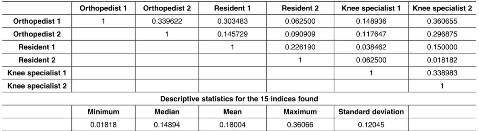
In relation to the classification proposed by the French Society of Arthroscopy, we found the follo-wing data. The inter-observer kappa was k = 0.45166, thus resulting in p = 0, among all the six observers. Among the different groups, the index was greatest for the orthopedists, with k = 0.339623, followed by the knee surgeons, with k = 0.338983. For the resi-dents, the concordance coefficient was k = 0.22619. These results are presented in Table 3.
ASSESSMENT OF THE REPRODUCIBILITY OF THE OUTERBRIDGE AND FSA CLASSIFICATIONS FOR CHONDRAL LESIONS OF THE KNEE
Rev Bras Ortop. 2011;46(3):266-69
arthroscopy of the knee, in cases with normal radio-logical findings(2).
There are some radiographic classifications for os-teoarthrosis of the knee, but they have been shown to be imprecise, especially in the initial stages.
Arthroscopic evaluation of the knee presents the characteristic of direct viewing of the joint surface, thus enabling greater detailing of the chondropathy, such as size, depth, consistency and lesion location(3).
Some arthroscopic classification systems for chon-dral lesions of the knee have been described in the literature. Among these is the Outerbridge system, which was originally described through direct viewing using arthrotomy, and the classification of the French Society of Arthroscopy(4).
In 1961, Outerbridge classified patellar chondral lesions into four grades, such that grade I was softe-ning; grade II was fragmentation/fissure of 1.25 cm or less; grade III was fragmentation/fissure greater than 1.25 cm; and grade IV was bone erosion(5).
In 1994, the French Society of Arthroscopy pro-posed the classification: grade I, softening; grade II, superficial fissure; grade III, deep fissure; and grade IV, bone exposure(6).
Reproducibility between observers is essential for any type of classification(6), and arthroscopy should be
considered to be an important measurement method for assessing osteoarthrosis of the knee(7).
The purpose of this study was to assess the repro-ducibility of the Outerbridge and French Society of Arthroscopy classifications between observers.
MAtEriAlS ANd MEthOdS
268
table 1 – Inter-observer kappa concordance index for the Outerbridge classification.
table 2 – Intra-observer concordance index for the Outerbridge
classification.
Orthopedist 1 Orthopedist 2 resident 1 resident 2 knee specialist 1 knee specialist 2
Orthopedist 1 1 0.165379 0.363636 0.217791 0.049296 0.437500
Orthopedist 2 1 0.010989 0.041553 0.220183 0.269406
resident 1 1 0.395973 0.127907 0.361702
resident 2 1 0.122807 0.212963
knee specialist 1 1 0.140127
knee specialist 2 1
descriptive statistics for the 15 indices found
Minimum Median Mean Maximum Standard deviation
0.01099 0.21296 0.20915 0.43750 0.13408
intra-observer kappa concordance index (calculated by comparing the observer’s first and second evaluations
on the same 30 patients)
Observer kappa coefficient (k) p-value
Orthopedist 1 0.509002 2.5207e-05 Orthopedist 2 0.40458 0.000145
resident 1 0.233227 0.026750 resident 2 -0.064516 0.700497 knee specialist 1 0.244604 0.044259 knee specialist 2 0.447853 3.48437e-05
Orthopedist 1 Orthopedist 2 resident 1 resident 2 knee specialist 1 knee specialist 2
Orthopedist 1 1 0.339622 0.303483 0.062500 0.148936 0.360655
Orthopedist 2 1 0.145729 0.090909 0.117647 0.296875
resident 1 1 0.226190 0.038462 0.150000
resident 2 1 0.062500 0.018182
knee specialist 1 1 0.338983
knee specialist 2 1
descriptive statistics for the 15 indices found
Minimum Median Mean Maximum Standard deviation
0.01818 0.14894 0.18004 0.36066 0.12045 table 3 – Inter-observer kappa concordance index for the classification of the french Society of Arthroscopy.
intra-observer kappa concordance index (calculated by comparing the observer’s first and second evaluations
on the same 30 patients)
Observer kappa coefficient (k) p-value
Orthopedist 1 0.540034 2.55338e-05 Orthopedist 2 0.39441 0.000306
resident 1 0.170732 0.084768 resident 2 0.033742 0.382217 knee specialist 1 0.440299 0.001714 knee specialist 2 0.322581 0.00428 table 4 - Intra-observer concordance index for the classification of the french Society of Arthroscopy.
Rev Bras Ortop. 2011;46(3):266-69
The highest value for the intra-observer concor-dance index was for orthopedist 1 (k = 0.540034), and the lowest value was for resident 2 (k = 0.033742), as shown in Table 4. The mean kappa index for the intra-observer evaluation was k = 0.3165.
diScUSSiON
Several authors have expressed the opinion that in assessing the reliability of inter-observer concordan-ce, there is a need to incorporate concordance due to chance in the evaluation(9-10). Kappa is a coefficient
of concordance that corrects for the error due to chance and is used to determine the intra and inter- observer variation. It is used when two observers separately classify a sample of objects using the same category scale.
Landis classified concordance as: poor (less than 0), slight (0-0.2), weak (0.21-0.4), moderate (0.41-0.6), substantial (0.61-0.8), almost perfect (0.81-1.0).
269
that the Outerbridge classification is moderately accura-te when used to grade chondral lesions arthroscopically.
In the present study on the Outerbridge and French Society of Arthroscopy classifications, we found kappa values of 0.434411 and 0.45166 respec-tively. According to Landis, this indicates that the classifications have moderate concordance.
In comparing the means within the groups, we could see that in the groups using the French Society of Arthroscopy classification, the means were higher than in the other groups.
cONclUSiON
On the basis of the present assessment, it can be concluded that the French Society of Arthroscopy and Outerbridge classifications have moderate concordan-ce between observers.
Comparing the two classifications, the proposal from the French Society of Arthroscopy was shown to be more reproducible, and the present authors suggest that this classification should be used preferentially for assessing chondral lesions of the knee in clinical practice.
rEFErENcES
1. Fife RS, Brandt KD, Braunstein EM, Katz BP, Shelbourne KD, Kalasinski LA, et al. Relationship between arthroscopic evidence of cartilage damage and ra-diographic evidence of joint space narrowing in early osteoarthritis of the knee. Arthritis Rheum. 1991;34(4):377-82.
2. Brismar BH, Wredmark T, Movin T, Leandersson J, Svensson O. Observer relia-bility in the arthroscopic classification of osteoarthritis of the knee. J Bone Joint Surg BR. 2002;84(1):42-7.
3. Brandt KD, Fife RS, Braunstein EM, Katz B. Radiographic grading of the severity of knee osteoarthritis: relation of the Kellgren and Lawrence grade to a grade based on joint space narrowing, and correlation with arthroscopic evidence of articular cartilage degeneration. Arthritis Rheum. 1991;34(11):1381-6. 4. Collins DH. The pathology of articular and spinal diseases. London: Edward
Arnold; 1949.
5. Outerbridge RE. The aetiology of chondromalacia patellae. J Bone Joint Surg BR. 1961;43:752-7.
6. Flikkilä T, Nikkola-Sihto A, Kaarela O, Pääkkö E, Raatikainen T. Poor interobserver
reliability of AO classification of fractures of the distal radius. J Bone Joint Surg Br. 1998;80(4):670-2.
7. Dougados M, Ayral X, Listrat V, Gueguen A, Bahuaud J, Beaufils P, et al. The SFA system for assessing articular cartilage lesions at arthroscopy of the knee. Arthroscopy. 1994;10(1):69-77.
8. Fleiss JL, Cohen J. The equivalence of weighted kappa and the intraclass correla-tion coefficient as measures of reliability. Educ Psychol Meas. 1973;33(3):613-9. 9. Felson DT, Naimark A, Anderson J, Kazis L, Castelli W, Meenan RF. The preva-lence of knee osteoarthritis in the elderly. The Framingham Osteoarthritis Study. Arthritis Rheum. 1987;30(8):914-8.
10. 10-Dillon CF, Rasch EK, Gu Q, Hirsch R. Prevalence of knee osteoarthritis in the United States: arthritis data from the Third National Health and Nutrition Examination Survey 1991-94. J Rheumatol. 2006;33(11):2110-2.
11. Cameron ML, Briggs KK, Steadman JR. Reproducibility and Reliability of the Outerbridge Classification for Grading Chondral Lesions of the Knee Arthros-copically. Am J Sports Med. 2003;31(1): 83-6.
Rev Bras Ortop. 2011;46(3):266-69