ABSTRACT
http://dx.doi.org/10.1590/1678-775720150224
Cone beam computed tomography study of apical
root resorption induced by Herbst appliance
João Paulo SCHWARTZ1, Taísa Boamorte RAVELI1, Kélei Cristina de Mathias ALMEIDA1, Humberto Osvaldo SCHWARTZ-FILHO2, Dirceu Barnabé RAVELI1
1- Universidade Estadual Paulista, Faculdade de Odontologia, Departamento de Clínica Infantil, Disciplina de Ortodontia, Araraquara, SP, Brazil. 2- Universidade de Santo Amaro, Departamento de Odontologia, São Paulo, SP, Brazil.
Corresponding address: João Paulo Schwartz - Rua Rio Grande do Sul, 368 - apt 203 - Curitiba - PR - Brazil - 80620-080 -
Phone: 55 14 9682-8920 - e-mail: [email protected]
6XEPLWWHG0D\0RGL¿FDWLRQ-XO\$FFHSWHG-XO\
O
bjective: This study evaluated the frequency of root resorption during the orthodontic treatment with Herbst appliance by Cone Beam Computed Tomography (CBCT). Material and Methods: The sample comprised 23 patients (11 men, 12 women; mean ages 15.76±1.75 years) with Class II division 1 malocclusion, treated with Herbst appliance. CBCT was obtained before treatment (T0) and after Herbst treatment (T1). All the dental roots, except third molars, were evaluated, and apical root resorption was determined using the axial guided navigation method. Paired t-tests and Wilcoxon T Test were used to compare the dependent samples in parametric and nonparametric cases, respectively. Chi-Square Test with Yates’ correction was used to evaluate the relationship between apical root resorption and gender. 5esults were considered at a signi¿cance level of 5. Results: Apical resorption was detected by CBCT in 57.96% of 980 roots that underwent Herbst appliance treatment. All patients had minimal resorption and there was no statistical signi¿cance between the genders. Conclusion: CBCT three-dimensional evaluation showed association between Herbst appliance and minimal apical root resorption, mostly in the anchoring teeth, without clinical signi¿cance.Ke y w or ds: Root resorption. Activator appliances. Cone-beam computed tomography.
I N TROD UCTI ON
InÀammatory root resorption orthodontically induced is a side effect of tooth movement related to the biological tissue response, and this correlation was reported in literature for the ¿rst time in 191413. It is a pathological or physiological
localized condition, characterized by loss of the cells layer surface that protects the tooth roots, associated with a structure loss caused by clastic cells10.
Apical root resorption is usually classi¿ed as minor, moderate or severe4. Minor root resorption
can be repaired with secondary cementum once orthodontic movement ceases. However, when root resorption is suf¿ciently severe to exceed the reparative capacity of the root, a cementum island may separate from the root surface, resulting in an irreversible root resorption mostly in the apical part of the root10,20.
Orthodontic treatment has some risk factors for root resorption such as treatment duration, tooth movement direction, amount of apical displacement, and type and magnitude of the force applied28. Herbst is a ¿xed functional appliance
with a bilateral telescopic mechanism that keeps the mandible in an anterior position and induces skeletal and dentoalveolar changes such as upper molars distalization8,15,17-19,21, mesial movement
of the lower molars8,18,19,21,26,and protrusion
of lower incisors2,9,17-19,21,26,27,29. Emil Herbst, in
1934, published his experience with the device in Zahnärztliche Rundschau, and, in the same journal, Martin Schwarz criticized the Herbst appliance, stating that the device could result in overloading the anchorage teeth with periodontal damage as a consequence20.
was not found in upper and lower premolars. However, regarding lower premolars, the ¿rst premolars showed more resorption than the second premolars16. The second study supports
that the teeth more affected by the apical root resorption was the upper ¿rst molars and the lower
¿rst premolars11.
These orthodontic studies have evaluated apical root resorption by conventional radiography, i.e., two dimension radiographies, periapical and panoramic, respectively. However, conventional radiographs may underestimate or overestimate the amount of root structure loss5. Panoramic
radiography underestimates apical root resorption when compared with microtomography, and overestimates it when compared with periapical radiography24. Periapical radiographs were
considered less accurate than the cone beam computed tomography (CBCT) to evaluate the root length25. The apical root resorption is a
three-dimensional change which can affect the root surface as a whole. The ability of CBCT to obtain distortion-free and reproducible images of single roots provides excellent possibilities to evaluate apical root resorption13. Different voxel sizes are
frequently found in the literature to evaluate the apical root resorption induced by orthodontic tooth movement by CBCT6,7,12,14,30.
Despite the common use of the Herbst appliance, there is still a lack of literature about the consequences of its use. To our knowledge, there are no studies assessing root resorption through the evaluation by CBCT. This research aimed to evaluate the apical root resorption induced by the Herbst device with CBCT.
M ATERI AL AN D M ETH OD S
This retrospective study was reviewed and approved by the Ethics Committee of Araraquara Dental School, Univ. Estadual Paulista (FOAr-UNESP). The subjects consisted of 23 patients, sequentially treated (11 men, 12 women; mean age 15.76±1.75 years), who had skeletal Class II division 1 malocclusion.
The inclusion criteria were bilateral Class II molar relationship, overjet bigger than 5 mm and complete permanent dentition, except third molars. The exclusion criteria were syndrome patients, extreme vertical grow pattern and prior orthodontics treatment. Face and occlusion were clinically analyzed to determine skeletal Class II division 1 malocclusion. Facial analysis observed the convex pro¿le, straight nasolabial angle, and short mentocervical line, and occlusion analysis observed the molar and canines in Class II, equal to or higher than the half of a cusp, and overjet equal to or greater than 5 mm.
Patients used banded Herbst appliance until they complete eight months of treatment (mean 8.50±0.70 months) with one step mandibular advancement until the incisor edge to edge relationship19
.
The telescopic mechanism used wasFlip-Lock Herbst® (Tp Orthodontics, Inc.) model,
which was constituted by connectors, tubes and pistons.
To upper anchorage, transpalatal ¿xed bar at the ¿rst molars was used, made with 1.2 mm steel wire, distant 2 mm from the palate and with an extension of 1.2 mm steel wire to second molar. In the lower arch, the Nance lingual arch modi¿ed for Herbst was used in the ¿rst molars, made with 1.2 mm steel wire and distant 3 mm from lingual face incisors. Anchor appliances were constructed only by one prosthetic.
To evaluate the apical root resorption induced by Herbst appliance, CBCT images were obtained before treatment (T0) and after treatment (T1) with Herbst appliance. Patients were scanned in an upright position with maximum intercuspation using tomography i-CAT® Classic (Imaging
Sciences International, Hat¿eld, PA, USA) with a 17x13.3 cm of ¿eld of vision, 120 k9p tube voltage, 18.45 mA tube current and 0.4 mm isometric voxel. CBCT images were examined using Dolphin® Imaging software (Dolphin Imaging and
Management Solutions, Chatsworth, Calif., USA) by multiplanar reconstruction (axial, sagittal and coronal).
All upper and lower teeth, except third molars, were evaluated by tooth length (apical root resorption). The coronal and sagittal cursors were adjusted in the tooth long axis (incisal edge center or cusp to root apex) according to the tooth root of interest6,7
. The length measurement was
linear between two points, one at the root apex and other at the incisal edge or tooth cusp. To precise the localization of root apex and incisal edge or cusp, the axial guided navigation (AGN) method was used. This method used the axial cursor movement at the sagittal and coronal multiplanar reconstruction6,7
.
Thereference pointto determine the root apex, incisal or cusp tooth is the intersection between axial and sagittal or coronal cursors (Figure 1).
distobuccal root of upper molar (sagittal section); mesiolingual cusp tip to apex of lingual root of upper molar (coronal section); mesiobuccal cusp tip to apex of mesial root of lower molar (sagittal section); distobuccal cusp tip to apex of distal root of lower molar (sagittal section) (Figure 2).
Measurements were randomly reevaluated after two weeks by the same examiner. The error of the method was evaluated by Intraclass Correlation Coef¿cient (ICC). Shapiro-Wilk Test was used to assess normal distribution, and Student’s t-Test and Wilcoxon t-Test were used to compare dependent samples in parametric and non-parametric cases, respectively. Chi-Square Test with Yates’ correction was used to evaluate the relationship between apical root resorption and gender. Results were considered at a signi¿cance level of 5%. Statistical analysis was performed using SPSS® (SPSS Inc,
Chicago, III) and GraphPad Prism® (GraphPad
Prism Inc, San Diego).
RESULTS
Systematic intra-examiner error indicated excellent reliability (ICC=0.91). There was statistical difference for mesiobuccal root of right upper ¿rst molar, distobuccal root of left upper ¿rst molar, distobuccal root of left upper second molar, root of left lower second premolar, distal root of left lower ¿rst molar, mesial root of right lower
¿rst molar, mesial root of right lower second molar and distal root right lower second molar (Table 1). However, the apical root resorption detected was minimal, with mean smaller than 0.5 mm (Table 1).
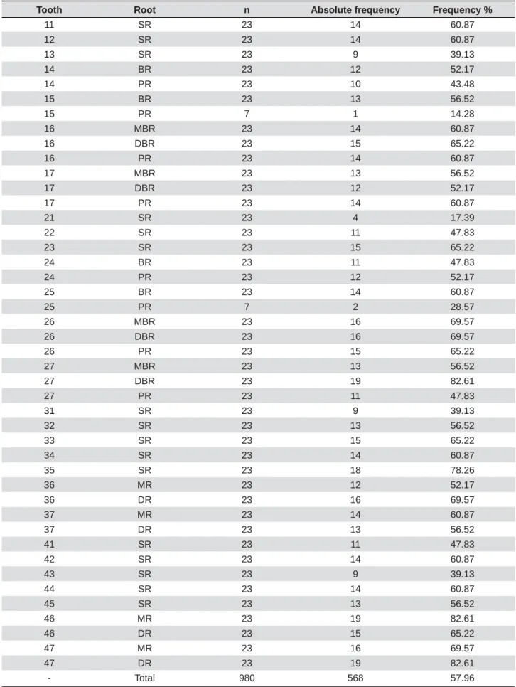
All subjects and 57.96% of 980 roots showed apical resorption. The root resorption frequency for each tooth can be observed in Table 2. Apical root resorption analyses between genders do not show statistical difference (Table 3).
D I SCUSSI ON
This CBCT study evaluated the apical root resorption induced by Herbst appliance. Patients with mean age of 15.76 years composed the group to simulate the post pubertal period, stage in which Class II treatment with Herbst appliance shows more dentoalveolar than skeletal response23.
CBCT images were used to analyze 980 tooth roots, and 568 (57.96%) presented resorption after the Herbst appliance treatment. Previous studies showed a high frequency of apical root resorption after the orthodontic treatment by two-dimensional radiographs analisys1,24. As
already mentioned, there are no literature reports assessing apical root resorption induced by Herbst appliance by CBCT, therefore, there are no parameters for comparison of our results. It is
noteworthy that a more accurate assessment can be obtained by the use of CBCT, which allows a more accurate analysis of the treatment results12.
In T1 the roots were longer than at T0 for tooth 21, 22, buccal root of 15 and palatal root of 14, 15, 25, and 27 (Table 1). This occurs probably by the accuracy of CBCT with voxel resolutions 0.4 mm to linear measurement of apical root resorption used in this study. However, in a previous similar study with voxel resolutions 0.25 mm the same problem was observed7, and this may be attributed
to the incomplete development of the tooth apices, except for teeth 21 and 22, or by the CBCT issues of methods sensitivity and speci¿city for apical root resorption assessment.
There was no statistical difference in apical root resorption between genders, which is a data similar to those related in literature1,3. There was
signi¿cant statistical difference for mesiobuccal root of right upper ¿rst molar, distobuccal root of left upper ¿rst molar, distobuccal root of left upper second molar, root of left lower second premolar, distal root of left lower ¿rst molar, mesial root of right lower ¿rst molar, mesial root of right lower second molar and distal root right lower second molar. The Nance lingual arch modi¿ed to Herbst distant from lingual face of lower incisors, and the transpalatal ¿x bar in the upper arch may further the apical root resorption in the anchorage teeth.
Such resorption can be justified by the fact that banded Herbst appliance might deliver unphysiologic forces to immediate anchor teeth, thereby exposing these to a higher risk of root resorption than the other teeth incorporated into the anchorage either directly via bands or indirectly via occlusal and proximal contacts11.
Another study showed no signi¿cant statistical difference in the roots morphology after the Herbst appliance treatment, however, in relation to lower premolars, the ¿rst showed more root resorption than the second16.Apical root resorption associated
to Herbst appliance showed a mean of teeth length reduction smaller than 0.5 mm (Table 1). This root shortening is classi¿ed as minor root resorption, and is repaired with secondary cementum once orthodontic movement ceases10,20. Despite the
apical root reduction, this minimal resorption has no clinical signi¿cance.
This CBCT study evaluated the amount of apical root resorption and showed that the forces delivered by the propulsion mechanism have no clinical signi¿cance to anchor teeth. Our results are in accordance with Nasiopoulos, et al.16
(2006) and in disagreement with Kinzinger, et al.11 (2011), however, these studies evaluated
Tooth Root T0, xࡃ ± SD T1, xࡃ ± SD T1-T0, xࡃ ± SD P Value
11 SR 23.92 ± 2.18 23.86 ± 2.24 -0.06 ± 0.04 0.518
12 SR 22.80 ± 2.15 22.68 ± 2.40 -0.12 ± 0.08 0.355
13 SR 26.94 ± 2.44 26.90 ± 2.51 -0.04 ± 0.02 0.725
14 BR 21.24 ± 1.62 21.15 ± 1.54 -0.09 ± 0.06 0.586
14 PR 19.68 ± 1.54 19.95 ± 1.64 0.27 ± 0.19 0.308 15 BR 20.96 ± 1.40 20.97 ± 1.53 0.01 ± 0.00 0.968
15 PR 19.82 ± 1.82 20.20 ± 1.77 0.38 ± 0.26 0.068
16 MBR 19.35 ± 1.55 19.09 ± 1.45 -0.26 ± 0.18 0.009**
16 DBR 19.02 ± 1.53 18.87 ± 1.47 -0.15 ± 0.10 0.066
16 PR 20.99 ± 1.34 20.91 ± 1.44 -0.08 ± 0.05 0.446
17 MBR 19.02 ± 1.66 18.86 ± 1.72 -0.16 ± 0.11 0.195
17 DBR 18.65 ± 1.38 18.41 ± 1.35 -0.34 ± 0.16 0.117
17 PR 20.39 ± 1.22 20.38 ± 1.24 -0.01 ± 0.00 0.948 21 SR 23.93 ± 1.92 24.19 ± 1.92 0.26 ± 0.18 0.019
22 SR 22.55 ± 2.32 22.67 ± 2.33 0.12 ± 0.08 0.278
23 SR 26.80 ± 2.41 26.73 ± 2.32 -0.07 ± 0.04 0.727
24 BR 21.29 ± 1.73 21.19 ± 1.83 -0.10 ± 0.07 0.591
24 PR 19.83 ± 1.82 19.81 ± 1.73 -0.02 ± 0.01 0.924
25 BR 21.46 ± 2.00 21.33 ± 1.63 -0.13 ± 0.09 0.472
25 PR 20.80 ± 1.52 20.81 ± 0.79 0.01 ± 0.00 0.974
26 MBR 19.37 ± 1.67 19.25 ± 1.86 -0.12 ± 0.08 0.174 26 DBR 18.87 ± 1.60 18.67 ± 1.68 -0.20 ± 0.14 0.043*
26 PR 21.22 ± 1.60 20.99 ± 1.58 -0.23 ± 0.16 0.125
27 MBR 19.17 ± 1.54 18.95 ± 1.32 -0.22 ± 0.15 0.152
27 DBR 18.81 ± 1.51 18.47 ± 1.37 -0.34 ± 0.24 0.002**
27 PR 20.30 ± 1.20 20.38 ± 1.32 0.08 ± 0.05 0.585
31 SR 21.19 ± 1.33 20.94 ± 1.28 -0.25 ± 0.17 0.107
32 SR 22.48 ± 1.53 22.24 ± 1.61 -0.24 ± 0.16 0.174
33 SR 25.48 ± 1.86 25.08 ± 1.92 -0.40 ± 0.28 0.064 34 SR 21.88 ± 1.81 21.78 ± 1.65 -0.10 ± 0.07 0.526
35 SR 22.20 ± 2.08 21.86 ± 2.12 -0.34 ± 0.24 0.017*
36 MR 20.65 ± 1.14 20.52 ± 1.57 -0.13 ± 0.09 0.467
36 DR 19.95 ± 1.31 19.66 ± 1.36 -0.29 ± 0.20 0.028*
37 MR 20.28 ± 1.43 20.12 ± 1.74 -0.16 ± 0.11 0.337
37 DR 19.46 ± 1.18 19.41 ± 1.56 -0.05 ± 0.03 0.718
41 SR 20.98 ± 1.52 20.88 ± 1.42 -0.10 ± 0.07 0.518
42 SR 22.31 ± 1.49 22.14 ± 1.48 -0.17 ± 0.12 0.153 43 SR 25.26 ± 2.05 25.25 ± 2.20 -0.01 ± 0.00 0.929
44 SR 21.97 ± 1.72 21.89 ± 1.87 -0.08 ± 0.05 0.593
45 SR 22.08 ± 1.94 21.84 ± 1.93 -0.24 ± 0.16 0.079
46 MR 20.94 ± 1.46 20.44 ± 1.39 -0.50 ± 0.35 0.012*
46 DR 19.92 ± 1.20 19.74 ± 1.30 -0.18 ± 0.12 0.078
47 MR 20.45 ± 1.18 20.05 ± 1.49 -0.40 ± 0.28 0.011*
47 DR 19.81 ± 1.31 19.47 ± 1.07 -0.34 ± 0.24 0.013*
a SR, single root; BR, buccal root; PR, palatal root; MBR, mesiobuccal root; DBR, distobuccal root; MR, mesial root; DR,
GLVWDOURRW33
Table 1- Mean (xࡃ VWDQGDUGGHYLDWLRQ6'DQGOHYHORIVLJQL¿FDQFH3RIWKHLQLWLDO7DQG¿QDO7PHDVXUHPHQWV
Tooth Root n Absolute frequency Frequency %
11 SR 23 14 60.87
12 SR 23 14 60.87
13 SR 23 9 39.13
14 BR 23 12 52.17
14 PR 23 10 43.48
15 BR 23 13 56.52
15 PR 7 1 14.28
16 MBR 23 14 60.87
16 DBR 23 15 65.22
16 PR 23 14 60.87
17 MBR 23 13 56.52
17 DBR 23 12 52.17
17 PR 23 14 60.87
21 SR 23 4 17.39
22 SR 23 11 47.83
23 SR 23 15 65.22
24 BR 23 11 47.83
24 PR 23 12 52.17
25 BR 23 14 60.87
25 PR 7 2 28.57
26 MBR 23 16 69.57
26 DBR 23 16 69.57
26 PR 23 15 65.22
27 MBR 23 13 56.52
27 DBR 23 19 82.61
27 PR 23 11 47.83
31 SR 23 9 39.13
32 SR 23 13 56.52
33 SR 23 15 65.22
34 SR 23 14 60.87
35 SR 23 18 78.26
36 MR 23 12 52.17
36 DR 23 16 69.57
37 MR 23 14 60.87
37 DR 23 13 56.52
41 SR 23 11 47.83
42 SR 23 14 60.87
43 SR 23 9 39.13
44 SR 23 14 60.87
45 SR 23 13 56.52
46 MR 23 19 82.61
46 DR 23 15 65.22
47 MR 23 16 69.57
47 DR 23 19 82.61
- Total 980 568 57.96
a SR, single root; BR, buccal root; PR, palatal root; MBR, mesiobuccal root; DBR, distobuccal root; MR, mesial root; DR,
GLVWDOURRW33
Regarding the acquisition of tomographic image, the accuracy of CBCT with different voxel resolutions (0.2 and 0.4 mm) to linear measurement of apical root resorption was evaluated. There was no signi¿cant statistical difference between these voxel protocols, and both are more accurate than the periapical radiograph to quantify the resorption22. More studies must
be performed with a larger sample size, including control group, with others protocols of tomography images acquisitions (smaller voxel size and ¿eld of vision to increase spatial resolution and decrease scatter noise) and with changes in the Herbst appliance anchorage, a fact that may inÀuence the force distribution on anchorage teeth.
CON CLUSI ON
According to this study, three-dimensional evaluation of dental roots by CBCT showed an association between Herbst appliance and orthodontically induced inflammatory root resorption mostly in the anchoring teeth, however, root structure loss was minimal and clinically insigni¿cant.
REFEREN CES
1- Apajalahti S, Peltola JS. Apical root resorption after orthodontic treatment - a retrospective study. Eur J Orthod. 2007;29:408-12. 2- Barnett GA, Higgins DW, Major PW, Flores-Mir C. Immediate skeletal and dentoalveolar effects of the crown- or banded type Herbst appliance on Class II division 1 malocclusion. Angle Orthod. 2008;78:361-9.
3- Blake M, Woodside DG, Pharoah MJ. A radiographic comparison of apical root resorption after orthodontic treatment with the edgewise and Speed appliances. Am J Orthod Dentofacial Orthop. 1995;108:76-84.
4- Brin I, Tulloch JFC, Koroluk L, Philips C. External apical root resorption in Class II malocclusion: a retrospective review of 1- versus 2-phase treatment. Am J Orthod Dentofacial Orthop. 2003;124:151-6.
5- Campos MJ, Silva KS, Gravina MA, Fraga MR, Vitral RW. Apical root resorption: the dark side of the root. Am J Orthod Dentofacial Orthop. 2013;143:492-8.
6- Castro I, Valladares-Neto J, Estrela C. Contribution of cone beam computed tomography to the detection of apical root
resorption after orthodontic treatment in root-¿lled and vital
teeth. Angle Orthod. 2014. Epub ahead of print.
7- Castro IO, Alencar AH, Valladares-Neto J, Estrela C. Apical root resorption due to orthodontic treatment detected by cone beam computed tomography. Angle Orthod. 2013;83:196-203.
8- Croft RS, Buschang PH, English JD, Meyer R. A cephalometric and tomographic evaluation of Herbst treatment in the mixed dentition. Am J Orthod Dentofacial Orthop. 1999;116:435-43. 9- El-Fateh T, Ruf S. Herbst treatment with mandibular cast splints - revisited. Angle Orthod. 2011;81:820-7.
10- Estrela C, Bueno MR, Alencar AH, Mattar R, Valladares Neto
J, Azevedo BC, et al. Method to evaluate inÀammatory root
resorption by using cone beam computed tomography. J Endod. 2009;35:1491-7.
11- Kinzinger GS, Savvaidis S, Gross U, Gülden N, Ludwig B, Lisson J. Effects of Class II treatment with a banded Herbst appliance on root lengths in the posterior dentition. Am J Orthod Dentofacial Orthop. 2011;139:465-9.
12- Lund H, Gröndahl K, Gröndahl HG. Cone beam computed tomography for assessment of root length and marginal bone level during orthodontic treatment. Angle Orthod. 2010;80:466-73.
13- Lund H, Gröndahl K, Gröndahl HG. Cone beam computed tomography evaluations of marginal alveolar bone before and after orthodontic treatment combined with premolar extractions. Eur J Oral Sci. 2012;120:201-11.
14- Lund H, Gröndahl K, Hansen K, Gröndahl HG. Apical root resorption during orthodontic treatment. A prospective study using cone beam CT. Angle Orthod. 2012;82:480-7.
15- McNamara JA Jr, Howe RP, Dischinger TG. A comparison of the Herbst and Fränkel appliances in the treatment of Class II malocclusion. Am J Orthod Dentofacial Orthop. 1990;98:134-44. 16- Nasiopoulos AT, Athanasiou AE, Papadopoulos MA, Kolokithas G, Ioannidou I. Premolar root changes following treatment with the banded herbst appliance. J Orofac Orthop. 2006;67:261-71. 17- Obijou C, Pancherz H. Herbst appliance treatment of Class II, division 2 malocclusions. Am J Orthod Dentofacial Orthop. 1997;112:287-91.
18- Pancherz H. Treatment of class II malocclusions by jumping the bite with the Herbst appliance. A cephalometric investigation. Am J Orthod. 1979;76:423-42.
19- Pancherz H. The mechanism of Class II correction in Herbst appliance treatment. A cephalometric investigation. Am J Orthod. 1982;82:104-13.
20- Pancherz H. History, background, and development of the Herbst appliance. Semin Orthod. 2003;9:3-11.
21- Pancherz H, Hansen K. Occlusal changes during and after Herbst treatment: a cephalometric investigation. Eur J Orthod. 1986;8:215-28.
22- Ponder SN, Benavides E, Kapila S, Hatch NE. 4uanti¿cation
of external root resorption by low- vs high-resolution cone-beam computed tomography and periapical radiography: a volumetric and linear analysis. Am J Orthod Dentofacial Orthop. 2013;143:77-91.
23- Ruf S, Pancherz H. When is the ideal period for Herbst therapy - early or late? Semin Orthod. 2003;9:47-56.
24- Sameshima GT, Asgarifar KO. Assessment of root resorption
and root shape: periapical vs panoramic ¿lms. Angle Orthod.
2001;71:185-9.
25- Sherrard JF, Rossouw PE, Benson BW, Carrillo R, Buschang PH. Accuracy and reliability of tooth and root lengths measured on cone-beam computed tomographs. Am J Orthod Dentofacial Orthop. 2010;137:100-8.
Gender Number of roots with resorption Number of roots without resorption P Value
Absolute Frequency
Frequency, % Absolute
Frequency
Frequency, %
Female 274 57.93 199 42.07 0.963
Male 294 57.99 213 42.01
-Total 568 57.96 412 42.04
26- Valant JR, Sinclair PM. Treatment effects of the Herbst appliance. Am J Orthod Dentofacial Orthop. 1989;95:138-47. 27- Von Bremen J, Pancherz H, Ruf S. Reduced mandibular cast splints an alternative in Herbst therapy? A prospective multicentre study. Eur J Orthod. 2007;29:609-13.
28- Weltman B, Vig KW, Fields HW, Shanker S, Kaizar EE. Root resorption associated with orthodontic tooth movement: a systematic review. Am J Orthod Dentofacial Orthop. 2010;137:462-76.
29- Weschler D, Pancherz H. Ef¿ciency of three mandibular
anchorage forms in Herbst treatment: a cephalometric investigation. Angle Orthod. 2005;75:23-7.