Ana Laura Pion Carvalho(a) Cacio Moura-Netto(b)
Abilio Albuquerque Maranhão de Moura(b)
Márcia Martins Marques(b) Harry Davidowicz(a)
(a) Department of Endodontics, Universidade Paulista, São Paulo, SP, Brazil
(b) Department of Endodontics, Universidade de São Paulo (USP), São Paulo, SP, Brazil
Corresponding author:
Cacio Moura-Netto
R. Prof. Lineu Prestes, 2227 - Depto de Dentistica
Cidade Universitária São Paulo - SP - Brasil CEP: 05508-000 E-mail: [email protected]
Received for publication on May 25, 2010 Accepted for publication on Aug 27, 2010
Accuracy of three electronic apex locators
in the presence of different irrigating
solutions
Abstract: The present study compared the accuracy of three electronic apex locators (EALs) – Elements Diagnostic, Root ZX and Apex DSP – in the presence of different irrigating solutions (0.9% saline solution and 1% sodium hypochlorite). The electronic measurements were car-ried out by three examiners, using twenty extracted human permanent maxillary central incisors. A size 10 K ile was introduced into the root canals until reaching the 0.0 mark, and was subsequently retracted to the 1.0 mark. The gold standard (GS) measurement was obtained by combining visual and radiographic methods, and was set 1 mm short of the apical foramen. Electronic length values closer to the GS (± 0.5 mm) were considered as accurate measures. Intraclass correlation coeficients (ICCs) were used to verify inter-examiner agreement. The comparison among the EALs was performed using the McNemar and Kruskal–Wal-lis tests (p < 0.05). The ICCs were generally high, ranging from 0.8859 to 0.9657. Similar results were observed for the percentage of electronic measurements closer to the GS obtained with the Elements Diagnostic and the Root ZX EALs (p > 0.05), independent of the irrigating solu-tions used. The measurements taken with these two EALs were more accurate than those taken with Apex DSP, regardless of the irrigating solution used (p < 0.05). It was concluded that Elements Diagnostic and Root ZX apex locators are able to locate the cementum-dentine junc-tion more precisely than Apex DSP. The presence of irrigating solutions does not interfere with the performance of the EALs.
Descriptors: Dental Instruments; Odontometry; Root Canal Therapy; Endodontics.
Introduction
Correct determination of working length is a key factor that can in-luence the outcome of root canal treatment.1-5
The cementum-dentine junction (CDJ) is thought of as the ideal limit for endodontic instrumentation because of the small diameter of the root canal at that point. Moreover, it has been established that endodontic procedures should take place inside the root canal and should not affect the cementum, thereby preserving the remaining apical periodontal tis-sue.6-9
the distance between the CDJ and the apical fora-men varied from 0.5 to 0.6 mm. In addition, studies have concluded that the location of the apical fora-men is also variable, usually 0.3-3.80 mm distal to the anatomic apex of the tooth root.11-13 The use of
radiographs is associated with several limitations, such as exposure of patients to radiation, dificulties in image interpretation due to the superpositioning of anatomic structures, and the frequent impossibil-ity of determining the exact location of the CDJ or apical foramen.14-15
The use of electronic apex locators (EALs) in clinical practice is well established, and several stud-ies have compared the results obtained from differ-ent models of EALs to others obtained from radio-graphic indings.1,3-4,7,11
On the other hand, few studies have focused on the inluence of different irrigating solutions used along with such devices.16-17 Therefore, the objective
of this laboratory study was to compare the accu-racy of three different EALs (Elements Diagnostic, Root ZX and Apex DSP) in the presence of two irrigating solutions (0.9% saline solution and 1% sodium hypochlorite).
Materials and Methods
Sample selection and preparationThe study was approved by the Research Ethics Committee of the School of Dentistry at Universi-dade Paulista, São Paulo, Brazil. Twenty extracted human permanent maxillary central incisors were randomly selected from the tooth bank of the School of Dentistry at Universidade Paulista. Specimens were immersed in 10% formalin for 48 h, and then washed with 1% sodium hypochlorite and brushed under running water. Teeth were placed in labeled vials containing saline solution, numbered 1 to 20, and stored at 37°C for 72 h.
Gold standard root canal length
The Gold standard (GS) measurement of root canal length was carried out using a visual method followed by buccolingual radiographs to assess the position of the ile within the working length of the root canal. Aided by the use of an operative micro-scope under 10 magniication, a size 10 K ile was
inserted into each canal until the tip became vis-ible at the major apical foramen and a rubber stop was then positioned at the coronal reference point. The working length was then established as 1.0 mm shorter than the measured distance. The teeth were radiographed again and the measurements were re-conirmed.
Examination methods
Subsequently, teeth were ixed in a sponge, soaked in 0.9% saline solution at 37°C, in a plastic container. Canals were illed with 0.9% saline solu-tion and electronic measurements were taken using the following EALs: Elements Diagnostic (Sybro-nEndo, Redmond, WA, USA), Root ZX (J. Morita, Tokyo, Japan) and Apex DSP (Septodont, Saint Maur, France).
After reaching the 0.0 mark on the display, the ile was retracted to the 1.0 mark on the display and the measurement was recorded in millimeters. Three different examiners carried out this procedure for electronic measurements with each EAL. The mean of the three measurements was used for the statisti-cal analysis. After one week, 0.9% saline solution was replaced by 1% sodium hypochlorite and mea-surements were repeated.
Statistical analysis
Measurement agreements between examiners were calculated for each method, using the actual values of the devices, by intraclass correlation co-eficient (ICC) analysis. Then, the gold standard value was subtracted from the distance determined by each method. Values closer to the gold standard (± 0.5 mm) were considered as acceptable measures. Negative values, less than 0.5 mm, were classiied as shorter than the GS; positive values, greater than 0.5 mm, were categorized as longer than the GS. Measurements shorter or longer than the GS were collapsed and the analyses were performed compar-ing the methods in pairs, uscompar-ing the McNemar test (different EALs in the same solution, and each EAL in different solutions).
(Kolmogorov-Smirnov test), the comparisons were carried out using Kruskal-Wallis test. All analyses were per-formed using BioEstat 4.0 statistical software (Man-uel Ayres, Pará, Brazil) and the level of signiicance for all tests was P < 0.05.
Results
The intraclass correlation coeficient (ICC) analysis showed high levels of agreement between examiners for all methods, ranging from 0.8859 to 0.9657.
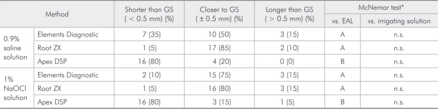
The distributions and the percentages of short-er than, closshort-er to, or longshort-er than the gold standard measurements are presented in Table 1.
Independent of the irrigating solutions used (0.9% saline solution and 1% sodium hypochlorite), the majority of measurements taken with the Ele-ments Diagnostic and Root ZX EALs were closer to those of the gold standard, whereas the major-ity of measurements taken with the Apex DSP EAL
were shorter than those of the gold standard. For both irrigating solutions used, the mean of the absolute differences from the gold standard ob-tained with the Apex DSP were signiicantly higher than those obtained with the other two EALs used (Table 2).
Discussion
As pointed out by several authors, the apical region may present important anatomic variations which make it dificult to locate the CDJ and the apical foramen.10-13 Determination of the apical
fo-ramen location, or working length analysis, has tra-ditionally been performed using radiographic meth-ods. However, because of the limitations associated with image interpretation in such methods, the use of electronic apex locators has become increasingly prevalent in clinical practice.16,18-20 Nevertheless,
studies have shown differences in the accuracy of EALs.6,8,16,19-20 Thus, in order to compare the
accu-Table 1 - Distribution and percentage of electronic measurements: shorter than, closer to and longer than those obtained using the gold standard method.
Method Shorter than GS
( < 0.5 mm) (%)
Closer to GS ( ± 0.5 mm) (%)
Longer than GS ( > 0.5 mm) (%)
McNemar test*
vs. EAL vs. irrigating solution
0.9% saline solution
Elements Diagnostic 7 (35) 10 (50) 3 (15) A n.s.
Root ZX 1 (5) 17 (85) 2 (10) A n.s.
Apex DSP 16 (80) 4 (20) 0 (0) B n.s.
1% NaOCl solution
Elements Diagnostic 2 (10) 15 (75) 3 (15) A n.s.
Root ZX 1 (5) 16 (80) 3 (15) A n.s.
Apex DSP 16 (80) 3 (15) 1 (5) B n.s.
*Significance obtained through McNemar test comparing the methods in pairs. Different letters indicate statistically significant differences among the EALs (P < 0.05).
Irrigants/EALs Absolute difference Kruskal–Wallis test * Mean SD vs. EAL vs. irrigating solution
0.9% saline solution
Elements Diagnostic 0.57 0.39 A n.s.
Root ZX 0.33 0.34 A n.s.
Apex DSP 1.15 0.80 B n.s.
1% NaOCl solution
Elements Diagnostic 0.53 0.55 A n.s.
Root ZX 0.37 0.29 A n.s.
Apex DSP 1.30 0.90 B n.s.
*Significance obtained through Kruskal–Wallis test. Different letters indicate statistically significant differences among the EALs (P < 0.05).
racy of three currently used electronic apex locators, Root ZX, Elements Diagnostic, and Apex DSP, this study was performed using human, single-root-ed teeth.
There are various ways to try to reproduce in
vivo conditions. Some authors use an alginate
mod-el or gmod-elatin, others use a sponge soaked in a solu-tion chosen by the authors.21-22 In the present study,
the in vitro model employed aimed to reproduce
in vivo conditions using a sponge soaked in saline
solution. Saline solution has properties similar to those of alveolar bone and its luids, according to well-established protocols.16 Regarding the
irrigat-ing solutions, manufacturers have provided infor-mation that the presence of luids or irrigating solu-tions such as sodium hypochlorite does not interfere with the accuracy of the devices. In fact, this inding was conirmed in this study because the irrigating solutions did not interfere with the results obtained with any of the EALs used. Jenkins et al.18 achieved
similar results testing Root ZX in different solu-tions, as did Kang and Kim17 comparing different
EALs under various conditions.
Using the visual and radiographic methods as the gold standard for locating the CDJ, we have shown that the data obtained with Elements Diag-nostic and Root ZX locators were more accurate than those of Apex DSP, independent of the irri-gating solution. In fact, most of the measurements
obtained with Elements Diagnostic and Root ZX locators were closer to the GS measurements.
Many studies on the performance of the Elements Diagnostic and Root ZX EALs have shown high percentages of measurements close to those of the gold standards, with no signiicant differences be-tween these two EALs.4,8,19,23-24 In other studies, the
high accuracy of the Root ZX was conirmed.6,16,20
Moreover, the Apex DSP showed low accuracy in a recent study by de Camargo et al.,24 in which the
au-thors measured the accuracy of this EAL in function of the prelaring of root canals. These indings are consistent with our study in pointing out the impor-tance of choosing the best possible EAL system, as different devices give measurements with different levels of accuracy.
Conclusions
Within the limitations of this in vitro study, we concluded that Elements Diagnostic and Root ZX apex locators were able to locate the cementum-dentine junction more precisely than Apex DSP. Moreover, the presence of irrigating solutions does not interfere with the performance of the EALs.
Acknowledgments
We would like to thank Prof. Dr. Fausto Me-deiros Mendes (Universidade de São Paulo) for his support with statistical analysis.
References
1. Pallares A, Faus V. An in vivo comparative study of two apex locators. J Endod. 1994 Dec;20(12):576-9.
2. Nguyen HO, Kaufman AY, Komorowski RC, Friedman S. Electronic length measurement using small and large files in enlarged canals. Int Endod J. 1996 Nov;29(6):359-64. 3. Nekoofar MH, Ghandi MM, Hayes SJ, Dummer PM. The
fun-damental operating principles of electronic root canal length measurement devices. Int Endod J. 2006 Aug;39(8):595-609. 4. Plotino G, Grande NM, Brigante L, Lesti B, Somma F. Ex vivo
accuracy of three electronic apex locators: Root ZX, Elements Diagnostic Unit, Apex Locator and ProPex. Int Endod J. 2006 May;39(5):408-14.
5. ElAyouti A, Dima E, Ohmer J, Sperl K, von Ohle C, Löst C. Consistency of apex locator function: a clinical study. J Endod. 2009 Feb;35(2):179-81.
6. Hassanien EE, Hashem A, Chalfin H. Histomorphometric study of the root apex of mandibular premolar teeth: an at-tempt to correlate working length measured with electronic and radiograph methods to various anatomic position in the apical portion of the canal. J Endod. 2008 Apr;34(4):408-12. 7. Venturi M, Breschi L. A comparison between two elec-tronic apex locators: an in vivo investigation. Int Endod J. 2005 Jan;38(1):36-45.
8. Brisenõ-Marroquín B, Frajlich S, Goldberg F, Willershausen B. Influence of instrument size on the accuracy of different apex locators: an in vitro study. J Endod. 2008 Jun;34(6):698-702.
10. Kuttler Y. Microscopic investigation of root apexes. J Am Dent Assoc. 1955 May;50(5):544-52.
11. Gutierrez JH, Aguayo P. Apical foraminal openings in human teeth (number and location). Oral Surg Oral Med Oral Pathol Oral Radiol Endod. 1995 Jun;79(6):769-77.
12. Green D. Stereomicroscopic study of 700 root apices of maxil-lary and mandibular posterior teeth. Oral Surg Oral Med Oral Pathol. 1960 Jun;13(6):728-33.
13. Burch JG, Hulen S. The relationship of the apical foramen to the anatomic apex of the tooth root. Oral Surg Oral Med Oral Pathol. 1972 Aug;34(2):262-8.
14. Lucena-Martin C, Robles-Gijon V, Ferrer-Luque CM, de Mondelo JM. In vitro evaluation of the accuracy of three electronic apex locators. J Endod. 2004 Apr;30(4):231-3. 15. ElAyouti A, Kimionis I, Chu AL, Lost C. Determining the
apical terminus of root-end resected teeth using three modern apex locators: a comparative ex vivo study. Int Endod J. 2005 Nov;38(11):827-33.
16. Goldberg F, Marroquin BB, Frajlich S, Dreyer C. In vitro evaluation of the ability of three apex locators to deter-mine the working length during retreatment. J Endod. 2005 Sep;31(9):676-8.
17. Kang JA, Kim SK. Accuracies of seven different apex locators under various conditions. Oral Surg Oral Med Oral Pathol Oral Radiol Endod. 2008 Oct;106(4):e57-62.
18. Jenkins JA, Walker WA 3rd; Schindler WG, Flores CM. An in vitro evaluation of the accuracy of the Root ZX in the presence of various irrigants. J Endod. 2001 Mar;27(3):209-11. 19. Tselnik M, Baumgartner JC, Marshall JG. An evaluation of
Root ZX and Elements Diagnostic Apex Locators. J Endod. 2005 Jul;31(7):507-9.
20. D’Assunção FL, Albuquerque DS, Ferreira LC. The ability of two apex locators to locate the apical foramen: an in vitro study. J Endod. 2006 Jun;32(6):560-2.
21. Pascon EA, Marrelli M, Congi O, Ciancio R, Miceli F, Ver-siani MA. An ex vivo comparison of working length deter-mination by 3 electronic apex locators. Oral Surg Oral Med Oral Pathol Oral Radiol Endod. 2009 Sep;108(3):e147-51. 22. Guise GM, Goodell GG, Imamura GM. In vitro comparison of
three electronic apex locators. J Endod. 2010 Feb;36(2):279-81.
23. Bernardes RA, Duarte MA, Vasconcelos BC, Moraes IG, Bernardineli N, Garcia RB, et al. Evaluation of precision of length determination with 3 electronic apex locators: Root ZX, Elements Diagnostic Unit and Apex Locator, and Ro-miAPEX D-30. Oral Surg Oral Med Oral Pathol Oral Radiol Endod. 2007 Oct;104(4):e91-4.