Article
ISSN 0102-695X http://dx.doi.org/10.1590/S0102-695X2012005000028
Received 27 Jul 2011 Accepted 16 Dec 2011 Available online 3 Feb 2012
of aqueous extract from aerial parts of Linaria
genistifolia subsp. genistifolia
Recep Liman,
*Uğur Gürol Gökçe, Dilek Akyıl, Yasin Eren,
Muhsin Konuk
Biology Department, Faculty of Science and Literatures, Afyon Kocatepe University, Afyonkarahisar, Turkey.
Abstract: Genotoxic and mutagenic effects of aqueous extract from aerial parts Linaria genistifolia (L.) Mill. subsp. genistifolia, Plantaginaceae (Lg-ext) were investigated by using both Allium cepa root meristematic cells and bacterial reverse mutation assay in
Salmonella typhimurium TA98 and TA100 with or without metabolic activation system (S9), respectively. In Allium root growth inhibition test, EC50 value was determined approximately 15 g/L and 0.5xEC50, EC50 and 2xEC50 concentrations of Lg-ext were introduced to onion tuber roots and distilled water and methyl methane sulfonate (MMS, 10 ppm) used as a negative and positive control, respectively. The characteristic effect caused by tested preparations was an increase of mitotic index (MI) in 7.5 g/L and 15 g/L (except 24 and 96 h) and simultaneous decrease of MI in 30 g/L and in MMS. While stickiness, bridges, chromosome laggards and disturbed anaphase-telophase were observed in anaphase-telophase cells, c-metaphase, pro-metaphase, polyploidy and binuclear cells were observed in other cells. Lg-ext was not found to be mutagenic on
S. typhimurium TA 98 and TA100 with or without S9. The results were also analyzed statistically by using SPSS for Windows, and Duncan’s multiple range tests were performed respectively. These results indicate that Lg-ext exhibits genotoxic activity in
A. cepa root meristematic cells but not mutagenic activity in Ames test system.
Keywords:
Allium
Ames chromosome aberration
Linaria genistifolia
Plantaginaceae
Introduction
The genus Linaria, a well known genus of the family Plantaginaceae, is widely distributed throughout the northern hemisphere with its centre of distribution in the Mediterranean basin and eastern Asia. It comprises currently around 200 species, twenty of which are in Turkey popularly known as “nevruz otu” and nine of them endemics (Davis, 1978; Seçmen et al., 2000). Some Linaria species have been used in folk remedies for various purposes such as hemorrhoid, skin eruptions, sores, ulcers, diuretic, laxative, tonic, antiscorbutic, antidiabetic and to treat some vascular disorders. Dosage is critical and it should not be given to pregnant women, since the plant might be slightly toxic (San Feliciano et al., 1993; Baytop, 1999). Linaria species are reported to contain alkaloids, iridoid glucosides, l avonoids, aurones and diterpenoids (Otsuka, 1992; Ilieva et al., 1993; Bianco et al., 1996; Hua et al., 2002; Ahmad et al., 2006; Tundis et al., 2008; Ferhat et al., 2010). They contains a poisonous glucoside that is reported to be mildly poisonous to cattle (Moroshita, 1991).
Vicia faba, Tradescantia paludosa, Pisum sativum, Hordeum vulgare, Crepis capillaris and A. cepa
are used to study of cytogenetic and genotoxic effect of plant extract or chemical(s). Among them, Allium test is one of the best-established test systems in order to determine the toxicity in the laboratories (Fiskesjö, 1985; Grant, 1992; Rank, 2003; Saxena et al., 2005; Konuk et al., 2007; Liman et. al., 2011). Because onions are easy to store and to handle, and also macroscopic and microscopic parameters can be observed easily. Moreover this system is well correlated with the data obtained from eukaryotic and prokaryotic systems (Fiskesjö, 1988).
Examining the mutagenicity of plant extract or chemical(s), Ames test, or the so-called Salmonella/ microsome test, is widely used (Konuk et al., 2008; Uysal et al., 2010; Liman et al., 2010). This test can be carried out rapidly and cheaply and it is also one of the most reliable short-term bacterial test systems (Maron & Ames, 1983; Mortelmans & Zeiger, 2000). In this system, S. typhimurium mutant strains, obtained from S. typhimurium LT2 parental line in vitro, are employed (Ames et al., 1975).
with or without S9, respectively.
Materials and Methods
Organisms
The S. typhimurium test strains TA98 and TA 100 were obtained from Nuran Diril, Hacettepe University, Turkey. While TA98 were used for determining the frame shift, TA100 was used to determine the base pair exchange type of mutations. Allium cepa (2n=16) onion bulbs, 25-30 mm diameter, without any treatment, were purchased from a local supermarket.
Chemicals
S9 from Liver of rat (Sprague-Dawley), Bacto agar, nutrient broth no.2 (Oxoid), 2-aminoanthracene (2AA), β-nicotinamide-adenine dinucleotide phosphate (β-NADP), glucose-6-phosphate (G6P), mitomycin-C (MMC), ampicillin and histidine were obtained from Sigma-Aldrich. Sodium azide (SA), citric acid monohydrate, sodium hydroxide, potassium chloride, sodium chloride, and dimethyl sulphoxide (DMSO) were purchased from Riedel. 4-Nitro-O-phenylenediamine (NPD) and 2-aminoluorene (2AF) were purchased from Fluka. Magnesium chloride, crystal violet, potassium phosphate and sodium ammonium phosphate were obtained from Merck.
Plant collection and extraction
The aerial parts of Linaria genistifolia (L.) Mill. subsp. genistifolia, Plantaginaceae, were collected from Başören Village, Afyonkarahisar, Turkey, in late-May 2010. The taxonomic identification of plant materials was confirmed by Dr Mehmet Temel, Department of Biology, Afyon Kocatepe University, Turkey. A voucher specimen was deposited at the Herbarium of Afyon Kocatepe University (number Kala 1410). Air dried and powdered aerial parts of the L. genistifolia subsp. genistifolia (80 g) were extracted with 1 L boiling water for 10 min (Sofowora, 1999). Both the decoctions and squeezed extracts were filtered with a 2.5-µm filter (Whatman® no. 42) to remove the suspended particles
and stored at 4 °C until usage. These were considered as the stock solutions and freshly prepared extracts were applied daily.
Allium cepa anaphase-telophase test
Prior to initiating the test, the outer scales of the bulbs and the dry bottom plate were removed without destroying the root primordia. In order to determine effective concentration (EC50), for each water sample,
a series of six bulbs were placed in distilled water for 24 h and afterwards the best growing ive bulbs exposed for four days (d) to the Lg-ext solutions (10, 20, 40, 60 and 80 g/L, respectively) at room temperature (~21±4 °C). The test concentrations were renewed at every 24 h during the experiments. On the 5th day, root lengths (lengths of
ten roots from each bulb) were measured from both Lg -ext exposed bulbs, and control group. EC50 value was considered as the concentration which retards the growth of root 50% when compared to the control.
In the determination of application doses, 2xEC50, EC50 and 0.5x EC5, positive (MMS, 10 ppm) and negative control group were used for 12, 24, 48, 72 and 96 h. Fixation and staining of the root tip cells were carried out as reported earlier (Yıldız et al., 2009; Liman et al., 2010). The MI and the frequencies of chromosomal aberrations (CA) were carried out according to Saxena et al. (2005). For each test group, ive slides (1 root tip/slide) were prepared by squashing root tips with 45% acetic acid. Slides were randomly coded and scored blindly. For MI, the different stages of mitosis were counted in a total of 5000-6000 cells (1000 cells/slide) per concentration, and expressed as a percentage. In chromosome aberration test, 100 cells in anaphase or telophase were examined for aberrations per slide if it is possible.
Ames plate incorporation test
Cytotoxic doses of Lg-ext (3000, 1500, 750, 375, 187.5 and 93.75 mg/plate) were determined by following the method of Dean et al. (1985).
Ames test was performed as a standard plate incorporation assay with S. typhimurium strains TA98 and TA100 with or without S9 (Maron & Ames, 1983). Selection of the strains was based on the testing and strain selection strategies of Mortelmans & Zeiger (2000). These strains were tested on the basis of associated genetic markers. For each tester strain, a speciic positive control was always used to test the experimental laws, if any. While 4-nitro-O-phenylenediamine (NPD) for TA 98 and sodium azide (SA) for TA100 were used as positive controls without S9, 2-aminoluorene (2AF) and 2-aminoanthracene (2AA) were used as positive controls with S9, respectively.
S9 mix (500 μL) (or 500 μL phosphate buffer), the test solution (100 μL) for each concentration and a cell suspension (100 μL) from an overnight culture (1-2x10⁹ cells/mL) were added to 2 mL top agar (kept
Statistical analysis
The data of root length, MI, mitotic phases, CA expressed as percentages, and the levels of signiicance in different treatment groups were analyzed statistically. For these, Duncan multiple range tests were performed by using one-way analysis of variance (ANOVA) on SPSS 15.0 version for Windows software both A. cepa anaphase-telophase and Ames test.
Results and Discussion
Herbal medicine is still the mainstay of about 75-80% of the whole population, mainly in developing countries, for primary health care because of better cultural acceptability, better compatibility with the human body and fewer side effects. However, an increase in the usage of these plants in the developed world has been observed for a few years (Parekh et al., 2005). Therefore, knowledge of mutagenic and toxic effects of these plants become very important. Because many plants synthesize toxic substances for defense against viruses, bacteria and fungi etc. and these compounds could have potentially deleterious effects in humans. Neither phytochemical nor biological studies of L. genistifolia subsp. genistifolia have been previously reported on the topic studied.
Test systems to determine the genotoxicity and/ or mutagenicity can be divided into groups based on the biological systems employed and their genetic endpoint detected. Bioassays with prokaryotes enable the detection of agents that induce gene mutation and primary DNA damages. On the other hand, analyses with eukaryotes enable the detection of a greater damage extent, varying from gene mutations to chromosome damages and aneuploidies (Houk, 1992; Leme & Marin-Morales, 2009). Using both pro- and eukaryotic test system make the results both strengthen and correlate to verify if the chemical(s) has/have really any bad effects on the genes.
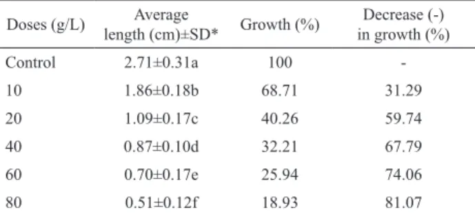
Allium root growth test results are shown in Table 1. The EC50 was found to be approximately 15 g/L. It can be easily seen that the effect of Lg-ext in Allium root growth was dose-dependent. Root growth decreased at all concentrations tested and yielded with statistically signiicant results (p<0.05). In the meantime over 40 g/L concentration, roots became dark colored, thicker and gel like formations. The inhibition of root growth generally related to apical meristematic activity (Webster & Macleod, 1996), and to cell elongation during differentiation (Fusconi et al., 2006) and the appearance of stunted roots indicate the retardation of growth and cytotoxicity (Yıldız et al., 2009). As reported earlier, neo-clerodane diterpenoids and lavonoids were isolated from
Linaria saxatilis var. glutinosa by showing cytotoxic activity in different neoplastic cell cultures (Gordaliza et al., 1997). Tundis et al. (2005) determined the
antiproliferative action of several lavones isolated from
Linaria relexa Desf., Plantaginaceae, against the large cell lung carcinoma cell line COR-L23, hepatocellular carcinoma cell line HepG-2, renal adenocarcinoma cell line ACHN, amelanotic melanoma cell line C32 and colorectal adenocarcinoma cell line Caco-2, and reported that pectolinarigenin and some lavonoid glycosides like pectolinarin exhibited strong cytotoxic activity on COR-L23 cell line with an IC50 value of 5.03 and 4.07 μM, respectively. Akkol & Elçi (2009) suggested that the extracts of Linaria species (L. grandilora, L. genistifolia subsp. confertilora and L. aucheri) had analgesic and anti-inlammatory effects without toxicity.
Table 1. Results of the Allium root growth inhibition test. Doses (g/L) Average
length (cm)±SD* Growth (%)
Decrease (-) in growth (%)
Control 2.71±0.31a 100
-10 1.86±0.18b 68.71 31.29
20 1.09±0.17c 40.26 59.74
40 0.87±0.10d 32.21 67.79
60 0.70±0.17e 25.94 74.06
80 0.51±0.12f 18.93 81.07
*Means with the same letter do not differ statistically at the level of 0.05. SD: Standard deviation.
Table 2 summarizes the effect of Lg-ext on MI and mitotic phase in the root meristematic cells of A. cepa treated for 12, 24, 48, 72 and 96 h. While at all concentrations used in the incubations of root in 7.5 g/L and 15 g/L (except 24 and 96 h) increased MI, applications of 30 g/L and MMS decreased MI compared to negative control at each exposure time. The highest values were obtained from 12 h examination of 7.5 g/L concentrations of Lg-ext (45.18±0.75), and the lowest one in 96 h applications of MMS (20.14±0.62). The increased and decreased MI showed statistically signiicant results (p<0.05). As stated by Fernandes et al. (2007), the cytotoxicity levels of an agent can be determined by the increase or decrease in the MI. MI lower than the negative control may indicate that the growth and development of exposed organisms have been affected by test compounds. On the other hand, MIs above those of the negative control are result of the induction of increased cell division, which may characterize an event detrimental to cells, leading to uncontrolled proliferation and even tumor formation (Hoshina et al., 2002). The increased cell proliferation activity can be the consequence of a reduction of the time necessary for DNA repair (Evseeva et al., 2003).
and simultaneous decrease of anaphase index except in 72 and 96 h at 7.5 g/L and telophase index except in 12 h at 30 g/L. This might be an indication of the blockage of chfr point (control point between prophase/metaphase). Scolnic & Halazonetis (2000) reported that chrf deines a checkpoint that delays entry into metaphase in response to mitotic stress. Most of the increased and decreased phase index showed statistically signiicant results (p<0.05).
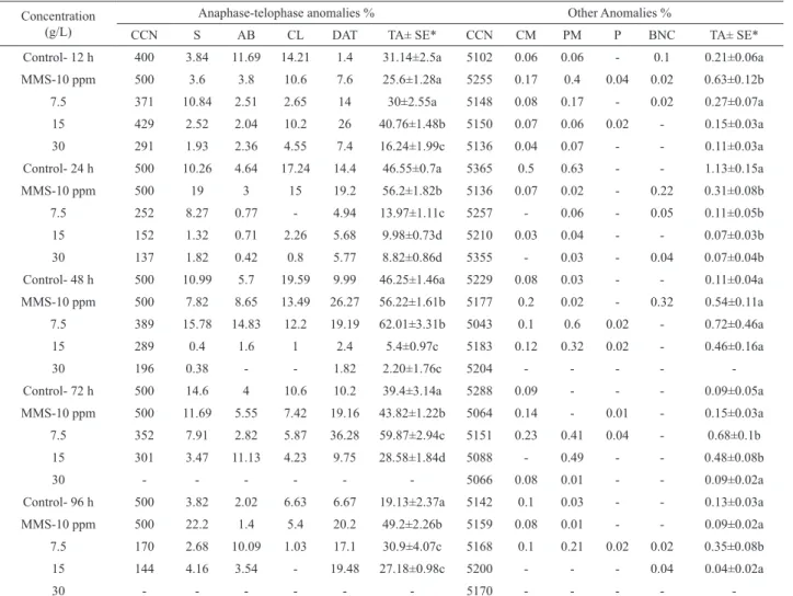
In the A. cepa anaphase-telophase chromosome aberration test conducted with root meristematic cells of A. cepa as shown in Table 3. The most frequent abnormalities were stickiness, anaphase bridges, chromosome laggards and disturbed anaphase-telophase in anaphase-telophase cells. The effect of Lg-ext concentration on CA was signiicantly different (p<0.05) except in 12 h at 7.5 g/L compared to the negative control. Analysis of the chromosomes showed that 7.5 g/L of Lg-ext concentration except in 12 and 24 h and 15 g/L of Lg-ext concentration in 12 and 96 h induced chromosomal aberrations but other Lg-ext concentrations decreased chromosomal aberrations. No aberration was recorded in the chromosome of A. cepa exposed to the 30 g/L of Lg-ext concentration in 72 and
96 h. Stickiness (especially at 7.5 g/L in 48 h) indicates highly irreversible type toxic effect of Lg-ext, and its occurrences during the study could be because of sub-chromatid linkage between chromosomes (Mc-Gill et al., 1974; Chauhan et al., 1986; Kovalchuk et al., 1998; Ajay & Sarbhoy, 1988). Anaphase bridges could happen during the translocation of the unequal chromatid exchange or due to dicentric chromosome presence or due to the breakage and fusion of chromosomes and chromatids. This bridges cause structural chromosome mutations (El-Ghamery et al., 2000; Luo et al., 2004). Disturbed anaphase-telophase (especially at 15 g/L in 12 h and at 7.5 g/L in 72 h) and chromosome laggards could occur by the effect of Lg-ext on microtubule formations (Amer & Ali, 1986; Kumari et al., 2009). Such spindle malfunctioning may arise due to inhibition of tubulin polymerization (Kuriyama & Sakai, 1974). The occurrence of chromosome laggards at anaphase was due to the failure of the chromosomes or acentric chromosome fragments to move to either of the pole.
In addition to these anomalies, others (c-metaphase, pro-metaphase, polyploidy and binuclear Table 2. The effects of Lg-ext on MI and mitotic phase of A. cepa root meristem cells.
Concentration (g/L) Counting cell number MI±SE* Mitotic Phases (%)±SE*
Prophase Metaphase Anaphase Telophase
Control- 12 h 5102 30.04±0.59a 88.92±1.31a 1.49±0.28a 3±0.68a 6.59±1.05ab
MMS-10 ppm 5255 27.08±1.56b 75.26±2.44b 4.75±0.5b 10.48±1.45b 9.51±1.27bc
7.5 5148 45.18±0.75c 91.49±1.36a 1.73±0.39a 1.4±0.23a 5.38±1.65a
15 5150 33.34±0.47d 93.29±0.54a 1.4±0.09a 1.24±0.36a 4.07±0.81a
30 5136 24.31±0.6e 84.55±0.53c 3.35±0.38c 1.58±0.41a 10.52±1.11c
Control- 24 h 5365 32.76±1.07a 87.99±1.7ab 4.72±1.48a 1.14±0.3a 6.15±0.83a
MMS-10 ppm 5136 26.67±0.63b 86.89±1.27a 3.57±0.31a 3.24±0.42b 6.3±1.08a
7.5 5257 38.29±0.99c 95.76±0.9d 0.56±0.19b 0.78±0.21a 2.9±0.8b
15 5210 28.49±0.53b 91.71±1.55bc 1.45±0.38b 0.75±0.17a 6.09±1.17a
30 5355 28.12±0.69b 93.16±0.49cd 0.4±0.09b 0.88±0.37a 5.56±0.62ab
Control- 48 h 5229 29.14±1.04a 84.43±0.67a 3.25±0.56a 4.69±0.5a 7.63±0.42a
MMS-10 ppm 5177 24.27±0.83b 86.91±0.93a 3.45±0.46a 3.86±0.36a 5.78±0.69ab
7.5 5043 32.71±0.63c 92.79±1.06b 3.52±0.95a 2.24±0.93b 1.45±0.72c
15 5183 33.96±1.03c 97.05±1.3c 1.17±0.37b 0.51±0.23c 1.27±0.86c
30 5204 25.56±1.23b 95.78±0.69bc - 0.13±0.03c 4.09±0.73b
Control- 72 h 5288 29.37±1.52a 84.44±0.64a 3.27±0.15ab 2.98±0.56a 9.31±0.47a
MMS-10 ppm 5064 25.18±1.37b 82.79±0.49a 4.71±0.12b 3.21±0.67a 9.29±0.44a
7.5 5151 37.75±0.36c 89.4±0.61b 4.14±0.49ab 4.72±0.25b 1.74±0.22b
15 5088 33.01±0.81c 93.67±0.97c 2.55±0.58a 0.67±0.22c 3.11±0.26b
30 5066 22.54±1.19b 93.56±1.86c 2.73±1.11a 1.32±0.37c 2.39±1.06b
Control- 96 h 5142 31.58±1.04a 85.45±1.15ab 2.76±0.42a 3.01±0.46a 8.77±1.1a
MMS-10 ppm 5159 20.14±0.62b 82.92±1.62a 4.61±0.32b 3.53±0.51ab 8.92±1.16a
7.5 5168 35.45±1.38c 88.69±0.77b 2.79±0.46a 4.85±0.92b 3.65±0.66b
15 5200 26.27±0.63d 94.54±1.1c 1.03±0.34c 0.66±0.27c 3.76±0.96b
30 5170 24.82±0.85d 96.61±0.53c - 0.24±0.16c 3.13±0.39b
Table 3. Percentage of chromosome aberrations of Lg-ext in different times and concentrations obtained for the A. cepa anaphase-telophase test.
Concentration (g/L)
Anaphase-telophase anomalies % Other Anomalies %
CCN S AB CL DAT TA± SE* CCN CM PM P BNC TA± SE*
Control- 12 h 400 3.84 11.69 14.21 1.4 31.14±2.5a 5102 0.06 0.06 - 0.1 0.21±0.06a
MMS-10 ppm 500 3.6 3.8 10.6 7.6 25.6±1.28a 5255 0.17 0.4 0.04 0.02 0.63±0.12b
7.5 371 10.84 2.51 2.65 14 30±2.55a 5148 0.08 0.17 - 0.02 0.27±0.07a
15 429 2.52 2.04 10.2 26 40.76±1.48b 5150 0.07 0.06 0.02 - 0.15±0.03a
30 291 1.93 2.36 4.55 7.4 16.24±1.99c 5136 0.04 0.07 - - 0.11±0.03a
Control- 24 h 500 10.26 4.64 17.24 14.4 46.55±0.7a 5365 0.5 0.63 - - 1.13±0.15a
MMS-10 ppm 500 19 3 15 19.2 56.2±1.82b 5136 0.07 0.02 - 0.22 0.31±0.08b
7.5 252 8.27 0.77 - 4.94 13.97±1.11c 5257 - 0.06 - 0.05 0.11±0.05b
15 152 1.32 0.71 2.26 5.68 9.98±0.73d 5210 0.03 0.04 - - 0.07±0.03b
30 137 1.82 0.42 0.8 5.77 8.82±0.86d 5355 - 0.03 - 0.04 0.07±0.04b
Control- 48 h 500 10.99 5.7 19.59 9.99 46.25±1.46a 5229 0.08 0.03 - - 0.11±0.04a
MMS-10 ppm 500 7.82 8.65 13.49 26.27 56.22±1.61b 5177 0.2 0.02 - 0.32 0.54±0.11a
7.5 389 15.78 14.83 12.2 19.19 62.01±3.31b 5043 0.1 0.6 0.02 - 0.72±0.46a
15 289 0.4 1.6 1 2.4 5.4±0.97c 5183 0.12 0.32 0.02 - 0.46±0.16a
30 196 0.38 - - 1.82 2.20±1.76c 5204 - - - -
-Control- 72 h 500 14.6 4 10.6 10.2 39.4±3.14a 5288 0.09 - - - 0.09±0.05a
MMS-10 ppm 500 11.69 5.55 7.42 19.16 43.82±1.22b 5064 0.14 - 0.01 - 0.15±0.03a
7.5 352 7.91 2.82 5.87 36.28 59.87±2.94c 5151 0.23 0.41 0.04 - 0.68±0.1b
15 301 3.47 11.13 4.23 9.75 28.58±1.84d 5088 - 0.49 - - 0.48±0.08b
30 - - - 5066 0.08 0.01 - - 0.09±0.02a
Control- 96 h 500 3.82 2.02 6.63 6.67 19.13±2.37a 5142 0.1 0.03 - - 0.13±0.03a
MMS-10 ppm 500 22.2 1.4 5.4 20.2 49.2±2.26b 5159 0.08 0.01 - - 0.09±0.02a
7.5 170 2.68 10.09 1.03 17.1 30.9±4.07c 5168 0.1 0.21 0.02 0.02 0.35±0.08b
15 144 4.16 3.54 - 19.48 27.18±0.98c 5200 - - - 0.04 0.04±0.02a
30 - - - 5170 - - - -
-*Means with the same letter do not differ statistically at the level of 0.05 in each group. SE: Standard Error, CCN: Counting Cell Numbers, S: Stickiness, AB: Anaphase Bridge, CL: Chromosome Laggards, DAT: Disturbed Anaphase-Telophase, TA: Total Anomalies, PM: Pro-metaphase, CM: c-metaphase, P: Polyploidy, BNC: Binuclear cell.
cell) were also observed. While the lowest anomalies were observed 0.04±0.02% at the 15 in 12 h, the highest ones were observed 0.72±0.46% at 7.5 g/L in 48 h. Statistically signiicant (p<0.05) frequencies of other anomalies were recorded in 12 h at 7.5 g/L, all 24 h applications, in 72 h at 15 and 30 g/L, and also in 96 h at 7.5 g/L. It was not observed any anomalies in 48 and 96 h at 30 g/L. C-metaphase, a possibly reversible effect, might occur due to disturbed microtubules by Lg-ext and could be induced aneuploidies (Fiskesjö, 1988; Shahin & El-Amodi, 1991; Odeigah et al., 1997). Polyploidy cells can occur due to the lack of fragmoplast that prevents the formation of the daughter cells (Vidakovivé-Cifrek et al., 2002; Fernandes et al., 2007). Binuclear cells are accepted as the inhibition of cytokinesis in any control points of the cellular cycle (Ateeq et al., 2002).
The results from the Ames test are shown in Table 4. The interpretation of the Ames test results
(SA, 2AF, 2AA and NPD) showed signiicant increases relative to the spontaneous mutation rate in the two tested strains. While the highest value observed was in the TA100 with S9 at 750 µg/plate concentration of Lg-ext (216.4±11.78), and the lowest value was in the TA98 with S9 at 750 µg/plate concentration of Lg-ext (25.6±1.14). Most of the results, increasing or decreasing relative to negative control group, were not statistically signiicant at p<0.05 (Duncan test) in examined strains. In order to establish a dose-response relationship, 5 different concentrations of Lg-ext were tested, and no induced revertants were observed along the dose range tested in either with or without S9 with two strains. The results of the present study showed that all applications of Lg-ext were not found to be mutagenic S. typhimurium TA98 and TA100 with or without S9.
In conclusion, our results indicate that Lg-ext was found cytotoxic and genotoxic in Allium test but not found mutagenic in Ames test. Although some Linaria species have been used in folk remedies for various purposes, they should be used with caution, always following the traditional methods of preparation exactly. Further studies are necessary to determine the active ingredient or compound(s) of Linaria genistifolia subsp. genistifolia and should be tested in other biological test systems to obtain deinitive conclusions on their cytotoxic, genotoxic and mutagenic potential.
Acknowledgements
The authors wish to thank Afyon Kocatepe University Scientiic Research Committee for supporting this study inancially (Project No: 11.FEN.BIL.16) and Dr. M. Temel for the identiication of the plant.
References
Ahmad VU, Kousar F, Zubair M, Khan A, Ali MS, Choudhary
MI, Sener B 2006. A new iridoid glycoside from Linaria genestifolia. Fitoterapia 77: 12-14.
Ajay KL, Sarbhoy RK 1988. Cytogenetic studies on the effect of some chlorinated pesticides. Cytologia 53: 427-436.
Akkol EK, Elci D 2009. Antinociceptive and anti-inlammatory
activities of some Linaria species from Turkey. Pharm. Biol 47: 188-194.
Amer SM, Ali EM 1986. Cytological effects of pesticides. XVII.
Effect of the insecticide dichlorvas on root mitosis of
Vicia faba. Cytologia 51: 21-25.
Ames B, Mccann J, Yamasaki E 1975. Methods for detecting carcinogens and mutagens with the Salmonella/ mammalian microsome mutagenicity test. Mutat Res 31: 347-364.
Ateeq B, Adul Farrah M, Ali MN, Ahmad W 2002. Clastogenicity of pentachlorophenol, 2-4-D and butachlor evaluated by Allium Root Tip Test. Mutat Res 514: 105-113.
Baytop T 1999. Therapy with Medicinal Plants in Turkey, Past and Present, Nobel Tip Kitapevi, Istanbul.
Bianco A, Guiso M, Mazzei RA, Piccioni F, Nicoletti M, Seraini
M, Ballero M 1996. 69-O-Acetylantirrhinoside, a new iridoid glucoside from Linaria lava subsp. sardoa. Fitoterapia 67: 364-366.
Chauhan LKS, Dikshith TSS, Sundararaman V 1986. Effect of
deltamethrin on plant cells. I. cytological effects on the root meristem cells of Allium cepa. Mutat Res 171: 25-30.
Davis PH 1978. Flora of Turkey and the East Aegean Island. vol. 6. University press, Edinburgh, 825 pp.
Dean BJ, Brooks TM, Hodson-Walker G, Hutson DH 1985.
Table 4. Mutagenicity of Lg-ext towards S. typhimurium TA98 and TA100 strain with or without S9.
Agent Amount (µg/plate)
No of His+ Revertants/plate Mean±SD*
TA98 TA100
- S9 + S9 - S9 + S9
Lg-ext
3000 30.2±3.42a 28.6±1.81a 110.6±8.56a 214±10.41a
1500 31±3.67a 28±1.58a 111.4±4.33a 184±7.44a
750 30±2.73a 25.6±1.14a 105,2±2.38a 216,4±11.78a
375 31.6±1.81a 27.2±0.83a 109±8.09a 177,8±6.3a
187.5 29.4±3.2a 27±1.58a 108±6.72a 183,6±21a
93.75 31.2±2.38a 26.2±3.56a 102.2±4.43a 112,4±10.6a
Control 32±3.31a 34.8±1.48a 105±7.21a 147.6±12.05a
SA 10 1646.2±112.34b
2AA 5 2279.2±108.72b
2AF 200 885.6±81.15b
NPD 200 1380.4±114.61b
*Means with the same letter do not differ statistically at the level of 0.05. SD Standard deviation, SA: Sodium azide, 2AA: 2-aminoanthracene,
Genetic toxicology testing of 41 industrial chemicals.
Mutat Res 153: 57-77.
El-Ghamery AA, El-Nahas AI, Mansour MM 2000. The action of atrazine herbicide as an indicator of cell division on chromosomes and nucleic acid content in root meristems of Allium cepa and Vicia faba. Cytologia 65: 277-287.
Evseeva TI, Stanislav A, Geras’kin I, Shuktomova I 2003. Genotoxicity and toxicity assay of water sampled from a radium production industry storage cell territory by means of Allium-Test. J Environ Radioactiv 68: 235-248.
Ferhat M, Harkat H, Lavaud C, Haba H, Long C, Benkhaled M
2010. Iridoids and lavonoid from Linaria aegyptiaca
(L.) Dum. subsp. fruticosa. Biochem Sys Ecol 38: 833-835.
Fernandes TCC, Mazzeo DEC, Marin-Morales MA 2007. Mechanism of micronuclei formation in polyploidizated cells of Allium cepa exposed to triluralin herbicide. Pestic Biochem Phys 88: 252-259.
Fiskesjö G 1985. The Allium Test as standart in enviromental monitoring. Hereditas 102: 99-112.
Fiskesjö G 1988. The Allium Test-an alternative in environmental studies: The relative toxicity of metal ions. Mutat Res 197: 243-260.
Fusconi A, Repetto O, Bona E, Massa N, Gallo C, Dumas-Gaudot E, Berta G 2006. Effect of cadmium on meristem activity and nucleus ploidy in roots of Pisum sativum L. cv. Frisson seedlings. Env Exp Bot 58: 253-260. Gordaliza M, Miguel Del Corral JM, Luz de la puente M,
Garcia-Grávalos MD, San Feliciano A 1997. Cytotoxic activity of neo-clerodane diterpenoids. Bioorg Med Chem Lett 7: 1649.
Grant WF 1992. Cytogenetics studies of agricultural chemicals in plants in: genetic toxicology an agricultural perspective. Plenum Pres, Newyork, p. 335-378. Hoshina MM 2002. Evaluation of a possible contamination
of the waters of the Claro River-Municipality of Rio Claro, part of the Corumbataí River Basin, with the mutagenicity tests using Allium cepa. State University
of São Paulo, Rio Claro, SP. (İn Portuguese).
Houk VS 1992. The genotoxicity of industrial wastes and efluents - A review. Mutat Res 277: 91-138.
Hua H, Cheng M, Li X, Pei Y 2002. A new pyrroloquinazoline alkaloid from Linaria vulgaris. Chem Pharm Bull 50: 1393-1394.
Ilieva E, Handjieva N, Spassov S, Popov S 1993. 5-O-allosylantirrhinoside from Linaria species.
Phytochemistry 32: 1068-1070.
Konuk M, Akyıl D, Liman R, Özkara A 2008. Examination
of the mutagenic effects of some pesticides. Fresen Environ Bull 17: 439-442.
Konuk M, Liman R, Cigerci IH 2007. Determination of genotoxic effect of boron on Allium cepa root meristematic cells. Pakistan J of Bot 39: 73-79.
Kovalchuk O, Arkhipov I, Telyuk A, Hohn P, Kovalchuk L 1998. The Allium cepa chromosome aberration test reliably measures genotoxicity of soils of inhabited areas in the Ukraine contaminated by the Chernobyl accident. Mutat Res 415: 47-57.
Kumari M, Mukherjee A, Chandrasekaran N 2009. Genotoxicity of silver nanoparticles in Allium cepa. Sci Total Environ 407: 5243-5246.
Kuriyama R, Sakai H 1974. Role of Tubulin-Sh groups in polymerization to microtubules. Functional-Sh groups in tubulin for polymerization. J Biochem 76: 651-654. Leme DM, Marin-Morales MA 2009. Allium cepa Test in
environmental monitoring: A review on its application.
Mutat Res 682: 71-81.
Liman R, Akyıl D, Eren Y, Konuk M 2010. Testing of the
mutagenicity and genotoxicity of metolcarb by using both Ames/Salmonella and Allium Test. Chemosphere 80: 1056-1061.
Liman R, Cigerci IH, Akyıl D, Eren Y, Konuk M 2011.
Determination of genotoxicity of fenaminosulf by
Allium and Comet Tests. Pestic Biochem Phys 99: 61-64.
Luo LZ, Werner KM, Gollin SM, Saunders WS 2004. Cigarette smoke induces anaphase bridges and genomic imbalances in normal cells. Mutat Res 554: 375-385. Maron DH, Ames BN 1983. Revised methods for the Samonella
typhimurium mutagenicity test. Mutat Res 113: 173-215.
Mc-Gill M, Pathak S, Hsu TC 1974. Effects of ethidium bromide on mitosis and chromosomes: A possible materyal basis chromosomes stickiness. Chromosoma 47: 157-167.
Moroshita DW 1991. Dalmatian toadlax, yellow toadlax, black
henbane, and tansymustard: importance, distribution, and control. p. 399-408. In: James LF, Evans JO, Ralphs MH, Child RD (eds), Noxious range weeds. Westview Press, Boulder, San Francisco and Oxford.
Mortelmans K, Zeiger E 2000. The Ames Salmonella/ microsome mutagenicity assay. Mutat Res 445: 29-60.
Odeigah PGC, Nurudeen O, Amund OO 1997. Genotoxicity
of oil ield wastewater in Nigeria, Hereditas 126: 161-167.
Otsuka HJ 1992. Isolation of isolinariin A and B, new lavonoid
glycosides from Linariajaponica. J Nat Prod 55: 1252-1255.
Parekh J, Jadeja D, Chanda S 2005. Eficacy of aqueous and
methanol extracts of some medicinal plants for potential antibacterial activity. Turk J Biol 29: 203-210.
Rank J 2003. The method of Allium anaphase-telophase chromosome aberration assay. Ekologiia 1: 38-42. San Feliciano A, Gordaliza M, Miguel del Corral JM, de la
Saxena PN, Chauhan LKS, Gupta SK 2005. Cytogenetic effects of commercial formulation of cypermethrin in root meristem cells of Allium sativum: Spectroscopic basis of chromosome damage. Toxicology 216: 244-252.
Scolnic D, Halazonetis T 2000. Chfr deines a mitotic stress
checkpoint that delays entry into metaphase. Nature 406: 430-435.
Seçmen Ö, Gemici Y, Görk G, Bekat L, Leblebici E 2000. Tohumlu Bitkiler Sistematiği, Ege Ünv. Fen Fak. Kitaplar Serisi No 116, VI. Baskı, Ege Ünv. Basımevi, İzmir.
Shahin SA, El-Amoodi KHH 1991. Induction of numberical chromosomal aberrations during DNA synthesis using fungicides nimrod and rubigan-4 in root tips of Vicia faba L. Mutat Res 261: 169-176.
Sofowora A 1999. The state of medicinal plants research in Nigeria. In: Proceeding of workshop. Held at Ile-Ife, Nigeria; 1-373.
Tundis R, Deguin B,. Loizzo MR, Bonesi M, Statti GA, Tillequin F, Menichini F 2005. Potential antitumor agents: Flavones and their derivatives from Linaria
relexa Desf. Bioorg Med Chem Lett 15: 4757-4760. Tundis R, Deguin B, Dodaro D, Statti GA, Tillequin
F, Menichini F 2008. Iridoid glycosides from
Linaria multicaulis (L.) Miller subsp. multicaulis
(Scrophulariaceae). Biochem Syst Ecol 36: 142-145. United States Environmental Protection Agency (USEPA)
1996. United States Environmental Protection Agency
(USEPA), Health Effects Tests Guidelines: OPPTS.
Uysal A, Durak Y, Aladağ MO 2010. Investigation of mutagenic
effects of some plant growth regulators on Salmonella/ Microsome Test System. Fresen Environ Bull 19: 2170-2175.
Vidakovivé-Cifrek Z, Pavlica M, Regula I, Papers D 2002.
Cytogenetic damage in shallot (Allium cepa) root meristems induced by oil industry “High-Density Brines”. Environ Cont Toxicol 43: 284-291.
Webster PL, Macleod RD 1996. The root apical meristem and
its magrin, In: Waishel Y, Eshel A, Kafkai U (eds.),
Plant roots. the hidden half (2nd ed.), Marcel Dekker, New York, p. 51-76.
Yıldız M, Cigerci IH, Konuk M, Fidan AF, Terzi H 2009.
Determination of genotoxic effects of copper sulphate and cobalt chloride in Allium cepa root cells by chromosome aberration and comet assays. Chemosphere 75: 934-938.
*Correspondence
Recep Liman
Biology Department, Faculty of Science and Literatures, Afyon Kocatepe University,
ANS Campus, Gazligol Yolu, 03200-Afyonkarahisar/Turkey [email protected]