Received from Hospital Oswaldo Cruz de Palmas (HOCP), TO, Brazil, and Hospital Geral de Palmas Universidade de Brasília – UnB, Brazil.
1. Responsible for the Anesthesiology Department of HOCP, Tocantins
2. PhD; Professor of the Genetics and Morphology Department, Instituto de Biologia of UnB
Submitted on July 06, 2010 Approved on August 16, 2010
Correspondence to: Dra. Luiza Alves de Castro Arai 404- Sul Alameda -02 QR-07 Lote 35 Plano Diretor Sul
77021-618 – Palmas, TO, Brazil E-mail: [email protected] Scientiic Article
contamination of Anesthesia circuits by Pathogens
Luiza Alves de Castro Arai
1, Ricardo Bentes Azevedo
2Summary: Arai lAc, Azevedo RB – contamination of Anesthesia circuits by Pathogens.
Background and objectives: evaluation of contamination of anesthesia circuits by collecting 56 culture samples from the circular system; previously reprocessed tracheas by disinfection with 1% hypochlorite or 2% glutaraldehyde after being washed in non-sterile water and soap and dried by using compressed air and stored in surgical grade paper; and from other places of the non-reprocessed respiratory circuit, before anesthetic procedures.
Methods: Samples from the inspiratory and expiratory branches of the tracheas, canister, soda-lime, and collector jar (drain) through swab in Stuart medium and streaked in Agar blood, Mac conkey, and Sabouraud growth media.
Results: the level of contamination with fungus and bacteria in the inspiratory and expiratory branches of tracheas reached up to 39.3% in some sites; in some cases, more than one microorganism was present, 75% fungal and 25% bacterial contamination. cultures were positive for Candida sp., Dermatophytus sp., Penicillium sp., Aspergillus sp., Staphylococcus aureus, Staphylococcus saprophyticus, and Staphylococcus epider-midis. contamination was observed in 25% of the canisters with growth of Candida sp., Penicillium sp., Dermatophytus sp., Aspergillus sp., and
Fusarium sp. in the collector jar, a contamination rate of 36% was observed with growth of Candida sp., Dermatophytus sp., S. saprophyticus
and Acinetobacter baumannii. Microorganisms did not grow in soda-lime cultures.
Conclusions: in all sites investigated except for soda-lime growth of microorganisms was observed with the possibility of cross infection.
Keywords: equipment contamination; Anesthesia, closed-circuit; Ventilators, Mechanical; cross infection.
[Rev Bras Anestesiol 2011;61(1): 50-59] ©elsevier editora ltda.
INTRODUCTION
Soda-lime is an integral part of the anesthesiologist daily practice 1,2. it absorbs carbon dioxide in the respiratory circuit
of the anesthesia equipment, allowing the use of low low of fresh gases to reduce anesthetic consumption, maintain the body temperature of the patient, airways humidity, and avoid pollution of the operating room 3. Due to lower consumption of
gases and anesthetics, it is an economic ally of the hospital, besides maintaining the humidity of inhaled air, avoiding the harmful effects of dry gases, such as drying, inlammation, and loss of ciliary movement, it decreases low, reducing pul-monary complacency 4. Absorption of carbon dioxide
repre-sents an exothermic chemical reaction. in the closed circuit, water and heat from the reaction contribute for humidiication and warming of the mixture. Besides water from the chemical reaction, water also comes from the loss of liquid from the expiratory phase, which accumulates in the expiratory branch of the respiratory circuit, in the canister, and throughout the circuit therefore creating an ideal culture medium for growth of microorganisms 5.
Although soda-lime has a bactericidal effect, besides its cytolytic effect5, when there is direct contact between the
mi-croorganism and the soda-lime granule, experimental studies have reported survival of Pseudomonas aeruginosa, for 18
hours, and Mycobacterium tuberculosis, for 48 hours, in a
soda-lime solution 6, and that those microorganisms crossed
the soda-lime-containing canister.
in this ideal culture medium (heat and humidity) for micro-organisms in the respiratory circular system of the anesthetic device, Gram-negative bacteria such as Flavobacterium and Pseudomonas 7 have been isolated. Also, a case of
transmis-sion of hepatitis c, whose culprit was most likely the reutiliza-tion of the circular circuit 8 has been reported, as well as other
studies demonstrating the growth of several microorganisms in the circular system of the anesthesia equip ment 5,7,9-13.
currently, several steps have been used to resolve the con-tamination and possibility of cross transmission such as disin-fection and sterilization of corrugated tubings, sterilization of tubings, use of non-reusable circuits, and the use of bacterial ilters in the circuit.
through bacteriologic tests of the material from the inside of the expiratory branch of the valvular circuit, after prior dis-infection for 30 minutes with 2% glutaraldehyde, bacterial growth was observed despite the disinfection routine used 9.
Revista Brasileira de Anestesiologia 51 Vol. 61, no 1, January-february, 2011
interior of the inspiratory branch, growth of two Gram-negative bacteria that cause pulmonary infection, besides other from the saprophytic lora of the human skin was observed 10. the
objective of the present study was to evaluate the contamin-ation of reprocessed tubings and non-reprocessed parts of anesthesia circuits.
METHODS
After approval by the study nucleus and the General Board of the Hospital Geral de Palmas and Hospital Oswaldo Cruz de Palmas, to, Brazil, 56 samples were collected from takaoka, Dräger, and intermédica anesthesia equipment: 25 samples from takaoka, 20 samples from Dräger, and 11 samples from intermédica anesthesia equipment. Samples were collected before each anesthesia and they were divided in groups ac-cording to collection sites: inspiratory branch, expiratory bran-ch, canister, collector jar, and soda-lime (for each sample, three granules of soda-limes were collected).
collection was performed with a swab with Stuart medium, a transport and conservation culture medium, and streaked in culture media for growth and isolation, i.e., Agar blood, Mac conkey, Mannitol, and Sabouraud.
initially, the person responsible for the operating room was asked whether tracheas of the anesthesia circuits had been changed; if affirmative, samples were collected.
Samples were collected using a swab with a transport me-dium, from within the inspiratory and expiratory branches, from their proximal and distal portions, with circular movements in their internal surface. Material was also collected from the in-ternal walls of the canister before anesthesia.
collections were performed in the circulatory circuits of Dräger, intermédica, and takaoka anesthesia equipment.
in the intermédica anesthesia equipment, shortly after the canister in the circular circuit, there is a recipient used to drain (collector jar) the liquid accumulated during surgery.
in all cases, soda-lime granules were collected before anesthesia; therefore, for each anesthesia, there were at least four samples, and when the intermédica was used, ive samples.
Samples were collected with a swab containing a transport media, so samples remained viable; although it is not a nutri-tive medium, the Stuart medium, a semi-solid medium cont-aining thiglycolate, glycerol phosphate, and sodium chloride, was chosen since it preserves the viability of most pathogens.
the routine of washing corrugated tracheas and how em-ployees cleaned them was followed-up. it was observed that after each anesthesia, corrugated tracheas were collected and sent to the surgical instrument cleaning service. there, they were washed on tap water and placed in a solution of 1% sodium hypochlorite for 30 minutes. After this proce dure, those corrugated tracheas were washed in running water, dried with compressed air, and packed in non-sterile surgical grade paper and sent along with sterile material to be stored. After some time, when approximately 50% of samples were collected in the Hospital Geral de Palmas, by determination of
the ccih, there was a change in protocol, and 2% glutaralde-hyde17 was used in place of the hypochlorite.
in some occasions, it was observed that tracheas in the anesthesia equipment were not changed, but employees still conirmed they were changed.
in Hospital Oswaldo Cruz, a change in protocol according to the ccih was also done. however, tracheas were steril-ized after removal of dirty material; this change in protocol was done after approximately 80% of samples had been col-lected.
in those hospitals, an attempt to standardize the soda-lime was made; it would only be changed after a standard retention of co2, according to capnography had been achieved16;
how-ever, anesthesiologists’ staff change all the time, and some of them ordered the change of soda-lime whenever its color changed from white to purple, while others order its change whenever their shift began. it was observed that, in the majo-rity of samples, it was replaced whenever its color changed, with a mean time of three days. in three cases, the soda-lime was changed after, approximately, 15 days. And in some ca-ses, the change in soda-lime was not recorded.
in one of the hospitals cleaning of the canister was standard ized. it was washed with water and soap, disinfected with an alcohol solution and/or with sodium hypochlorite; in the other hospital the soda-lime was changed, but the canister was not cleaned.
With a total of 235 samples, which were streaked in culture media for Gram-positive and Gram-negative bacteria and fun-gus, a total of 705 cultures were performed. of those, only the cultures of soda-lime did not show any growth.
During collection, it was evaluated whether the samples did not touch any other part of the anesthesia equipment to avoid false-positive results.
it was observed that anesthetic procedures lasted a mean of two hours and thirty minutes. At the end of the procedure, the circuits were changed and the external part of the equip-ment was disinfected with 70% alcohol. it was attempted to establish a mean time between the use of the equipment and a new collection; however, due to the rotation of anesthesiolo-gists and the variety in anesthetic techniques used it was not possible. therefore, the time among collections, the reuse of the equipment, and a new collection varied.
the software excel® for Windows® was used for
proces-sing the data. the software SPSS® (Statistic Package for the
Social Sciences, chicago, il, uSA) version 13 for Windows®
was used for all statistical analysis.
RESULTS
Samples were collected from 56 anesthetic procedures. for-ty-ive of those samples were collected from the inspiratory and expiratory branches of the trachea, canister, and soda-lime from takaoka and Dräger anesthesia equipment.
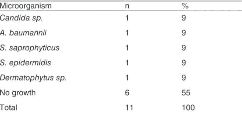
Table IV – Results of cultures form collecting Jars
Microorganism n %
Candida sp. 1 9
A. baumannii 1 9
S. saprophyticus 1 9
S. epidermidis 1 9
Dermatophytus sp. 1 9
no growth 6 55
total 11 100
Table I – Results of cultures of the Samples from the inspiratory Branches in Anesthetic Procedures
Microorganism Before
n %
Candida sp 4 7.1
Dermatophytus sp. 5 8.9
Penicillium sp. 5 8.9
other fungus 2 3.6
S. saprophyticus 4 5.4
S. aureus 1 1.8
no growth 35 64.3
total 56 100
Table II – Results of cultures of Samples from the expiratory Branches in Anesthetic Procedures
Microorganism Before
n %
Candida sp. 7 12.5
Dermatophytus sp. 6 10.7
Penicillium sp. 1 1.8
Aspergillum sp. 1 1.8
other fungus 2 3.6
S. aureus 2 3.6
S. epidermidis 1 1.8
S. saprophyticus 2 3.6
no growth 34 60.7
total 56 100
Table III – Results of cultures from canisters in Anesthetic Procedures
Microorganism Before
n %
Candida sp. 6 10.7
Dermatophytus sp. 3 5.4
Penicillium sp. 2 3.6
Aspergillum sp. 1 1.8
Fusarium sp. 1 1.8
no growth 41 76.7
total 56 100
Soda-lime: Growth of fungus or positive and Gram-negative bacteria was not observed in all cultures of 56 sam-ples of soda-lime granules.
inspiratory Branch: table i shows the results from the sam-ples of this site. in 19 samsam-ples (33.9%) collected before the procedure, the presence of at least one microorganism was demonstrated. Besides, more than one microorganism was detected in two of those samples (3.6%), Staphylococcus aureus, in one of the cases, and Staphylococcus saprophyti-cus, in the other. in four samples, growth of S. saprophyticus
was observed, while in one sample, the growth of S. aureus
was observed. Regard ing fungal growth, it was positive in 16 samples with the growth of Candida sp., Penicillium sp., and Dermatophytus sp.
expiratory Branch: Results of the samples are shown in table ii. twenty-two samples (39.3%) collected before the procedure showed growth of microorganisms. More than one organ ism was not detected in any of the samples. in ive sam-ples, the growth of S. aureus, S. saprophyticus, and Staphylo-coccus epidermidis was observed while seventeen samples
showed growth of fungus, Candida sp., Penicillium sp., Der-matophytus sp., and Aspergillus sp.
canister: fourteen samples (25%) collected before the procedure were positive (table iii) with growth of Candida sp., Penicillium sp., Dermatophytus sp., Aspergillus sp., and
Fusarium sp. Growth of more than one microorganism was
not observed.
collector jar: five samples were positive (45%). in two anesthetic procedures, more than one organism was ob-served, S. saprophyticus, in one of the cases (9.1%), and a Gram-negative bacterium, Acinetobacter baumannii, in the
other (9.1%). in another culture, growth of S. saprophyticus, as well as S. epidermidis, was observed. in two samples, the
growth of two types of fungus was observed in fungal-speciic media: Candida sp. and Dermatophytus sp. (table iV).
DISCUSSION
Since the antimicrobial activity of glutaraldehyde depends on the conditions of use, such as dilution, contents of orga-nic material 17, and inadequate use it can lead to the
deve-lopment of a bioilm, a microbial mass containing cell and extracellular material on the surface of objects immersed in liquids (including blood) 17. Similarly, aqueous chloride
solu-tions are bactericidal with good germicide action, although spore-forming microorganisms are 10 to 1000 times more resistant to chlor ide than vegetative bacteria, and organic material and alkaline detergents can reduce the efficacy of chloride compounds. chloride compounds are indicated for medium level disinfection of articles and surfaces as well as decontamination of surfaces 17. therefore, removal of
Revista Brasileira de Anestesiologia 53 Vol. 61, no 1, January-february, 2011
it was observed that initially corrugated tubes were washed with water and soap and placed in 1% hypochlorite or 2% glu-taraldehyde solution for 30 minutes, and later washed on tap not sterile water, and dried with compressed air. When this non-sterile air from a non-iltered humid system reaches the internal surface of those tubings, they are actually placing new microorganisms in the inspiratory and expiratory branches.
When the compressed air jet reaches the internal surfaces of the inspiratory and expiratory branches of the circular circuit of the equipment, in sites with dirt or bioilm residues, there is the possibility that the compressed air jet will remove the dirt spot and spread it in the interior of the tube therefore repopu-lating a greater area on the internal lumen of the tubing with still viable microorganisms due to the bioilm.
Since the compressed air jet, that is humid, does not have a bacterial or luid ilter, at the moment those corrugated tubes, dried, in theory, are packed in surgical-grade paper they are still humid due to the compressed air used with a ine layer of luid. At this moment, a proper environment for the growth of microorganisms was created.
in processed corrugated tubes, called cleaned, we obtai-ned cultures with growth of Candida sp., Dermatophytus sp., Aspergillus sp., and Penicillium sp., as well as the growth of bacteria, such as S. aureus, S. epidermidis, and S. sapro-phyticus.
even after being reprocessed, inspiratory and expiratory tubings showed a high index of contamination 4-6,9-13,28,19,
sug-gesting that the way reprocessing is being performed it is not effective and it did not achieve its objective.
Besides, there are parts of the respiratory circuit of the anesthesia equipment that are not cleaned between anes-thesias, such as the soda-lime canister, which, most times, is changed whenever the soda-lime is changed, without prior cleaning of the canister, after several anesthetic pro-cedures.
Directional valves, pop off valves, and reservoir balloons are also part of the anesthesia equipment through which ga-ses enter and leave the patient during mechanical ventilation; those gases are hot and humid, may it be by the process of respiration or release of heat and water from soda-lime during absorption of expired co2. this causes accumulation of liquid throughout the respiratory circuit, therefore creating a proper environment for the growth of microorganisms not only in the corrugated tubes but also in other places increasing the pos-sibility of crossed-contamination 6-8,11-13.
Although in the present study soda-lime cultures did not show growth of microorganisms, there are studies such as those of langevin who reported the survival of M. tuberculo-sis for more than 48 hours in a highly alkaline medium, and
the potential for its spread through the respiratory circuit of the anesthesia equipment 6,19.
in the cultures of the canister only the growth of viable fun-gus was observed, despite the alkaline medium it was not re-gularly washed. We obtained positive culture for Aspergillus sp., which can cause pulmonary infection 20.
Similar to Murphy et al. 13, that observed contamination in
several sites of the respiratory circuit of the anesthesia equip-ment, in the present study contamination was also observed. in the study conducted on the viability of bacteria after passing through the circular circuit of the anesthesia equipment, they recommended recommended the change of disposable parts and sterilization of all other parts of the circuit.
We questioned whether other parts of the respiratory cir-cular circuit of the anesthesia equipment from which samples were not collected could show a high degree of contamination.
the high index of contamination in the parts of the respira-tory circuit of the anesthesia equipment that were analyzed shows that reprocessing is ineffective in disinfecting. how-ever, even if those parts had been sterilized the whole pro-cess becomes invalid when parts of the same circuit are not clean ed between each anesthetic procedure, generating the possibility of crossed contamination among patients 6,8.
in a group of 520 patients undergoing inhalational general anesthesia the efficacy of low-resistance 0.22-micron bacte-rial ilters in the prevention of postoperative pneumonia14 was
evaluated. A difference in the development of pneumonia bet-ween the group that used the ilter and the group that did not use the ilter was not observed. those results suggest that the use of bacterial ilters does not have any inluence on the incidence of postoperative pneumonia.
in a study to evaluate the efficacy of cleaning the anesthesia equipment to determine whether the use of bacterial ilters is needed, three groups of patients, one with symptoms of respi-ratory tract disease, one with secretions in the respirespi-ratory tract, and the last one with chronic bronchitis, were evaluated 15. out
of 550 cultures performed before and after anesthesia only ive cultures were positive for non-pathogenic bacteria, indicating that colonization of the equipment is low and it is controlled by adequate cleaning and sterilization after the use not justifying the use of bacterial ilters in anesthesia equipment.
Although the corrugated tracheas of the circular system underwent disinfection with 1% hypochlorite or 2% glutaral-dehyde before each anesthetic procedure, culture of the sam-ples from their internal walls showed the presence of viable microorganisms.
REFERÊNCIAS / REFERENCES
01. Kits RJ, Vandam lD – histórico e objetivos da Prática Anestésica, em: Miller RD - tratado de Anestesia. 2ª ed, São Paulo, Manole, 1999;3-22.
02. collins VJ – técnica de Absorção do Dióxido de carbono, em: collins VJ - Princípios de Anestesiologia. 2ª ed, Rio de Janeiro, Guanabara Koogan, 1976;218-223.
03. Saraiva RA – como evitar a formação de substâncias tóxicas durante a absorção de dióxido de carbono pela cal sodada com uso de anes-tésicos halogenados. Rev Bras Anestesiol, 2004;54:431-437. 04. Pires oc, lacerda MA, fonseca nM et al. – normatização da
limpe-za do aparelho de anestesia. SBA/comissão de normas técnicas, 2006. Disponível em: http://www.sba.com.br /educacao/par_cntsa. asp. Acesso em 01/09/07.
05. teuler Rc – l’eicàcia dels circuits anestèsics: un nou sistema d’anestesia. Acadèmia de ciències Mèdiques i de la Salut de cata-lunya i de Baleares.
06. langevin PB, Rand Kh, layon AJ – the potential for dissemination of
Mycobacterium tuberculosis through the anesthesia breathing circuit, chest, 1999; 115:1107-1114.
07. Body Sc, Philip Jh – Gram-negative rod contamination of an ohmeda anesthesia machine. Anesthesiology, 2000;92:911.
08. heinsen A, Bendtsen f, fomsgaard A – A phylogenetic analysis elu-cidating a case of patient-to-patient transmission of hepatitis c virus during surgery. J hosp infect, 2000;46:309-313.
09. Pinto PS, Romero Mn, Bruno neto A et al. – contaminação das tra-queias do aparelho de anesthesia. Rev Bras Anestesiol, 1999;49(supl 24):95.
10. Stabile Jr Sl, cicareli DD, Momo t et al. – Avaliação da contamina-ção do circuito respiratório do aparelho de anestesia. Rev Bras Anes-tesiol, 1998;48:492-500.
11. leitjen Dt, Rejger VS, Mouton RP – Bacterial contamination and the effect of ilters in anaesthetic circuits in a simulated patient model. J hosp infect, 1992;21:51-60.
12. harrison GR - the contamination of volatile anaesthetics in an in-cir-cle vaporizer with water during prolonged closed-cirin-cir-cle anaesthesia. Anaesthesia, 2000; 55:791-792.
13. Murphy PM, fitzgeorge RB, Barrett Rf – Viability and distribution of bacteria ofter passage through a circle anaesthetic system. Br J Ana-esth, 1991;66:300-304.
14. Garibaldi RA, Britt MR, Webster c et al. – failure of bacterial ilters to reduce the incidence of pneumonia after inhalation anesthesia. Anes-thesiology, 1981;54:364-368.
15. Ping fc, oulton Jl, Smith JA et al. – Bacterial ilters - are they neces-sary on anaesthetic machine? can Anaesth Soc J, 1979;26:415-419. 16. Sociedade de Anestesiologia do estado de São Paulo – limpeza,
desinfecção e esterilização de equipamentos utilizados em anestesia. Disponível em: <http//www.saesp.org.br>.
17. Brasil. Agência nacional de Vigilância Sanitária – Segurança e equi-pamentos médico-hospitalares. Bol inf tecnovigil, 2004(4). 18. Rathgeber J, Kietzmann D, Mergeryan h et al. – Prevention of
pa-tient bacterial contamination of anaesthesia-circle-systems: a clinical study of the contamination risk and performance of different heat and moisture exchangers with electret ilter (hMef). eur J Anaesthesiol, 1997;14:368-373.
19. American Association nurse Anesthetists – infection control Guides. Part iii - infection control Procedures for Anesthesia equipment. 20. Murray PR, Rosenthal KS, Kobayashi GS et al. – Princípios Básicos
da Microbiologia Médica, em: Murray PR, Rosenthal KS, Kobayashi GS et al. – Microbiologia Médica, 4ª ed, Rio de Janeiro, Guanabara Koogan, 2004;5-80.
21. Medeiros eAS – infecção hospitalar: Situação Atual e Medidas de Prevenção, em: Salomão R, Pignatari Acc – Guia de Medicina Ambulatorial e hospitalar de infectologia. unifeSP-ePM, Manole, 2004;535-566.
Resumen: Arai lAc, Azevedo RB – contaminación del Aparato de Anestesia por Agentes Patógenos.
Justiicativa y objetivos: evaluar la contaminación de los aparatos de anestesia a través de recolecciones de 56 muestras para cultivo en el sistema circular del aparato de anestesia, en traqueas previa-mente reprocesadas por desinfección con hipoclorito al 1% o gluta-raldehido al 2%, después del lavado con jabón y agua no estériles, secadas con chorro de aire comprimido y almacenadas en papel qui-rúrgico, y en otros locales del circuito respiratorio no reprocesados, antes de los procedimientos anestésicos.
Método: fueron realizados cultivos de muestras de las traqueas de los ramos inspiratorios, ramos espiratorios, caníster, cal soda-da y frasco recolector (dreno), a través de swab con medio Stuart, y sembradas entre los cultivos Agar sangre, Mac conkey y Sa-bouraud.
conta-Revista Brasileira de Anestesiologia 59 Vol. 61, no 1, Janeiro-fevereiro, 2011
minación en algunos sitios fue de hasta un 39,3%, con la presencia de hongos y bacterias, siendo que en algunos casos había más de un microorganismo, un 75% de la contaminación por hongos y un 25% de bacterias. Se encontró un cultivo positivo para candida sp., Dermatophytus sp., Penicillium sp., Aspergillus sp., Staphylococcus aureus, Staphylococcus saprophyticus y Staphylococcus epidermi-dis. en el caníster, hubo contaminación en un 25%, con el aumento de candida sp., Penicillium sp., Dermatophytus sp., Aspergillus sp. y fusarium sp. en el frasco recolector, se observó la contamina-ción de un 36% de las muestras analizadas, con un crecimiento de
candida sp., Dermatoitus sp., S. saprophyticcus y Acinetobacter baumannii. en los cultivos de la cal sodada no hubo crecimiento de microorganismos.
Conclusiones: en todos los puntos analizados, con excepción de la cal sodada, hubo un aumento de los microorganismos, con la posibi-lidad de contaminación cruzada.