Received from the Divisão de Anestesiologia e Terapia Cirúrgica do Instituto do Coração of Hospital das Clínicas of Faculdade de Medicina of Universidade de São Paulo (InCor-HC-FM-USP), Brazil.
1. Master’s Degree in Anesthesiology from USP, Assistant Physician at InCor-HC-FM-USP. 2. PhD in Anesthesiology from USP. Assistant Physician at Anesthesiology Service of InCor-HC-FM-USP.
3. Anesthesiologist, Certiied by Sociedade Brasileira de Anestesiologia. Assistant Physician at Anesthesiology Service of InCor-HC-FM-USP.
4. Anesthesiologist, Certiied by Sociedade Brasileira de Anestesiologia. Professor at Anes-thesiology Discipline - Surgery Service, FM-USP.
Submitted on July 28, 2010. Approved on August 12, 2010.
Correspondence to:
Dr. José Otávio Costa Auler Júnior
Av. Dr. Enéas de Carvalho Aguiar, 44 - 2º andar - bloco I Cerqueira César
05403-000 – São Paulo, SP, Brazil E-mail: auler.junior@incor.usp.br Scientiic Article
intraosseous Anesthesia in Hemodynamic Studies in children
with cardiopathy
Ana Cristina Aliman
1, Marilde de Albuquerque Piccioni
2, João Luiz Piccioni, TSA
3, José Luiz Oliva, TSA
3,
José Otávio Costa Auler Júnior, TSA
4Summary: Aliman Ac, Piccioni MA, Piccioni Jl, Oliva Jl, Auler Júnior JOc – intraosseous Anesthesia in Hemodynamic Studies in children with cardiopathy.
Background and objectives: intraosseous (iO) access has been used with good results in emergency situations, when venous access is not available for luids and drugs infusion. the objective of this study was to evaluate iO a useful technique for anesthesia and luids infusion during hemodynamic studies and when peripheral intravascular access is unobtainable. the setting was an university hospital hemodynamics unit, and the subjects were twenty one infants with congenital heart disease enrolled for elective hemodynamic study diagnosis.
Methods: this study compared the effectiveness of iO access in relation to iV access for infusion of anesthetics agents (ketamine, midazolam, and fentanyl) and luids during hemodynamic studies. the anesthetic induction time, procedure duration, anesthesia recovery time, adequate hydration, and iV and iO puncture complications were compared between groups.
Results: the puncture time was signiicantly smaller in iO group (3.6 min) that in iV group (9.6 min). the anesthetic onset time (56.3 second) for the iV group was faster than iO group (71.3 second). no signiicant difference between groups were found in relation to hydration (iV group, 315.5 ml vs iO group, 293.2 ml), and anesthesia recovery time (iO group, 65.2 min vs iV group, 55.0 min). the puncture site was reevaluated after 7 and 15 days without signs of infection or other complications.
Conclusions: Results showed superiority for iO infusion when considering the puncture time of the procedure. Due to its easy manipulation and efficiency, hydration and anesthesia by iO access was satisfactory for hemodynamic studies without the necessity of other infusion access. Keywords: infusions, intraosseous; Heart Defects, congenital; Anesthesia; Angiography; Hemodynamics.
[Rev Bras Anestesiol 2011;61(1): 41-49] ©elsevier editora ltda.
INTRODUCTION
intraosseous (iO) infusion is considered an useful technique for the administration of medications and luids in emergency situations when peripheral intravascular access is inacces-sible and it was used during the World War ii. in 1941, tocantins et al. 1 introduced the technique for clinical use of luid
infusion that allows immediate access to the vascular system. However, iO infusion was gradually substituted by intravenous catheters (1950-1960) 2. in 1977, from venography studies 3,
the interest in iO infusion was renewed and recent literature has referred to the use of iO infusion in emergency situations 4,5.
Since the iO space has rigid veins that do not collapse in case
of hypovolemia or systemic circulatory failure, it has been considered as an alternative in emergency situations when venous access is extremely difficult. the iO technique is in-cluded in standard protocols and training procedures such as the Advanced Pediatric life Support textbook 6 and it is
recommended by the American Heart Association 7 and the
American Academy of Pediatrics 8.
infants with congenital heart disease when undergoing to hemodynamic studies need general anesthesia and they are mainly underweight infants whose venous access is very dif-icult. the aim of this study was to demonstrate the feasibility of iO access and its efficiency for administering anesthetics agents and hydration luids during hemodynamic studies to infants with congenital cardiopathies anddifficult venous ac-cess by conventional means.
METHODS
AliMAn, PicciOni, PicciOni et Al.
42 Revista Brasileira de Anestesiologia
Vol. 61, no 1, January-february, 2011
PROTOCOL
the irst step in the preparation of infants eligible for the he-modynamic study was to inform the parents about the risks associated with anesthesia and about the hemodynamic study. in addition, the parents were informed that the infant should not drink any water 3 hours and no milk 4 hours be-fore surgery 9. Premedication was intramuscular (iM)
keta-mine 10 1 mg.kg-1 for either venous access or intraosseous
technique. infants were separated into intravenous group (iV) and intraosseous group (iO). Groups iV and iO consis-ted of 10 and 11 infants respectively. in the iO group the venous access was extremely difficult and would be obtai-ned only by profound venous puncture or dissection. the iO technique was preceded by rigorous asepsis and local anesthesia (lidocaine 1% without epinephrine) followed by the insertion of a 30x0,9 mm sterile needle in the tibia, con-necting a continuous infusion pump for the administration of anesthetic agents and luids. After local anesthesia, an iO puncture was performed 1 to 2 cm just below the tibial tube-rosity. needle should be held in a 45° to 60° inclination and introduced with gentle circular movements until slight resis-tance was overcome following a short cracking sound. there should be continuous aspiration with a 3 ml syringe contai-ning 2 ml of distilled water, which should make it easier to view the brown substance when aspired. When the infant started to wake up, it was time to inject the following anes-thetics: iV group received midazolam 0.20 mg.kg-1, fentanyl
2.24 μg.kg-1, and ketamine 2.24 mg.kg-1; and iO group received
midazolam 0.28 mg.kg-1, fentanyl 4.7μg.kg-1, and ketamine
4.7 mg.kg-1. the anesthetics agents were repeated in bolus
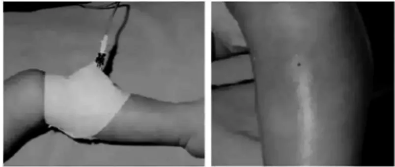
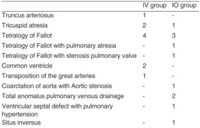
when necessary for maintenance of anesthesia. the iO pro-cedure was conirmed by radioscopy after injecting contrast (Hexabrix®) (figure 1). After the infant was anesthetized,
the needle set was protected with gauze and plaster forming a cushion, connecting the infusion pump (infusion Pump 670-SAMtROnic) for the administration of drugs and luids (figure 2). this technique was kept as a venous access for maintenance of anesthesia during hemodynamic study until infant recovered completely from anesthesia and the infu-sion was interrupted only after complete hydration of the in-fant. the hydration was accomplished with a mixed solution of glucose 5% (50 ml) and saline 0.9% (50 ml) on the dose of 20 ml.kg-1.h-1. the puncture spot was reevaluated after 7
and 15 days (figure 2). the infants were clinically evaluated by a pediatrician after one week, six months and one year and no complications regarding the techniques applied on this study were reported.
All results were reported as mean ± standard deviation. After inspecting for normality and homogeneity of vari ance among groups, differences between groups were calculated using the unpaired Student’s t test. Analysis included Wilcox-on’s test when appropriate. Statistical analysis was performed using PadGraph Prism software (GraphPad Software inc., San Diego, cA, U.S.A.).
Figure 1 – the intraosseous technique and infusion of Anesthetic Agents.
left, middle: the intraosseous technique for infusion of anesthetic agents. Right: radioscopy after injecting contrast (Hexabrix®) conir-med the needle’s position.
Figure 2 – connecting the infusion Pump.
left: infusion pump line connected to administer medications and lui-ds. Right: intraosseous puncture spot.
RESULTS
table i summarizes etiologic aspects of congenital heart di-seases enrolled in the study. the main age of infants was 7.9 ± 4.3 months (iV group) and 7.8 ± 3.8 months (iO group), mean weight was 7.2 ± 1.3 kg (iV group) and 6.9 ± 1.9 kg (iO group), six were female and four were male (iV group) and in the iO group four infants were female and seven were male. table ii summarizes the results of the technique of puncture in the iV group and iO group. the only signiicant difference for the technique between groups was the puncture time (p = 0.012). the mean anesthetic dose used in the iV group were midazolam (1.58 ± 0.03 mg.kg-1), ketamine (17.64 ± 0.25
mg.kg-1), and fentanyl (17.64 ± 0.25 mg.kg-1). in the iO group
those were midazolam (2.18 ± 0.08 mg.kg-1), ketamine (36.6 ±
0.80 mg.kg-1), and fentanyl (36.6 ± 0.80 mg.kg-1) (figure 3). the
onset time of anesthesia (56.3 second) in the iV group was fas-ter than in iO group (71.3 second) (p = 0.014) (figure 4). the hydration was 20 ml.kg-1.h-1 with the solution described and no
Table II – technique of Puncture in the intravenous Group and intraosseous Group
intravenous group intraosseuous group Puncture site
Peripheral veins cutdown
antecubital right 4
-antecubital left 4
-Proximal end of the tibia, just below the tibial tuberosity - 11
Puncture time (min) 9.6 ± 6.9 3.6 ± 2.2 p = 0.012
Punctures (n) 1.8 ± 0.9 1.3 ± 0,5 p = 0.127
Maintenance of puncture (min) 110.0 ± 65.7 98.0 ± 37.9 p = 0.610
complications p = 0.311
local extravasation of luids and blood 1
-local extravasation of luids - 4
Table I – etiologic Aspects of congenital Heart Diseases of Peripheral intravenous infusion Group or Anesthesia intraosseous infusion Group
iV group iO group
truncus arteriosus 1
-tricuspid atresia 2 1
tetralogy of fallot 4 3
tetralogy of fallot with pulmonary atresia - 1 tetralogy of fallot with stenosis pulmonary valve - 1
common ventricle 2
-transposition of the great arteries 1 -coarctation of aorta with Aortic stenosis - 1 total anomalus pulmonary venous drainage - 2 Ventricular septal defect with pulmonary
hypertension
- 1
Situs inversus - 1
iV group: intravenous group; iO group: intraosseous group.
Figure 4 – time of Anesthetic induction and Anesthesia Recovery time, Hydration Volume.
the induction time was faster for the iV group (*p = 0.0145).
275
250
225
200
175
150
125
100
75
50
25
0
intravenous group intraosseous group
*
anesthetic induction time (seconds)
anesthesia recoverytime (minutes)
hydration (ml) 50.0
47.5 45.0 42.5 40.0 37.5 35.0 32.5 30.0 27.5 25.0 22.5 20.0 17.5 15.0 12.5 10.0 7.5 5.0 2.5 0.0
ketamine (mg.kg) fentanyl (μg.kg) midazolam (mg.kg)
intravenous group intraosseous group
*
*
*
Figure 3 – Anesthetic Agentes used during Hemodynamic Study. Higher dose of anesthesic agents was observed: midazolam (p = 0.007), ketamine (p < 0.001) and fentany (p < 0.001) in the intraos-seous group when compared with the intravenous group.
DISCUSSION
AliMAn, PicciOni, PicciOni et Al.
44 Revista Brasileira de Anestesiologia
Vol. 61, no 1, January-february, 2011 access site is the saphenous vein above the medial
malleo-lus of the tibia, but antecubital, axillary, cephalic and femoral vessels are also suitable and improved procedures using Sel-dinger techniques have been reported 11, 12. the usual time to
achieve access by pediatric surgeons was 6 min in children aged 6-16 years, 8 min in those aged 1 month to 5 years, and 11 min in neonates 13. this time delay makes its use
unrealis-tic for most clinicians, and iO or percutaneous femoral access can be achieved more rapidly 11.
the iO method was compared to the peripheral iV method using the following anesthetic agents: midazolan, ketamine, fenatyl. the effectiveness of hydration on iO route has also been tested, demonstrating that this method could be an alternative when peripheral access is considered difficult or impossible (figures 1 and 3). this study demonstrated a quick systemic low during iO infusion (figure 2). the case reported and reviewed literature 15 of severe laryngospasm during inhalation induction of pediatric patients without in-travenous accesss. the speed and effectiveness in treating laryngospasm by intravenous route with suxamethonium agent used as a muscle relaxant was achieved when other iV routes had been inaccessible. Despite intramuscular rou-te being easier, the time response was much slower than the intraosseos route. clinical experience indicates that the iO route is probably superior to the intramuscular route, and comparable to the intravenous route in response time.
Helm et al. 15 demonstrated the other alternative to the
puncture of peripheral veins in emergency situations with chil-dren less than 6 years of age. in 37% of the cases (10/27) the iO infusion line was used for induction of general anesthesia; dosage and onset of administered drugs were described as being equivalent to a peripheral infusion line. in all cases, the iO needle was replaced in-hospital within 2 h by a central or peripheral iV line. no complications were observed, and the iO infusion technique was considered a simple, fast and safe alternative method for emergency access to the vascular sys-tem in children less than 6 years of age in the pre-hospital setting.
in another study it was observed that the greatest advan-tage of iO route is the high success rate (about 80%) and most experienced providers can reach an iO route within 1 to 2 minutes. A number of smaller studies and case studies have established the usefulness of this route for the delivery of all
resuscitation drugs. the most common side effect seen when using iO route is extravasation. it has been reported in 12% of patients. compartment syndrome, osteomyelitis, and tibial fracture are rare, but have been reported as well. the side effect of iO method was extravasation on 4 children that had a good recovery (table ii).
iO access can be used as an alternative line for medication/ volume expansion when umbilical or other direct venous access are not readily attainable (class iib, lOe 5) 17. two
prospecti-ve randomized trials in adults and children (lOe3) 18,19 and 6
other studies (lOe 4 20; lOe 5 21,22;lOe 7 23,24) have
sup-ported iO access as safe and effective for luid resuscitation, drug delivery, and blood sampling for laboratory evaluation. the consensus process (2006) that produced this document was sponsored by the international liaison committee on Re-suscitation (ilcOR) and demonstrated per patient iO success rates that were high despite a small number of attempts over a prolonged time period. these data suggest that successful iO access with a low complication rate can be accomplished despite its infrequent use.
in our study, it was observed that after having administra-ted anesthetics agents the onset time for iO infusion was sig-niicantly faster than the observed in the iV group however the mean mass of anesthetic agents were smaller for iV group. nevertheless, even though the higher anesthetics dose for iO group impaired anesthesia recovery time, no difference was observed between the groups for awakenings. the time for the cathether maintenance was similar in the groups. the fasting before procedure and the injection of hyperosmolar contrast were factors that may contribute to cause important dehydration during hemodynamic studies, therefore it is ne-cessary to promote the infusion of luids during this procedure. the volume of luid for hydration was similar in both groups, but in the iO group an infusion pump for administering luids was necessary because the high vascular iO resistance. con-sidering that since the 1830s luids have been administered intravenously, and iV access is not always possible to be es-tablished, the iO route provides rapid, safe and easy access to the vascular system.
REFERÊNCIAS / REFERENCES
01. tocantins lM, O’neill Jf, Jones HW – infusions of blood and
other luids via the bone marrow; applications in pediatrics. JAMA, 1941;117:1229-1234.
02. Cournand J – Technique for insertion of plastic catheter into saphe-nous vein. Pediatrics, 1941;24:631-637.
03. Begg AC – Intraosseous venography of the lower limb and pelvis. Br J Radiol, 1954;27:318-324.
04. Engle WA – Intraosseous access for administration of medications in neonates. Clin Perinatol, 2006;33:161-168.
05. Smith R, Davis N, Bouamra O et al. – The utilisation of intraosseous infusion in the resuscitation of paediatric major trauma patients. Injury, 2005;36:1034-1038.
Revista Brasileira de Anestesiologia 49 Vol. 61, no 1, Janeiro-fevereiro, 2011
AneSteSiA intRAóSSeA eM eStUDO HeMODinâMicO eM cRiAnçA cARDiOPAtA
transfusion, em: Mackway-Jones KMe , Molyneix e, Phillips B et al – Advanced Paediatric life Support. 3rd ed, london, BMJ Books, 2001;229-230.
07. American Heart Association – 2005 American Heart Association guidelines for cardiopulmonary resuscitation and emergency car-diovascular care. international consensus on science. circulation, 2005;112:iv12-iv211.
08. Brown K, lightfoot c – the 2005 Guidelines for cPR and emergency cardiovascular care: implications for emergency Medical Services for children. clin Ped emerg Med, 2006;7:105-113.
09. Goresky GV, Maltby JR – fasting guidelines for elective surgical pa-tients. can J Anaesth, 1990;37:493-495.
10. Green SM, Johnson ne – Ketamine sedation for pediatric procedu-res: Part 2 - Review and implications. Ann emerg Med, 1990;19:1033-1046.
11. Haas nA – clinical review: vascular access for luid infusion in chil-dren. Crit Care, 2004;8:478-484.
12. Iserson KV, Criss EA – Pediatric venous cutdowns: utility in emer-gency situations. Pediatr Emerg Care,1986;2:231-234.
13. Westfall MD, Price KR, Lambert M et al. – Intravenous access in the critically ill trauma patient: a multicentered, prospective, randomized trial of saphenous cutdown and percutaneous femoral access. Ann Emerg Med,1994;23:541-545.
14. Seah TG, Chin NM – Severe laryngospasm without intravenous access---a case report and literature review of the non-intravenous routes of ad-ministration of suxamethonium. Singapore Med J, 1998;39:328-330. 15. Helm M, Hauke J, Bippus N et al. – Die intraossare Punktion in der
praklinischen Notfallmedizin. 10-jahrige Erfahrungen im Luftrettungs-dienst. Anaesthesist, 2007;56:18-24.
16. Buck ML, Wiggins BS, Sesler JM – Intraosseous drug administra-tion in children and adults during cardiopulmonary resuscitaadministra-tion. Ann Pharmacother, 2007;41:1679-1686.
17. Banerjee S, Singhi SC, Singh S et al. – The intraosseous route is a suitable alternative to intravenous route for luid resuscitation in seve-rely dehydrated children. Indian Pediatr, 1994;31:1511 -1520. 18. Brickman KR, Krupp K, Rega P et al. – Typing and screening of blood
from intraosseous access. Ann Emerg Med, 1992;21:414-417. 19. Fiser RT, Walker WM, Seibert JJ et al. – Tibial length following
intra-osseous infusion: a prospective, radiographic analysis. Pediatr Emerg Care, 1997;13:186-188.
20. ummenhofer W, Frei FJ, urwyler A et al. – Are laboratory values in bone marrow aspirate predictable for venous blood in paediatric pa-tients? Resuscitation, 1994;27:123 -128.
21. Guy J, Haley K, Zuspan SJ – use of intraosseous infusion in the pe-diatric trauma patient. J Pediatr Surg, 1993;28:158-161.
22. Macnab A, Christenson J, Findlay J et al. – A new system for sternal intraosseous infusion in adults. Prehosp Emerg Care. 2000;4:173 -177. 23. Ellemunter H, Simma B, Trawoger R et al. – Intraosseous lines in
preterm and full term neonates. Arch Dis Child Fetal Neonatal Ed, 1999;80:F74 -F75.
24. International Liaison Committee on Resuscitation – The International Liaison Committee on Resuscitation (ILCOR) consensus on science with treatment recommendations for pediatric and neonatal patients: pediatric basic and advanced life support. Pediatrics, 2006;117(5):e955-977.
Resumen: Aliman AC, Piccioni MA, Piccioni JL, Oliva JL, Auler Ju-nior JOC – Anestesia Intraosea en Estudio Hemodinamico en Nino Cardiopata.
Justiicativa y objetivos: El acceso intraoseo (IO), se ha venido uti-lizando con buenos resultados en situaciones de emergencia, cuan-do no existe el acceso venoso disponible para la administracion de luidos y farmacos. El objetivo del presente estudio es evaluar si el acceso IO es una tecnica util para la administracion de anestesia y de luidos en el estudio hemodinamico cuando el acceso periferico es imposible de obtenerse. Ese estudio fue realizado en el laboratorio de hemodinamica de un hospital universitario, con 21 lactantes por-tadores de enfermedad cardiaca congenita que fueron seleccionados para un estudio hemodinamico diagnostico.
Métodos: Este estudio comparo la efectividad del acceso IO con re-lacion al EV para la infusion de anestesicos (quetamina, midazolam y fentanil), y de luidos durante el estudio hemodinamico. El tiempo de induccion anestesica, la duracion del procedimiento, el tiempo de recuperacion de la anestesia, la adecuada hidratacion y las compli-caciones de las punciones EV e IO se compararon entre los grupos.
Resultados: El tiempo de puncion fue signiicativamente menor en el grupo IO (3,6 minutos) que en el grupo EV (9,6 minutos). El tiempo de inicio de la accion de la anestesia fue mas rapido en el grupo EV (56,3 segundos) que en el grupo IO (71,3 segundos). No se observaron diferencias signiicativas entre los dos grupos con relacion a la hidra-tacion (grupo EV 315,5 mL vs. grupo IO 293,2 mL), y sobre el tiempo de recuperacion de la anestesia (grupo IO 65,2 min vs, grupo EV 55,0 min). El sitio de la puncion se evaluo nuevamente despues de 7 a 15 dias, y no presento senales de infeccion u otras complicaciones.
Conclusiones: Los resultados comparativos arrojaron una superiori-dad de la infusion IO con relacion al tiempo de puncion. Debido a su eiciencia y manipulacion bastante facil, la hidratacion y la anestesia que se hicieron por medio de la infusion IO demostraron ser satis-factorias para los estudios hemodinamicos sin la necesidad de otros accesos.