ABSTRACT
Tissue engineering is a contemporary ield of science, which aims to create conditions based on principles of cell and molecular biology, bioengineering and biomaterials to regenerate tissues. Mesenchymal stem cells present high proliferation rates and are able to differentiate into multilineages under certain conditions, suggesting that they have great potential to act in regeneration ield. Tooth derived stem cells are a suitable alternative source of mesenchymal cells once they are easily accessible and have poor morbidity to the donor. Studies showed that they have been isolated and characterized from diverse tissues such as dental pulp, exfoliated deciduous teeth, periodontal ligament, gingiva, dental follicle and apical papilla. However studies show that there is heterogeneity among these populations and there is no standard method to select the most appropriate tooth derived stem cells for regenerative procedures. The aim of this review is to present the current perspective of the multiple types of tooth-derived stem cells and to discuss the basis for their use in periodontal tissue engineering.
Indexing terms: Periodontics. Stem cells. Tissue engineering.
RESUMO
A engenharia de tecidos é um campo contemporâneo da ciência, que visa criar condições baseadas em princípios de biologia celular e molecular, bioengenharia e biomateriais para regenerar tecidos. As células tronco mesenquimais apresentam altas taxas de proliferação e são capazes de se diferenciar, sob certas condições, em multi-linhagens, sugerindo que elas têm grande potencial para atuar no campo da regeneração. As células tronco derivadas de tecidos dentais são uma fonte alternativa adequada de células mesenquimais uma vez que são de fácil acesso e têm baixa morbidade para o doador. Estudos demonstraram que elas já foram isoladas e caracterizadas a partir de diversos tecidos tais como polpa dentária, dentes decíduos esfoliados, ligamento periodontal, gengiva, folículo dental e papila apical. Entretanto, os estudos demonstram que há heterogeneidade entre essas populações e não existe um método padrão para selecionar as células-tronco dentais mais apropriadas para procedimentos regenerativos. O objetivo desta revisão é apresentar o conhecimento atual dos vários tipos de células-tronco derivadas de dentes e discutir as novas perspectivas para seu uso na engenharia de tecidos periodontais.
Termos de indexação: Periodontia.Células-tronco. Engenharia tecidual.
Mesenchymal stem cells in periodontics: new perspectives
Células mesenquimais indiferenciadas em periodontia: novas perspectivas
Bruna Rabelo AMORIM1
Enilson Antonio SALLUM2
Marcio Zaffalon CASATI2
Karina Gonzales Silverio RUIZ2
Renato Correa Viana CASARIN2
Kamila Rosamilia KANTOVITZ3
Francisco Humberto NOCITI JUNIOR2
1 Universidade de Brasília, Faculdade de Saúde, Laboratório de Histopatologia Bucal. Asa Norte, 70910900, Brasília, DF, Brasil. Correspondência para / Correspondence to: BR AMORIM. E-mail: <[email protected]>.
2 Universidade Estadual de Campinas, Faculdade de Odontologia, Departamento de Prótese e Periodontia. Piracicaba, SP, Brasil. 3 Faculdade São Leopoldo Mandic, Curso de Odontologia. Campinas, SP, Brasil.
from earliest stages of development to adult stage
2-3.
Embrionic SCs are a pluripotent cell type which can
differentiate into all cells of the body but ethical issues like
the using of human embryos for research purpose prevents
further development in the ield, thus Adult SCs are the cell
choice for investigation. They have differentiation abilities
but it is restricted to some cell types. Mesenchymal stem
cells (MSCs) are included on this group
4-5.
MSCs are widely studied within the medical ield
because of their therapeutic potential. They present high
proliferation rates and can be induced to differentiate
into multiple lineages
2. These populations are very
heterogeneous once there is no deined marker to identify
mesenchymal stem cells
6. International Society for cell
INTRODUCTION
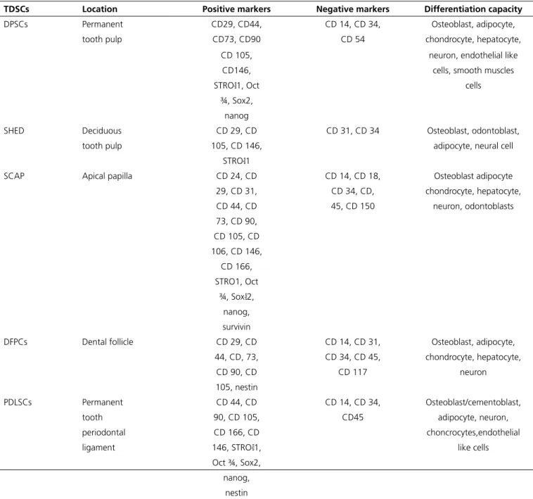
Table 1. Characterization of tooth- derived stem cells.
TDSCs Location Positive markers Negative markers Differentiation capacity
DPSCs Permanent CD29, CD44, CD 14, CD 34, Osteoblast, adipocyte,
tooth pulp CD73, CD90 CD 54 chondrocyte, hepatocyte,
CD 105, neuron, endothelial like
CD146, cells, smooth muscles
STRO- 1, Oct cells
¾, Sox2, nanog
SHED Deciduous CD 29, CD CD 31, CD 34 Osteoblast, odontoblast,
tooth pulp 105, CD 146, adipocyte, neural cell
STRO- 1
SCAP Apical papilla CD 24, CD CD 14, CD 18, Osteoblast adipocyte
29, CD 31, CD 34, CD, chondrocyte, hepatocyte,
CD 44, CD 45, CD 150 neuron, odontoblasts
73, CD 90, CD 105, CD 106, CD 146,
CD 166, STRO1, Oct
¾, Sox- 2, nanog, survivin
DFPCs Dental follicle CD 29, CD CD 14, CD 31, Osteoblast, adipocyte,
44, CD, 73, CD 34, CD 45, chondrocyte, hepatocyte,
CD 90, CD CD 117 neuron
105, nestin
PDLSCs Permanent CD 44, CD CD 14, CD 34, Osteoblast/cementoblast,
tooth 90, CD 105, CD45 adipocyte, neuron,
periodontal CD 166, CD choncrocytes,endothelial
ligament 146, STRO- 1, like cells
Oct ¾, Sox2, nanog,
nestin
dental pulp
8-12, exfoliated deciduous teeth
13-17, periodontal
ligament, dental follicle
18-20, apical papilla
21-24, periodontal
ligament of deciduous teeth
14-15,25-27and gingival tissue
stem cells
28. This review article proposes to summarize the
literature regarding the current knowledge about stem
cells from dental tissue, and their potential in regenerative
therapy.
TOOTH DERIVED STEM CELLS (TDSCS)
Variable methodologies are used to isolate and
characterize TDSCs. A summary is represented in Table 1.
therapy proposed a minimal criteria to deine mesenchymal
DePDL Deciduous CD105, CD CD 34, CD 45 Osteoblast adipocyte,
tooth 166, STRO- cementoblast ,
periodontal Oct4 chondrocyte
ligament
GSCs Gingival tissue CD 90, CD CD34, CD 38, Osteoblast, choncrocyte,
105, CD 73, CD 45, CD 54 adipocyte
CD 44, CD 13
Note: CD: Cluster of differentiation; DePDL: Periodontal ligament of deciduous teeth stem cells; DFPCs: Dental follicle progenitor cells; DPSCs: Dental pulp stem cells; GSCs: Gingival stem cells; Oct: Octamer; PDLScs: Periodontal ligament stem cells; SCAP: Stem cells from apical papilla; SHED: Stem cells from human exfoliated decidu-ous teeth; Sox2: SRY- box containing gene 2.
Dental Pulp Stem Cells (DPSCs)
In 2000, Gronthos and collaborators were the
pioneers on isolation and characterization of DPSC, the
irst TDSCs
8. When compared with human bone marrows
stem cells (BMSCs), DPSCs showed higher proliferation rate
and greater capacity to form mineral nodules, so they are
more appropriate for regeneration of mineralized tissues
than BMSCs. They are able to differentiate into osteoblast,
smooth muscle cells, adipocyte-like cells, neuron, dentin,
dentin-pup-like complex and endothelial like cells
8,29.
Stem cells from human exfoliated deciduous teeth (SHED)
SHEDs are progenitor cells irst isolated in 2003,
from the remnant pulp of exfoliated deciduous teeth
13.
They showed a higher proliferative rate, when compared
to BMSCs and DPSCs
13,15, and a higher capability to
differentiate in osteoblast and adipocyte-like cells when
compared to DPSCs in vitro
15. They also showed the
capability do differentiate into odontoblast, neural cells
15,30,.
Stem cells from apical papilla (SCAPs).
SCAPS are cells isolated from apical papilla located
on the root apex of developing teeth
31. It is distinct from
the pulp tissue
32. They presented a higher proliferation,
migration and telomerase activity. They are able to
differentiate into osteoblastic, odontoblastic, adopocyte
- like and neuron-like cells under speciic induction
22. A
cDNA microarray proiled comparative analysis between
SCAP and DPSCs concluded that genes such as CD24 and
survivin were highly expressed in SCAPS
22.
Dental follicle progenitor cells (DFPCs)
DFPCs are cells obtained from dental follicle
which is a condensation of cells originated from the
ectomesenchyma that surrounds the tooth germ in early
stages of tooth formation. It contains a heterogenic cell
population that forms the periodontium
18,33. They can
differentiate into osteoblast, adipocyte, chondrocyte and
neuronal cells, but they present differences on proliferation
and mineralization patters which suggests that they could
commit in distinct lineages
33.
Periodontal ligament stem cells (PDLSCs)
PDLSCs are a heterogeneous cell population with
neural crest cell origin. They have higher proliferation
rate but forms less mineralized nodules when compared
to BMSCs. They present the ability to differentiate into
osteoblasts, cementoblasts, adipocytes, chondrocytes and
endothelial like cells. In vivo experiments conirmed the
ability to form periodontal ligament and cementum-like
tissue
34.
Decidous periodontal ligament cells (DePDL)
Periodontal ligament cells also can be isolated from
decidous teeth. It showed higher proliferative rate than
PDLSCs, and share the same ability of differentiate into
osteoblasts, cementoblasts, adipocytes and chondrocytes,
but with a higher potential to differentiate into adipocytes
25.
Gingival tissue Stem cells (GSCs)
GSCs are obtained from gingival connective
tissue, so the sample must be deepithelialized, to leave
only connective tissue. They are able to differentiate into
osteogenic, chondrogenic and adipogenic lineages. It also
present an immunemodulatory capacity
28.
Induced pluripotent stem cells (iPSCs)
cells. Although iPS cells are an option without any ethical
concerns and it has a great potential towards regeneration
of periodontal ligament, alveolar bone, cementum and
dentin-pulp complex, issues like epigenetic memory,
viral-transduction, tumorgenesis and teratoma formation has to
be further investigated
35.
REGENERATIVE APPLICATIONS OF TDSCS
Basic components for tissue engineering includes
scaffolds, signal molecules and cells. Scaffolds are
tridimensional structures, which mimics the extracellular
matrix and must have physical, chemical and biological
characteristics to provide a microenvironment for cell
signaling activation, and stimulation of cellular growth,
differentiation, cell adhesion and migration. The cells
provide synthesis of extracellular matrix and tissue
regeneration. MSCs presents important characteristics
such as high proliferation rates and ability to differentiate
into multilineages, therefore they have a great potential
into tissue engineering ield.
Increasing amount of research presents TDSCs
applicability in diverse conditions, including myocardial
infarction
36, ischemic disease
37, neural regeneration
38,
inlammatory diseases
39, diabetes
40, muscular dystrophy
41,
bone and cartilage defects
42, hair follicle loss
43, skin
injuries
44, salivary gland defects
45, corneal reproduction
46,
and the regeneration of dental tissues
12,22,35,47-49.
Several studies demonstrated that TDSCs have
successfully regenerated dental tissues such as dentin,
pulp and periodontal ligament
12,22,47-49. In vivo experiments
demonstrated that human PDLSCs and SCAP were able
to generate periodontal ligament in minipigs
22. Also
DPSCs promoted complete pulp regeneration in dogs
12.
Combination of iPS cells with silk scalffold and enamel
matrix promoted PDL regeneration in mouse periodontal
fenestration defects
35.
Although the literature conirms the potential of
TDSCs in regeneration, there are some aspects that must
be discussed. First, proliferation capacity, clonogeniticy
and differentiation ability are different from each type of
TDSCs lineages suggesting that it has an association with
the type of original tissue. Even in the same population
there are heterogeneous cell subpopulation with different
behavior
50. Notwithstanding that International Society
for cell therapy deined a minimal phenotype criteria for
MSCs, speciic surface markers associated with TDSCs
commitment are not established.
CONCLUSION
Interest in regeneration topic has increased inside
scientiic community. TDSCs are potential actors for
regenerative procedures once they are an easy available
source that presents almost no morbidity to the donor.
Studies, which used TDSCs for regeneration, presented
promising results. However, MSCs populations obtained
from dental tissues are heterogeneous and, currently,
there is no standard method to select the most appropriate
TDSCs for regenerative procedures. Further studies must
be designed to conirm TDSC-based therapies as safe,
predictable and reproducible.
Collaborators
BR AMORIM, EA SALLUM, MZ CASATI, KGS
RUIZ, RCV CASARIN, KR KANTOVITZ and FH NOCITI
JUNIOR participated in all stages of the preparation of the
manuscript.
REFERENCES
1. Saito MT, Silvério KG, Casati MZ, Sallum EA, Nociti Jr FH. Tooth-derived stem cells: Update and perspectives. World J Stem Cells. 2015;7(2):399-407. doi: 10.4252/wjsc.v7.i2.399
2. Kaukua N, Shahidi MK, Konstantinidou C, Dyachuk V, Kaucka M, Furlan A, et al. Glial origin of mesenchymal stem cells in a tooth model system. Nature. 2014;513:551-54. doi: 10.1038/ nature13536
3. Gothard D, Roberts SJ, Shakesheff KM, Buttery LD. Engineering embryonic stem-cell aggregation allows an enhanced osteogenic differentiation in vitro. Tissue Eng Part C Methods. 2010;16:583-595. doi: 10.1089/ten.TEC.2009.0462
4. Watt FM, Driskell RR. The therapeutic potential of stem cells. Philos Trans R Soc Lond B Biol Sci. 2010;365:155-63. doi: 10.1098/ rstb.2009.0149
5. Fischbach GD, Fischbach RL. Stem cells: science, policy, and ethics. J Clin Invest. 2004;114:1364-70. doi:10.1172/JCI23549
6. Sanz AR, Carrión FS, Chaparro AP. Mesenchymal stem cells from the oral cavity and their potential value in tissue engineering. Periodontol 2000. 2015;67(1):251-67. doi: 10.1111/prd.12070
8. Gronthos S, Mankani M, Brahim J, Robey PG, Shi S. Postnatal human dental pulp stem cells (DPSCs) in vitro and in vivo. Proc Natl Acad Sci USA. 2000;97:13625-630. doi: 10.1073/pnas.240309797
9. Yang X, Walboomers XF, van den Beucken JJ, Bian Z, Fan M, Jansen JA. Hard tissue formation of STRO-1-selected rat dental pulp stem cells in vivo. Tissue Eng Part A. 2009;15:367-75. doi: 10.1089/ten. tea.2008.0133
10. Gronthos S, Brahim J, Li W, Fisher LW, Cherman N, Boyde A, et al. Stem cell properties of human dental pulp stem cells. J Dent Res. 2002;81:531-35. doi: 10.1177/154405910208100806
11. Zhang W, Walboomers XF, Wolke JG, Bian Z, Fan MW, Jansen JA. Differentiation ability of rat postnatal dental pulp cells in vitro. Tissue Eng. 2005;11:357-68. doi: 10.1089/ ten.2005.11.357
12. Iohara K, Imabayashi K, Ishizaka R, Watanabe A, Nabekura J, Ito M, et al. Complete pulp regeneration after pulpectomy by transplantation of CD105+ stem cells with stromal cell-derived factor-1. Tissue Eng Part A. 2011;17:1911-920. doi: 10.1089/ ten.TEA.2010.0615
13. Miura M, Gronthos S, Zhao M, Lu B, Fisher LW, Robey PG, et al. SHED: stem cells from human exfoliated deciduous teeth. Proc Natl Acad Sci USA. 2003;100:5807-812. doi: 10.1073/ pnas.0937635100
14. Fu X, Jin L, Ma P, Fan Z, Wang S. Allogeneic stem cells from deciduous teeth in treatment for periodontitis in miniature swine. J Periodontol. 2014;85:845-51. doi: 10.1902/ jop.2013.130254
15. Wang X, Sha XJ, Li GH, Yang FS, Ji K, Wen LY, et al. Comparative characterization of stem cells from human exfoliated deciduous teeth and dental pulp stem cells. Arch Oral Biol. 2012;57:1231-1240 doi: 10.1016/ j.archoralbio.2012.02.014
16. Zheng Y, Liu Y, Zhang CM, Zhang HY, Li WH, Shi S, et al. Stem cells from deciduous tooth repair mandibular defect in swine. J Dent Res. 2009;88:249-54. doi: 10.1177/0022034509333804
17. Behnia A, Haghighat A, Talebi A, Nourbakhsh N, Heidari F. Transplantation of stem cells from human exfoliated deciduous teeth for bone regeneration in the dog mandibular defect. World J Stem Cells. 2014;6:505-10. doi: 10.4252/wjsc
18. Morsczeck C, Götz W, Schierholz J, Zeilhofer F, K̈hn U, Möhl C, et al. Isolation of precursor cells (PCs) from human dental follicle of wisdom teeth. Matrix Biol. 2005;24:155-65. doi: 10.1016/j. matbio.2004.12.004
19. Silvério KG, Davidson KC, James RG, Adams AM, Foster BL, Nociti FH, et al. Wnt/ß-catenin pathway regulates bone morphogenetic protein (BMP2)-mediated differentiation of dental follicle cells. J Periodontal Res. 2012;47:309-19. doi: 10.1111/j.1600-0765.2011.01433.x
20. Guo L, Li J, Qiao X, Yu M, Tang W, Wang H, et al. Comparison of odontogenic differentiation of human dental follicle cells and human dental papilla cells. PLoS One. 2013;8:e62332. doi: 10.1371/journal.pone.0062332
21. Huang GT, Al-Habib M, Gauthier P. Challenges of stem cell-based pulp and dentin regeneration: a clinical perspective. Endod Topics. 2013;28:51-60. doi: 10.1111/etp.12035
22. Sonoyama W, Liu Y, Fang D, Yamaza T, Seo BM, Zhang C, et al. Mesenchymal stem cellmediated functional tooth regeneration in swine. PLoS One. 2006;1:e79. doi: 10.1371/journal.
pone.0000079
23. Zhang W, Zhang X, Ling J, Liu W, Zhang X, Ma J, et al. Proliferation and odontogenic differentiation of BMP2 gene-transfected stem cells from human tooth apical papilla: an in vitro study. Int J Mol Med. 2014;34:1004-12. doi: 10.3892/ijmm.2014.1862
24. Wang J, Liu B, Gu S, Liang J. Effects of Wnt/ß-catenin signaling on proliferation and differentiation of apical papilla stem cells. Cell Prolif. 2012;45:121-31. doi: 10.1111/ j.1365-2184.2012.00806.x
25. Silvério KG, Rodrigues TL, Coletta RD, Benevides L, Da Silva JS, Casati MZ, et al. Mesenchymal stem cell properties of periodontal ligament cells from deciduous and permanent teeth. J Periodontol. 2010;81:1207-215. doi: 10.1902/ jop.2010.090729
26. Fukushima H, Kawanabe N, Murata S, Ishihara Y, Yanagita T, Balam TA, et al. SSEA-4 is a marker of human deciduous periodontal ligament stem cells. J Dent Res. 2012;91:955-60. doi: 10.1177/0022034512458123
27. Ji K, Liu Y, Lu W, Yang F, Yu J, Wang X, et al. Periodontal tissue engineering with stem cells from the periodontal ligament of human retained deciduous teeth. J Periodontal Res. 2013;48:105-16. doi: 10.1111/j.1600-0765.2012.01509.x
28. Mitrano T, Grob M, Carrion F, Nova-Lamperti E, Luz P, Fierro F, et al. Culture and characterization of mesenchymal stem cells from human gingival tissue. J Periodontol. 2010;81:917-25. doi: 0.1902/jop.2010.090566
29. Park YJ, Cha S, Park YS. Regenerative applications using tooth derived stem cells in other than tooth regeneration: a literature review. Stem Cells Int. 2016;2016:9305986. doi: 10.1155/2016/9305986
30. Nourbakhsh N, Soleimani M, Taghipour Z, Karbalaie K, Mousavi SB, Talebi Rabiei F, et al. Induced in vitro differentiation of neural-like cells from human exfoliated deciduous teeth-derived stem cells. Int J Dev Biol. 2011;55:189-95. doi: 10.1387/ ijdb.103090nn
31. Sedgley CM, Botero TM. Dental stem cells and their sources. Dent Clin North Am. 2012;56:549-61. doi: 10.1016/ j.cden.2012.05.004
32. Sonoyama W, Liu Y, Yamaza T, Tuan RS, Wang S, Shi S, et al. Characterization of the apical papilla and its residing stem cells from human immature permanent teeth: a pilot study. J Endod. 2008;34:166-71. doi: 10.1016/j.joen.2007.11.021
33. Luan X, Ito Y, Dangaria S, Diekwisch TG. Dental follicle progenitor cell heterogeneity in the developing mouse periodontium. Stem Cells Dev. 2006;15:595-608. doi: 10.1089/scd.2006.15.595
34. Gay IC, Chen S, MacDougall M. Isolation and characterization of multipotent human periodontal ligament stem cells. Orthod Craniofac Res. 2007;10:149-60. doi: 10.1111/ j.1601-6343.2007.00399.x
35. Malhotra M. Induced Pluripotent Stem (iPS) cells in Dentistry: a review. Int J Stem Cells. 2016 Nov 30;9(2):176-185. doi: 10.15283/ijsc16029
with acute myocardial infarction. Stem Cells. 2008;26(3):638-45. doi:10.1634/stemcells.2007-0484
37. Shen CY, Li L, Feng T, Li JR, Yu MX, Lu Q, et al. Dental pulp stem cells derived conditioned medium promotes Angiogenesis in Hindlimb Ischemia. Tissue Eng Regen Med. 2015;12(1):59-68. doi:10.1007/s13770-014-9053-7
38. Fang CZ, Yang YJ, Wang QH, Yao Y, Zhang XY, He XH. Intraventricular injection of human dental pulp stem cells improves hypoxic-ischemic brain damage in neonatal rats. PLoS One. 2013;8(6):e66748. doi:10.1371/journal.pone.0066748
39. Yamaza T, Kentaro A, Chen C, Liu Y, Shi Y, Gronthos S, et al. Immunomodulatory properties of stem cells from human exfoliated deciduous teeth. Stem Cell Res Ther. 2010;1(1):5. doi:10.1186/scrt5
40. Kanai MM, Rajeshwari YB, Gupta S, Dadheech N, Nair PD, Gupta PK, et al. Transplantation of islet-like cell clusters derived from human dental pulp stem cells restores normoglycemia in diabetic mice. Cytotherapy. 2013;15(10):1228-236. doi: 10.1016/j. jcyt.2013.05.008
41. Yang R, Chen M, Lee CH, Yoon R, Lal S, Mao JJ. Clones of ectopic stem cells in the regeneration of muscle defects in vivo. PLoS One. 2010;5(10):e13547. doi: 10.1371/journal.pone.0013547
42. Graziano A, d’Aquino R, Laino G, Papaccio G. Dental pulp stem cells: a promising tool for bone regeneration. Stem Cell Rev. 2008;4(1):21-26. doi: 10.1007/s12015-008-9013-5
43. Reynolds AJ, Jahoda CA. Cultured human and rat tooth papilla cells induce hair follicle regeneration and iber growth. Differentiation. 2014;72(9-10):566-75. doi: 10.1111/j.1432-0436.2004.07209010.x
44. Nishino Y, Ebisawa K, Yamada Y, Okabe K, Kamei Y, Ueda M. Human deciduous teeth dental pulp cells with basic ibroblast growth factor enhance wound healing of skin defect. J Craniofac Surg.
2011;22(2):438-42. doi: 10.1097/SCS.0b013e318207b507
45. Yamamura Y, Yamada H, Sakurai T, Ide F, Inoue H, Muramatsu T, et al. Treatment of salivary gland hypofunction by transplantation with dental pulp cells. Arch Oral Biol. 2013;58(8):935-42. doi: 10.1016/j. archoralbio.2013.02.015
46. Gomes JA, Geraldes Monteiro B, Melo GB, Smith RL, Cavenaghi Pereira da Silva M, Lizier NF, et al. Corneal reconstruction with tissue-engineered cell sheets composed of human immature dental pulp stem cells. Invest Ophthalmol Vis Sci. 2010;51(3):1408-414. doi:10.1167/ iovs.09-4029
47. Cordeiro MM, Dong Z, Kaneko T, Zhang Z, Miyazawa M, Shi S, Smith AJ, Nör JE. Dental pulp tissue engineering with stem cells from exfoliated deciduous teeth. J Endod. 2008; 34: 962-969 doi: 10.1016/j.joen.2008.04.009
48. Suaid FF, Ribeiro FV, Rodrigues TL, Silvério KG, Carvalho MD, Nociti FH, et al. Autologous periodontal ligament cells in the treatment of class II furcation defects: a study in dogs. J Clin Periodontol. 2011;38:491-98. doi: 10.1111/ j.1600-051X.2011.01715.x
49. Suaid FF, Ribeiro FV, Gomes TR, Silvério KG, Carvalho MD, Nociti FH, et al. Autologous periodontal ligament cells in the treatment of Class III furcation defects: a study in dogs. J Clin Periodontol. 2012;39:377-84. doi: 10.1111/j.1600-051X.2012.01858.x
50. Okamoto T, Aoyama T, Nakayama T, Nakamata T, Hosaka T, Nishijo K, et al. Clonal heterogeneity in differentiation potential of immortalized human mesenchymal stem cells. Biochem Biophys Res Commun. 2002;295:354-61. doi: 10.1016/S0006- 291X(02)00661-7
Received on: 31/3/2017 Final version resubmitted on: 7/7/2017