Alan Araujo de Jesus*, Milena Botelho Pereira Soares**, Ana Prates Soares***, Renata Campos Nogueira****, Elisalva Teixeira Guimarães*****, Telma Martins de Araújo******, Ricardo Ribeiro dos Santos*******
» The authors report no commercial, proprietary, or inancial interest in the
products or companies described in this article.
Collection and culture of stem cells derived
from dental pulp of deciduous teeth:
Technique and clinical case report
Introduction: Stem cells (SCs) are capable of inducing tissue regeneration and are, there-fore, potentially therapeutic. Similarly to bone marrow and umbilical cords, dental pulp is one of the available sources of SCs. The fact that these cells are easily accessible and that deciduous teeth are not vital organs, and are normally discarded after exfoliation, make them particularly attractive for use in safety and viability tests. Objective: To de-scribe the collection, isolation and culture of SCs obtained from the pulp of deciduous teeth as well as their characterization by flow cytometry, and the induction of differen-tiation into osteogenic and adipogenic lineages. Methods: SCs were obtained in a rela-tively straightforward manner and showed good proliferative capacity, even from a small amount of pulp tissue. Results: Analysis by flow cytometry confirmed the characteristics of mesenchymal SCs with low expression of CD34 and CD45 antigens, which are mark-ers for hematopoietic cells, and high levels of expression of CD105, CD166, CD90 and CD73 antigens, which are markers for mesenchymal SCs. Cell plasticity was confirmed by identifying calcium deposits in cultures that received osteogenic medium, and in-tracellular lipid accumulation in adipogenic cultures that received adipogenic medium. Conclusions: SCs collected from deciduous teeth show promising potential for applica-tion in tissue regeneraapplica-tion. Therefore, it is important that knowledge about the existence and characteristics of this source of stem cells be disseminated among dentists and that the technique, its limitations and possible indications are highlighted and discussed.
Abstract
Keywords: Stem cells. Tissue therapy. Cell culture techniques. Deciduous teeth.
* PhD in Biotechnology, Federal University of Feira de Santana (PPGBiotec/UEFS) / Oswaldo Cruz Institute Foundation (Fiocruz). ** Head researcher, Fiocruz, Bahia. Head of the Tissue Engineering and Immunopharmacology Laboratory (LETI).
*** Dentistry graduate, UFB.
**** MSc in Biotechnology, UEFS / Fiocruz. ***** PhD in Pathology, Fiocruz, Bahia.
****** Head professor in Orthodontics, Federal University of Bahia (UFBA). President of the Brazilian Board of Orthodontics and Facial Orthopedics (BBO). ******* Head researcher of the Gonçalo Moniz Research Center, Fiocruz, Bahia. Head of Basic Research São Rafael Hospital (HSR).
IntROduCtIOn
Science has taken a keen interest in stem cells (SCs) given their ability to stimulate tissue regen-eration, which raises exciting and promising ther-apeutic prospects.1-13 This fact makes SCs a viable alternative in dentistry.14 However, there are still limitations in how they are obtained, grown and controlled in terms of proliferation and differ-entiation, which encourages the search for new sources, techniques and applications.
SCs can have an embryonic or adult origin.2,6,15 Adult SCs are present in a wide range of tissues such as the pancreas, bone marrow, adipose tis-sue and umbilical cord. Because they are obtained from the patients themselves, these cells have the advantages of not triggering immune rejection, re-sponding to growth factors inherent in the host, and not incurring ethical or moral objections.6 The foundation for cell therapy is the isolation of high quality adult human SCs from different sources as these cells feature peculiar characteris-tics, and there may be preferred SCs sources for each specific need.12,16,17
Recently, it was discovered a new source of SCs deriving from the pulp of deciduous teeth. The fact that these cells are easily accessible and that deciduous teeth are not vital organs, being normally discarded after exfoliation, make them attractive for safety and viability tests. Studies conducted with these cells un-derscore their outstanding capacity to prolif-erate and induce tissue regeneration,12,17,18,19 although limitations still exist regarding the amount of available cells as well as the tech-niques used for SC collection and culture.19,20
Although there is still the need to standardize techniques and to conduct clinical studies in order to determine their potential application, this study aims to disseminate among dentists knowledge about SCs obtained from the pulp of deciduous teeth, discussing the technique, its limitations and possible therapeutic potential through the de-scription of a technique and a clinical case report.
This will allow professionals to inform patients and/or their legal guardians that although this is still an experimental technique it shows promise and that this tissue — which is usually discarded — can be collected and cryopreserved for future use.
tECHnIQuE And CLInICAL CASE REPORt tissue collection
An eight-year-old female patient with mixed dentition has been monitored since five years old by an orthodontist and was selected as pulp tis-sue donor after authorization by her legal guard-ians. Radiographic and photographic exams were requested and, in conjunction with the orthodon-tist, teeth 6.3, 7.3, 7.5 and 8.3 were defined as tar-gets as they were in the phase of exfoliation and did not show any carious lesions.
The surgery was performed in two stages with a 15-day interval. The procedure requires strict control of the aseptic chain due to widespread presence of microorganisms in the oral environ-ment. Extraoral asepsis and intraoral prophylaxis were performed and 2% chlorhexidine rinses ap-plied. Next, infiltrating and gingival anesthesia was applied, followed by syndesmotomy and removal of the teeth with a pedodontic forceps as quickly as possible to avoid saliva contamination. Soft tis-sue remnants were removed and the teeth were immediately placed in individual containers filled with Dulbecco Modified Eagle medium (DMEM, Sigma Chemical Co. St. Louis, Mo, USA) culture medium with 50 mg/ml of gentamicin (Nova-farma, Anápolis, Brazil), stored under controlled temperature, between 4 ºC and 8 ºC, and sent to a laboratory for cell isolation.
Isolation, selection and expansion of mesenchymal SCs
B
D C
A
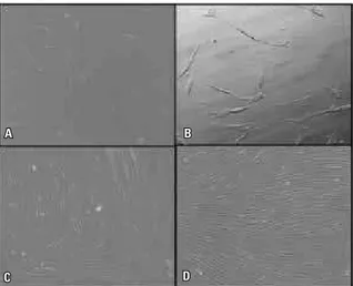
constant irrigation, and the pulp tissue removed with the aid of curettes and endodontic files. Only a small amount of pulp tissue was obtained. One of the four deciduous teeth extracted no longer contained pulp tissue and the others had a small amount of pulp. Therefore, few cells were availed to begin cultivation. Once collected, the tissue was immediately placed in culture bottles containing DMEM supplemented with 10% fetal bovine se-rum (SBF, Cultilab, Campinas, Brazil) and stored at 37 °C and 5% CO2 for cell proliferation and adherence to the bottle. The medium was com-pletely replaced every three days during a period of approximately 10 days, when culture reached about 80-90% confluence. The culture was moni-tored by means of an inverted optical microscope. After adherence to the plastic surface, SCs ini-tially exhibited an ovoid shape that evolved early during the first 24 hours to a fibroblastoid form, which remained until confluence (Fig 1). As the culture was replaced, the adherent mesenchymal SCs were selected while cells suspended in cul-ture medium were gradually discarded.
After culture confluence cells were released from the plastic surface to allow the continued pro-liferation and cryopreservation of part of the cells. To release the cells, the medium was replaced twice with sterile saline solution and, after removing the saline solution, 0.25% trypsin (Invitrogen, São Pau-lo, Brazil) was added for 2-5 minutes to allow the connections between cells and extracellular matrix to be broken, thereby enabling the detachment of cells from the surface of the plastic culture flask.
After ascertaining that cells had been released through observation with an inverted optical mi-croscope, the enzyme was inactivated by adding a complete medium. The medium of the bottle containing the cells was then collected using a pipette and centrifuged at 1500 rpm for 10 min-utes. The supernatant was discarded and the cell pellet resuspended in 1 mL of DMEM supple-mented with 10% fetal bovine serum (SBF). At this point, some of the cells were set aside for cryopreservation in liquid nitrogen for further studies and potential therapeutic uses, while the remaining was used for continuity of culture, al-lowing in vitro characterization.
For cryopreservation, the maximum concentra-tion of 106–107 cells per tube was observed, with a final volume of 1 mL being added, i.e., 900 µL of complete medium containing cells and 100 µL of dimethyl sulfoxide (DMSO). Cryopreservation tubes had their cryostatic temperature lowered, ranging from 4 to –80 °C for 24 h, with subse-quent storage in liquid nitrogen.
Characterization of mesenchymal SCs
Cell characterization was performed by anal-ysis of flow cytometry and confirmation of cell plasticity through the induction of differentiation into osteogenic and adipogenic lineages.
Flow cytometry
The sample cells were labeled with specific monoclonal antibodies linked to fluorochromes. Reading was performed in a flow cytometer.
The following antibodies were used: FITC man CD90, APC anti-human CD45, PE anti-hu-man CD166, PE anti-huanti-hu-man CD73 (BD Pharmin-gen), PE anti-human CD34 (Becton Dickinson) and FITC anti-human CD105 (R&D Systems).
Cells with less than 100% confluence were trypsinized as described before. Immediately after detachment, cells were resuspended in DMEM supplemented with 10% SBF and remained at rest in the oven for 2 hours. After the rest period, the cells were washed twice with saline at 4 °C, at 3000 rpm for 2 minutes at 10 °C and resuspended in 1 mL of saline solution. Two-hundred µL of the solu-tion were placed in tubes which received 2 µL of antibodies. The tubes were incubated at 4 ºC in the dark for 30 minutes and washed twice with saline at 4 °C (1 mL) by centrifugation at 2000 rpm, 10 °C for 2 minutes. After this procedure, CellQuest software was used for data acquisition and analysis with a FACSCalibur flow cytometer (Becton Dick-inson, San Diego, CA, USA). At least 50,000 events were collected and analyzed.
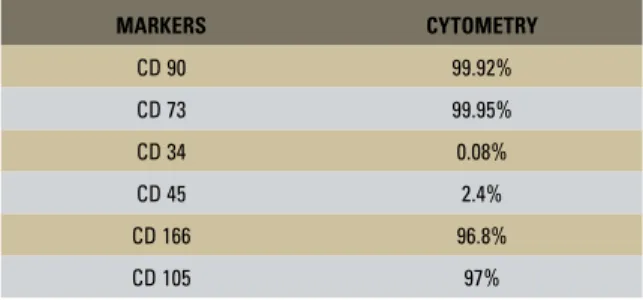
Results confirmed the characteristics of mes-enchymal SCs (Table 1) with low level expression of CD34 and CD45 antigens, which are markers for hematopoietic cells, and high levels of expres-sion of CD105, CD166, CD90 and CD73 anti-gens, which are markers for mesenchymal SCs.20
Osteogenic differentiation
The cells were cultivated in a 24-well plate in DMEM supplemented with 10% fetal bo-vine serum for two days until 50% confluence was reached. From this point on, osteogenesis inducing medium was employed, which is com-posed of: DMEM containing 10% fetal bovine serum, 100 nM dexamethasone, 0.05 mM of L-ascorbic acid 2-phosphate and 10 mM of β-glycerophosphate (Sigma-Aldrich, USA). Cul-ture time was 21 days. Control group cells were grown in DMEM supplemented with 10% fetal bovine serum. The experiments were conducted in triplicate, culture medium was changed every three days and the evolution of differentiation was monitored daily by optical microscopy.
Osteogenic differentiation capacity was veri-fied by alizarin red staining, which identifies the deposition of calcium in the culture. After 21 days of cultivation, the cultures were washed with PBS, fixed with 4% paraformaldehyde (Electron Mi-croscopy Sciences, USA) for 30 minutes, washed twice with distilled water, and 2% alizarin red staining was applied (Sigma–Aldrich, USA) for three minutes. After removing the alizarin, the cultures were washed three times with distilled water to remove residue and the stained areas were analyzed with optical microscopy to confirm color and quality evaluation.
In all cultures that received osteogenic me-dium, red calcium deposition was found due to staining with alizarin, whereas in the negative control group calcium deposits were not found in any of the wells, confirming the plasticity of the cultured cells (Fig 2).
Adipogenic differentiation
The cells were cultivated in a 6-well plate in DMEM supplemented with 10% fetal bovine se-rum until 100% confluence was reached. They were then stimulated for up to three weeks with modified DMEM containing: 10% fetal bovine serum, 60 µM of indomethacin (Sigma–Aldrich),
MARKERS CYTOMETRY
CD 90 99.92%
CD 73 99.95%
CD 34 0.08%
CD 45 2.4%
CD 166 96.8%
CD 105 97%
B
D C
A
dISCuSSIOn
The scientific community has conducted re-search that underscores the importance and pros-pects of therapy with SCs in several areas, such as medullary damage,2,3 neurological changes, such as in Parkinson’s8 and Alzheimer’s diseases,21 auto-immune diseases such as diabetes type 1,5,15 liver diseases,13 kidney damage,7 and retina degenera-tion.22 In some cases, such as coronary heart dis-ease, consistent clinical results have already been found,9,10 indicating SCs’ safety and viability.
In dentistry, experiments have focused on the use of cell therapy in oral tissue regeneration12,14,23 and on the collection, isolation, culture and char-acterization of SCs derived from the pulp of teeth.16-19,24 Some studies indicate that the SCs ob-tained from deciduous teeth have greater regenera-tive and proliferaregenera-tive potential when compared to permanent teeth,16,17,19 besides being more easily accessible. Furthermore, deciduous teeth are not vi-0.5 µM of isobutylmethylxanthine
drich) and 0.5 µM hydrocortisone (Sigma–Al-drich). Throughout the experiments a control group was maintained cultivated in DMEM sup-plemented with 10% fetal bovine serum. The cul-ture medium was replaced every three days.
The evolution of differentiation was moni-tored daily under an inverted microscope. To al-low observation of fat deposition the wells were washed with PBS, cells were fixed with 4% PFA for 1 hour at room temperature, stained with Oil Red solution (Sigma–Aldrich) (3 volumes of 3.75% Oil Red O in isopropanol and 3 volumes of distilled water) for 5 minutes and washed with distilled water to remove residue.
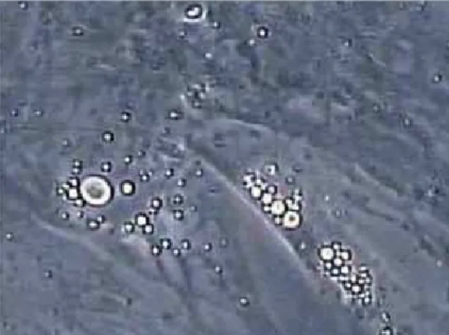
After 14 days intracellular lipid accumulation was found in the culture, confirming the plastic-ity of the cultured cells (Fig 3), whereas in the negative control group droplets were not found in any of the wells.
FIGURE 2 - Induction of differentiation of SCs collected from deciduous teeth cultured with osteogenic medium. Analysis by optical micros-copy of calcium deposits stained with alizarin red after 21 days of cul-ture. A) Negative control with DMEM, with no calcium deposits. B-D) Cell differentiation in osteogenic induction medium, confirmed by presence of calcium deposits stained with alizarin red.
tal organs and are usually disposed after exfoliation. It is conjectured that SCs from different sources may have different characteristics and therefore specific indications for therapeutic application.
Exfoliation of deciduous teeth, however, is one of the factors hindering their use as it limits the time during which deciduous teeth remain avail-able, i.e., from 6 to about 12 years of age.25 This hurdle can be circumvented by informing the pa-tient’s legal guardian that tissue can be collected during the exfoliation period while the cells can be cultivated and kept by cryopreservation in liq-uid nitrogen. This technique is well established and described in the literature, and allows the maintenance of SC characteristics.26,27
There have been recent attempts at cryo-preservation of dental pulp, or even of the whole tooth — instead of just SCs — with the purpose of thawing and growing them at a later date.28,29 Once proved that these cells do not change with time, either in quality or quantity in this process of tissue (not SCs) cryopreservation, storage would
be simpler and cheaper as lab cultures would only be performed when the use of cells is indicated, thereby saving time, reagents and personnel, re-quiring a simpler laboratory structure.
In this study, the technique for collecting and cultivating SCs obtained from deciduous teeth proved to be relatively simple and fast. However, there was only a small amount of pulp tissue to start the cell culture. This condition can pose an ob-stacle to the effective therapeutic use of SCs since a longer time may be required for cell culture and divisions, which can lead to changes in the charac-teristics of these cells.20 Thus, an alternative solu-tion to this shortcoming would be to request the evaluation and monitoring of patients with mixed dentition by an orthodontist, who would indicate a greater number of tooth extractions without inter-fering with the normal development of dentition.
1. Becker C, Jakse G. Stem cells for regeneration of urological structures. Eur Urol. 2007;51(5):1217-28. Epub 2007 Jan 18. 2. Christou YA, Mooret HD, Shaw PJ, Monk PN. Embryonic
stem cells and prospects for their use in regenerative medicine approaches to motor neuron disease. Neuropathol Appl Neurobiol. 2007;33(5):485-98.
3. Cummings BJ, Uchida N, Tamaki SJ, Anderson AJ. Human neural stem cell differentiation following transplantation into spinal cord injured mice: association with recovery of locomotor function. Neurol Res. 2006;28(5):474-81. 4. Krebsbach PH, Robey PG. Dental and skeletal stem cells:
potential cellular therapeutics for craniofacial regeneration. J Dent Educ. 2002;66(2):766-73.
5. Lechner A. Stem cells and regenerative medicine for the treatment of type 1 diabetes: the challenges lying ahead. Pediatr Diabetes. 2004;5 Suppl 2:88-93.
6. Prentice DA, Tarne G. Treating diseases with adult stem cells. Science. 2007;315(5810):328.
7. Ricardo SD, Deane JA. Adult stem cells in renal injury and repair. Nephrology (Carlton). 2005;10(3):276-82. 8. Rice CM, Halfpenny CA, Scolding NJ. Stem cells for
the treatment of neurological disease. Transfus Med. 2003;13(6):351-61.
9. Santos RR, Soares MBP, Carvalho ACC. Transplante de células da medula óssea no tratamento da cardiopatia chagásica crônica. Rev Bras Med Trop. 2004;37(6):490-5. 10. Soares MBP, Lima RS, Rocha LL, Takyia CM,
Pontes-de-Carvalho LC, Pontes-de-Carvalho ACC, et al. Transplanted bone marrow cells repair heart tissue and reduce myocarditis in chronic chagasic mice. Am J Pathol. 2004;164(2):441-7. 11. Zhan Y, Wang Y, Wei L, Chen H, Cong X, Fei R, et al.
Differentiation of hematopoietic stem cells into hepatocytes in liver ibrosis in rats. Transplant Proc. 2006;38(9):3082-5.
REFEREnCES
12. Yamada Y, Nakamura S, Ito K, Sugito T, Yoshimi R, Nagasaka T, et al. A feasibility of useful cell-based therapy by bone regeneration with deciduous tooth stem cells, dental pulp stem cells, or bone marrow-derived mesenchymal stem cells for clinical study using tissue engineering technology. Tissue Eng Part A. 2010;16(6):1891-900.
13. Lyra AC, Soares MB, da Silva LF, Fortes MF, Silva AG, Mota AC, et al. Feasibility and safety of autologous bone marrow mononuclear cell transplantation in patients with advanced chronic liver disease. World J Gastroenterol. 2007;13(7):1067-73. 14. Soares AP, Knop LAH, Jesus AA, Araújo TM. Células-tronco
em Odontologia. Rev Dental Press Ortod Ortop Facial. 2007;12(1):33-40.
15. Miszta-Lane H, Mirbolooki M, James Shapiro AM, Lakey JR. Stem cell sources for clinical islet transplantation in type 1 diabetes: embryonic and adult stem cells. Med Hypotheses. 2006;67(4):909-13. Epub 2006 Jun 9.
16. Miura M, Gronthos S, Zhao M, Lu B, Fisher LW, Robey PG, et al. Shed: Stem cells from human exfoliated deciduous teeth. Proc Natl Acad Sci USA. 2003;100(10):5807-12. Epub 2003 Apr 25.
17. Nakamura S, Yamada Y, Katagiri W, Sugito T, Ito K, Ueda M. Stem cell proliferation pathways comparison between human exfoliated deciduous teeth and dental pulp stem cells by gene expression proile from promising dental pulp. J Endod. 2009;35(11):1536-42. Epub 2009 Sep 20.
18. Koyama N, Okubo Y, Nakao K, Bessho K. Evaluation of pluripotency in human dental pulp cells. J Oral Maxillofac Surg. 2009;67(3):501-6.
19. Huang GT, Gronthos S, Shi S. Mesenchymal stem cells derived from dental tissues vs. those from other sources: their biology and role in regenerative medicine. J Dent Res. 2009;88(9):792-806.
inherent in cell culture techniques, it is necessary to observe criteria such as absence of extensive carious lesions in the selected teeth, control of the aseptic chain during surgical procedure, and pre-venting that the pulp from the removed decidu-ous teeth has contact with oral fluids. Selected teeth should be in an advanced stage of root re-sorption, but with intact junctional epithelium to avert prior contamination of the pulp tissue.
The American Academy of Pediatric Dentistry recently published a text30 advising dentists to monitor the progress of investigations published about SCs collected from deciduous teeth so they can educate parents about the collection, cultiva-tion, preservation and potential uses of these cells.
COnCLuSIOnS
20. Nogueira RC. Isolamento, caracterização e análise da estabilidade citogenética após expansão in vitro de células tronco mesenquimais derivadas do epitélio amniótico, tecido adiposo e polpa de dente decíduo humano [dissertação]. Feira de Santana (BA): Universidade Federal de Feira de Santana; 2009.
21. Maler JM, Spitzer P, Lewczuk P, Kornhuber J, Herrmann M, Wiltfang J. Decreased circulating CD34þ stem cells in early Alzheimer’s disease: evidence for a deicient hematopoietic brain support? Mol Psychiatry. 2006;11(12):1113-5. 22. Young MJ. Stem cells in the mammalian eye: a tool for
retinal repair. APMIS. 2005;113(11-12):845-57. 23. Batouli S, Miura M, Brahim J, Tsutsui TW, Fisher LW,
Gronthos S, et al. Comparison of stem-cell-mediated osteogenesis and dentinogenesis. J Dent Res. 2003;82(12):976-81.
24. Gronthos S, Brahim J, Li W, Fisher LW, Cherman N, Boyde A, et al. Stem cell properties of human dental pulp stem cells. J Dent Res. 2002;81(8):531-5.
25. Nolla CM. The development of the permanent teeth. J Dent Child. 1960;27:254-66.
26. Martinello T, Bronzini I, Maccatrozzo L, Iacopetti I, Sampaolesi M, Mascarello F, et al. Cryopreservation does not affect the stem characteristics of multipotent cells isolated from equine peripheral blood. Tissue Engineering: Part C. Forthcoming 2009.
27. Ding G, Wang W, Liu Y, An Y, Zhang C, Shi S, Wang S. Effect of cryopreservation on biological and immunological properties of stem cells from apical papilla. J Cell Physiol. 2010;223(2):415-22.
28. Perry BC, Zhou D, Wu X, Yang FC, Byers MA, Chu TM, et al. Collection, cryopreservation, and characterization of human dental pulp-derived mesenchymal stem cells for banking and clinical use. Tissue Eng Part C Methods. 2008;14(2):149-56. 29. Woods EJ, Perry BC, Hockema JJ, Larson L, Zhou D, Goebel
WS. Optimized cryopreservation method for human dental pulp-derived stem cells and their tissues of origin for banking and clinical use. Cryobiology. 2009;59(2):150-7. Epub 2009 Jun 16.
30. American Academy of Pediatric Dentistry. Policy on stem cell. Reference Manual. 2008;31(6):84.
Contact address Alan Araujo de Jesus
Av. ACM, 585, bloco A, sala 806
Zip code: 41.825-000 – Salvador / BA, Brazil E-mail: araujoalan@yahoo.com.br