XI
Radiol Bras. 2012 Jul/Ago;45(4):XI–XII
Paulo Moraes Agnollitto1, Marcello Henrique Nogueira-Barbosa2
0100-3984 © Colégio Brasileiro de Radiologia e Diagnóstico por Imagem
WHICH IS YOUR DIAGNOSIS?
Agnollitto PM, Nogueira-Barbosa MH. Which is your diagnosis? Radiol Bras. 2012 Jul/Ago;45(4):XI–XII.
A female, 22-year-old patient presented with a soft tissue lesion adjacent to the cal-varia. Skull radiography was performed as part of the preoperative investigation for the lesion excision (Figure 1). In order to supplement the evaluation of incidental radiographic findings, the patient was submitted to computed tomography (CT) of the facial sinuses (Figures 2 and 3).
Study developed at Hospital das Clínicas da Faculdade de Medicina de Ribeirão Preto da Universidade de São Paulo (HCFMRP-USP), Ribeirão Preto, SP, Brazil. 1. MD, Resident of Radiology at Hospital das Clínicas da Faculdade de Medicina de Ribeirão Preto da Univer-sidade de São Paulo (HCFMRP-USP), Ribeirão Preto, SP, Brazil. 2. PhD, Professor, Division
of Radiology, Department of Medical Practice, Faculdade de Medicina de Ribeirão Preto da Universidade de São Paulo (FMRP-USP), Ribeirão Preto, SP, Brazil. Mailing Address: Dr. Paulo Moraes Agnollitto. Avenida Caramuru, 2300, República. Ribeirão Preto, SP, Brazil, 14030-000. E-mail: [email protected]
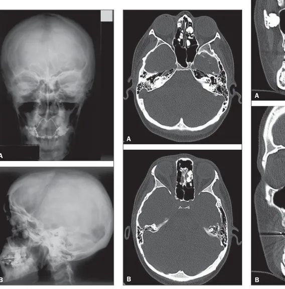
Figure 1. Skull radiography. Posteroanterior (A) and lateral (B) views.
A
B
Figure 2. Non-contrast-enhanced CT, bone window.
A
B
Figure 3. Coronal CT sections, bone window.
A
XII Radiol Bras. 2012 Jul/Ago;45(4):XI–XII Images Description
Figure 1. Skull radiography, posteroan-terior and lateral views demonstrating in-cidentally found osteoblastic lesion with projection into the facial sinuses and man-dible.
Figure 2. Non-contrast-enhanced axial CT sections, bone window, confirming the presence of multiple osteoblastic lesions in the facial sinuses. Such lesions project into the air spaces of the paranasal sinuses.
Figure 3. Coronal CT sections, bone window, demonstrating multiple osteoblas-tic lesions in the facial sinuses and man-dible. The lesion on the mandible shows exophytic growth. The findings are sugges-tive of multiple osteomas.
Diagnosis: Gardner’s syndrome.
COMMENTS
Gardner’s syndrome is a variant of fa-milial adenomatous polyposis which is a dominant autosomal disease characterized by the presence of multiple adenomatous polyps on the intestinal mucosa, particu-larly in the colon, with high potential for malignant transformation(1).
Gardner’s syndrome was originally de-scribed as a triad comprising: 1) adenoma-tous polyposis of the colon; 2) skull and
mandible osteomas; 3) epidermoid cysts. However, from the original description by Garder in 1953, the triad has been ex-panded to include other soft tissue abnor-malities such as desmoid tumors, seba-ceous cysts, lipomas, fibromas and sarco-mas(2). The incidence of this syndrome is estimated to be 1:14.000 births(3).
Osteomas are benign, slow-growing bone tumors, corresponding to the most common benign neoplasms of the nose and facial sinuses. Generally, they are asymp-tomatic and incidentally found at imaging studies, although they may run their course with symptoms, depending on their size and location. Typically, the growth of these bone tumors occurs towards inside the air space of a facial sinus or simply exophyt-ically in relation to the bone surface. In Gardner’s syndrome, osteomas presenta-tion is varied, generally preceding the on-set of colon polyps(4,5).
The genetic basis of this syndrome is re-lated to a mutation in the gene of colon adenomatous polyposis located in the long arm of the chromosome 5. The loss of the functions of this gene, which is a tumor suppressor, is considered to be the initial event in the development of adenomas(6,7). Differential diagnoses include pure fa-milial adenomatous polyposis, Turcot
syn-drome (an association of colon adenomas and encephalic tumors such as glioblas-toma multiforme and medulloblasglioblas-toma), Peutz-Jegher syndrome (consisting in mul-tiple hamartomatous polyps) and hereditary nonpolyposis colorectal cancer(6).
The management of these patients in-cludes total colectomy, since the malignant transformation of adenomas occurs in all of the cases. Familial screening is indi-cated(1–3).
REFERENCES
1. Juhn E, Khachemoune A. Gardner syndrome: skin manifestations, differential diagnosis and manage-ment. Am J Clin Dermatol. 2010;11:117–22. 2. Brodbeck AJ, Cruz JV, Camargo Filho SA, et al.
Síndrome de Gardner: apresentação de um caso e revisão da literatura. Rev Bras Colo-Proct. 1983; 3:95–7.
3. Costa JHG, Azevedo IF, Moreira H, et al. Síndrome de Gardner – descrição de um caso raro. Rev Bras Colo-Proct. 1986;6:131–5.
4. Alexander AAZ, Patel AA, Odland R. Paranasal sinus osteomas and Gardner’s syndrome. Ann Otol Rhinol Laryngol. 2007;116:658–62.
5. Wijn MA, Keller JJ, Giardiello FM, et al. Oral and maxillofacial manifestations of familial adenoma-tous polyposis. Oral Dis. 2007;13:360–5. 6. Half E, Bercovich D, Rozen P. Familial adenomatous
polyposis. Orphanet J Rare Dis. 2009;4:22. 7. Gómez-García EB, Knoers NVAM. Gardner’s