279 Mohan K et al. 18F-FDG PET and benign thoracic lesions
Radiol Bras. 2011 Set/Out;44(5):279–282
Impact of
18
F-FDG PET scan on the prevalence of benign
thoracic lesions at surgical resection
*
Impacto do estudo com 18F-FDG PET sobre a prevalência de achados de lesões torácicas benignas em ressecções cirúrgicas
Kamlesh Mohan1, James McShane2, Richard Page3, Klaus Irion4, Martin J. Ledson5, Martin J. Walshaw5
Objective: The main utility of 18-fluorodeoxyglucose positron emission tomography (FDG-PET) lies in the staging of lung cancer. However, it can also be used to differentiate indeterminate pulmonary lesions, but its impact on the resection of benign lesions at surgery is unknown. The aim of this study was to compare the prevalence of benign lesions at thoracotomy carried out for suspected lung cancer, before and after the introduction of PET scanning in a large thoracic surgical centre.
Materials and Methods: We reviewed our prospectively recorded surgical database for all consecutive patients undergoing thoracotomy for suspected or proven lung cancer and compared the prevalence of benign lesions in 2 consecutive 2-year groups, before (group I) and after (group II) the introduction of FDG-PET scan respectively. Results: Surgical resection was performed on 1233 patients during the study period. The prevalence of benign lesions at surgery in groups I and II was similar (44/626 and 41/607, both 7%), and also in group II between those who underwent FDG-PET scan and the remainder (21/301 and 20/306 respectively, both 7%). In group II, of the 21 patients with benign lesions, who underwent FDG-PET, 19 had a false positive scan (mean standardised uptake value 5.3 [range 2.6–12.7]). Of these, 13 and 4 patients respectively had non-diagnostic bronchoscopy and percutaneous transthoracic lung biopsy pre thoracotomy. There was no difference in the proportion of different benign lesions resected between group I and those with FDG-PET in group II.
Conclusion: The introduction of FDG-PET scanning has not altered the proportion of patients undergoing thoracotomy for ultimately benign lesions, mainly due to the avidity for the isotope of some non-malignant lesions. Such false positive results need to be considered when patients with unconfirmed lung cancer are contemplated for surgical resection.
Key words: Positron emission tomography; Computed tomography; Lung – benign or congenital lesions; Lung cancer – diagnosis and staging; Lung cancer – surgery.
Objetivo: A principal utilidade da tomografia por emissão de pósitrons com 18-fluordeoxiglicose (FDG-PET) está no es-tadiamento do câncer de pulmão. Porém, ela também pode ser utilizada para diferenciar lesões pulmonares indeter-minadas, mas seu impacto na ressecção cirúrgica de lesões benignas é desconhecido. O objetivo deste estudo foi com-parar a prevalência de lesões benignas em toracotomias feitas por suspeição de câncer de pulmão, antes e após a intro-dução do FDG-PET, em um centro de referência de cirurgia torácica. Materiais e Métodos: Os autores analisaram, pros-pectivamente, uma base de dados cirúrgicos de todos os pacientes consecutivos submetidos a toracotomia por câncer de pulmão suspeito ou comprovado e compararam a prevalência de lesões benignas em dois grupos ao longo de dois anos consecutivos, respectivamente antes (grupo I) e depois (grupo II) da introdução da FDG-PET. Resultados: Res-secção cirúrgica foi feita em 1.233 pacientes durante o período do estudo. A prevalência de lesões benignas na cirurgia nos grupos I e II foi similar (44/626 e 41/607, ambas correspondendo a 7%), e também no grupo II, entre aqueles sub-metidos a FDG-PET e os restantes (21/301 e 20/306 respectivamente, ambos correspondendo a 7%). No grupo II, dos 21 pacientes com lesões benignas submetidos a FDG-PET, 19 tiveram um estudo falso-positivo (valor médio padrão de captação 5.3 [faixa 2.6–12.7]). Desses, respectivamente 13 e 4 pacientes tiveram broncoscopia não diagnóstica e biópsia transtorácica percutânea de pulmão antes da toracotomia. Não houve diferença na proporção de lesões benig-nas diferentes ressecadas entre o grupo I e aqueles submetidos a FDG-PET no grupo II. Conclusão: A introdução da FDG-PET não alterou a proporção de pacientes submetidos a toracotomia por lesões benignas, principalmente devido à avidez pelo isótopo de algumas lesões não malignas. Tais resultados falsos-positivos devem ser considerados nos casos em que se contempla a possibilidade de ressecção cirúrgica em pacientes com câncer de pulmão não confirmado.
Unitermos: Tomografia por emissão de pósitrons; Tomografia computadorizada; Pulmão – lesões benignas ou congê-nitas; Câncer de pulmão – diagnóstico e estadiamento; Câncer de pulmão – cirurgia.
Abstract
Resumo
Mohan K, McShane J, Page R, Irion K, Ledson MJ, Walshaw MJ. Impact of 18F-FDG PET scan on the prevalence of benign thoracic lesions at surgical resection. Radiol Bras. 2011 Set/Out;44(5):279–282.
0100-3984 © Colégio Brasileiro de Radiologia e Diagnóstico por Imagem ORIGINAL ARTICLE • ARTIGO ORIGINAL
* Study developed at Liverpool Heart and Chest Hospital, Liverpool, United Kingdom.
1. MRCP, Department of Respiratory Medicine, Liverpool Heart and Chest Hospital, Liverpool, United Kingdom.
2. BSc, Department of Audit and Research, Liverpool Heart and Chest Hospital, Liverpool, United Kingdom.
3. FRCS, Department of Thoracic Surgery, Liverpool Heart and Chest Hospital, Liverpool, United Kingdom.
4. FRCR, Department of Radiology, Liverpool Heart and Chest Hospital, Liverpool, United Kingdom.
5. FRCP, Department of Respiratory Medicine, Liverpool Heart and Chest Hospital, Liverpool, United Kingdom.
Corresponding author: Dr. Martin J. Walshaw. Consultant Res-piratory Physician. Liverpool Heart and Chest Hospital. Thomas Drive, Liverpool L143PE, United Kingdom. E-mail: mwalshaw@ doctors.org.uk
280
Mohan K et al. 18F-FDG PET and benign thoracic lesions
Radiol Bras. 2011 Set/Out;44(5):279–282 INTRODUCTION
Despite advances in imaging and inter-ventional techniques, indeterminate pul-monary lesions are common and represent a diagnostic challenge in the evaluation of patients with suspected lung cancer. Al-though the majority will represent early malignancy, where resection is usually curative, separating these out from the re-mainder can be problematic. Accurate di-agnosis is therefore imperative to avoid unnecessary surgery. Recently, 18-fluoro-deoxyglucose positron emission tomogra-phy (FDG-PET), which measures tissue metabolic activity, has become available as an imaging modality to aid in the detection of malignancy in a number of organ sys-tems. Its main use in pulmonary malignancy is to stage non-small cell lung cancer (NSCLC) and thereby prevent futile thora-cotomies(1,2), but although it is superior to
computed tomography (CT) scanning in the diagnosis of indeterminate pulmonary lesions(3), its role in their management is
less clear and remains to be explored. In-deed, there have been no studies looking at its impact on thoracotomy rates for patients who ultimately have benign disease.
To investigate this further, we compared the prevalence of benign lesions at thorac-otomy carried out for suspected lung can-cer in two 2-year groups, before and after the introduction of PET scanning respectively, in a large thoracic surgical centre serving a catchment population of 2.5 million.
MATERIALS AND METHODS
We reviewed our prospectively re-corded database for all patients who under-went surgical resection for proven or sus-pected NSCLC over a four year period (1233 cases). During this time patients were referred from other hospitals to our tertiary centre, and with the exception of the FDG-PET scan, all pre surgical imag-ing, invasive tests and multidisciplinary team decisions to treatment were per-formed at the referring hospitals. We com-pared the prevalence of benign lesions at surgery for 2 consecutive 2-year groups of patients: those who underwent surgery April 2003 to March 2005 (before the FDG-PET scan was available [group I, 626 patients]) with those undergoing surgery April 2005 to March 2007 (after the
avail-ability of the FDG-PET scan [group II, 607 patients of which 301 (50%) underwent FDG-PET scan]). For patients with an ul-timately benign pathological diagnosis, data on demographics, imaging (CT and FDG-PET), lung pathology, methods of resection and perioperative mortality were reviewed. Information on the lesion size, location, margins, attenuation, presence of calcification and lymphadenopathy were recorded from the CT scan. The PET scan was performed using a dedicated PET scan-ner and interpreted by two experienced nuclear medicine radiologists. PET images were obtained 1 hour after intravenous in-jection of the 336 MBq FDG (227–400 MBq) in patients with blood glucose val-ues < 11.1 mmol/L. The avidity of a lesion for FDG was measured using the maximum standardised uptake value (SUV), where a score > 2.5 was considered indicative of malignancy(4). The study was approved by the local audit and research committee. Descriptive statistics in the form of percent-ages and mean ± standard deviation (SD) have been used to express the results. Chi-square and student’s t test were used to compare data between the two groups. A p value of < 0.05 was considered significant.
RESULTS
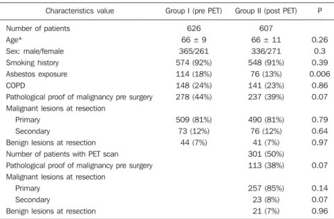
Similar numbers of patients underwent resection in each of the 2-year groups, and with the exception of asbestos exposure,
there was no difference in their clinical and pathological characteristics (Table 1). The number of patients with a definitive histo-logical diagnosis of malignancy pre-resec-tion was similar between groups I and II (278/626 [44%] vs 237/607 [39%] respec-tively; χ2 = 3.43, p = 0.07). Similarly, the
resection rate for ultimately benign lesions was unchanged between groups I and II (44 vs 41 respectively, both 7%), and also in group II when subdivided into those who underwent FDG-PET and the remainder (21/301 vs 20/306, both 7%).
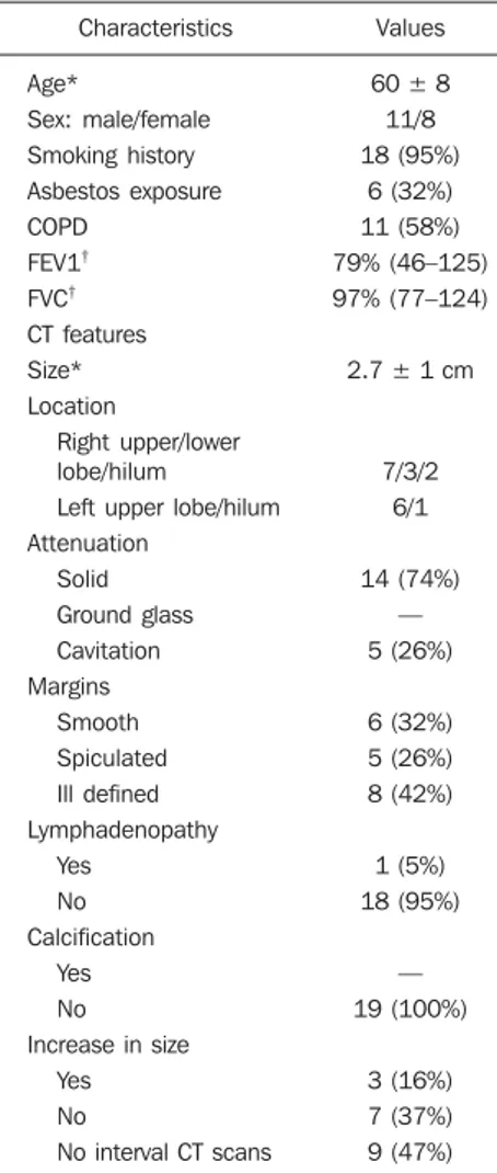
The 21 patients with resected benign lesions following FDG-PET were analysed in more detail: 19 of these had a (false) positive scan and the two with negative scans proceeded to thoracotomy due to preoperative inaccurate positive cytology in one and an increase in the size of the le-sion on interval CT scan in the other. Ex-cluding these two patients, there was no difference in the prevalence of benign le-sions in group I and group II who underwent FDG-PET scan (44/626 [7%] vs 19/301 [6.3%] respectively; χ2 = 0.17, p = 0.68). Table 2 shows the clinical and CT scan characteristics of the 19 patients with PET positive benign lesions: nearly all had a smoking history. On the CT scan, the av-erage size of the lesion was 2.7 cm (1.1-5.0 cm); two thirds were ≤ 3 cm. All were non-calcified, and the majority had a solid con-sistency and spiculated or irregular
mar-Table 1 Clinical and pathological characteristics of 1233 patients who underwent surgery from 2003–2007.
Characteristics value
Number of patients Age*
Sex: male/female Smoking history Asbestos exposure COPD
Pathological proof of malignancy pre surgery Malignant lesions at resection
Primary Secondary
Benign lesions at resection Number of patients with PET scan Pathological proof of malignancy pre surgery Malignant lesions at resection
Primary Secondary
Benign lesions at resection
Group I (pre PET)
626 66 ± 9 365/261 574 (92%) 114 (18%) 148 (24%) 278 (44%)
509 (81%) 73 (12%)
44 (7%)
Group II (post PET)
607 66 ± 11 336/271 548 (91%)
76 (13%) 141 (23%) 237 (39%)
490 (81%) 76 (12%)
41 (7%) 301 (50%) 113 (38%)
257 (85%) 23 (8%) 21 (7%)
P
0.26 0.3 0.39 0.006
0.86 0.07
0.79 0.64 0.97
0.07
0.14 0.07 0.96
281 Mohan K et al. 18F-FDG PET and benign thoracic lesions
Radiol Bras. 2011 Set/Out;44(5):279–282 Table 2 Clinical and CT scan characteristics of 19 patients with PET positive benign lesions.
Characteristics
Age*
Sex: male/female Smoking history Asbestos exposure COPD
FEV1† FVC† CT features Size* Location
Right upper/lower lobe/hilum
Left upper lobe/hilum Attenuation
Solid Ground glass Cavitation Margins
Smooth Spiculated Ill defined Lymphadenopathy
Yes No Calcification
Yes No Increase in size
Yes No
No interval CT scans
Values
60 ± 8 11/8 18 (95%)
6 (32%) 11 (58%) 79% (46–125) 97% (77–124)
2.7 ± 1 cm
7/3/2 6/1
14 (74%) — 5 (26%)
6 (32%) 5 (26%) 8 (42%)
1 (5%) 18 (95%)
— 19 (100%)
3 (16%) 7 (37%) 9 (47%)
*Mean (SD), † Mean (range). Percentages are given in
parenthesis. COPD, chronic obstructive pulmonary dis-ease; FEV1, forced expiratory volume in one second; FVC, forced vital capacity.
Table 3 Histopathology of resected benign lesions in 19 patients along with the PET-SUV values.
Diagnosis
Fibrosis Fibrosis Fibrosis Fibrosis
Chronic inflammation and fibrosis Tuberculosis
Tuberculosis Tuberculosis Aspergilloma Aspergilloma Rheumatoid nodule Rheumatoid nodule Organisisng pneumonia Organising pneumonia Infarct
Necrosis Granuloma Hamartoma
Bronchiolitis with interstitial lung disease
SUV
7.9 3.9 4.5 4.5 4.6 5.7 5.2 3.1 12.7 4.8 3.7 3.0 4.2 6.8 2.6 3.5 11.5 3.0
5.0
though CT scanning can suggest cancer based on the morphology of a lesion, it cannot demonstrate increased metabolic activity, which is a hall mark of malignant disease. In this respect, a metabolic imag-ing technique such as the FDG-PET scan should be superior to CT in differentiating benign from malignant lesions(5). Although
FDG-PET has a major role in lung cancer in defining metastatic or local spread and thereby reducing futile thoracotomies, no studies have evaluated its impact on the prevalence of unexpected benign disease at resection for apparently malignant lone pulmonary lesions. We therefore looked at the prevalence of benign lesions in patients with isolated suspected malignant pulmo-nary lesions undergoing resection for a period of 2 years following the introduction of the FDG-PET scan and compared it with that in the preceding 2 years.
Our results show that FDG-PET scan did not reduce the resection rate for benign lesions. No previous studies have specifi-cally addressed the issue of FDG-PET scans on the incidence of unexpected be-nign lesions at thoracotomy, but rather they have been targeted at preventing futile tho-racotomies by identifying patients with otherwise unknown disseminated dis-ease(7,8). Of these, the PET in lung cancer
staging (PLUS) study showed that PET scan reduced the number of futile thorac-otomies, by identifying patients with lo-cally advanced disease, post operative re-currence and death(7). Of the 9 cases where
benign lesions were discovered at thorac-otomy, although 2 of these underwent FDG-PET scan, it is not clear whether these were falsely positive or merely reassured the clinician that there was no disseminated disease. Our study is therefore the largest in the literature, which specifically
ad-Table 4 Prevalence of common benign lesions resected pre and post PET scan.
Benign lesions
Tuberculosis Hamartoma
Organising pneumonia Aspergilloma Fibrosis
Rheumatoid nodule Others
Prevalence
Pre PET (44/626)
9 8 6 3 2 1 15
Post PET (19/301)
3 1 2 2 5 2 4
P value
0.76 0.29 0.96 0.60 0.49 0.25 0.46 histological diagnosis and then staging the
disease in order to decide the best treatment strategy. However, in those patients with isolated pulmonary lesions where defini-tive histology can only be obtained at tho-racotomy, imaging techniques are crucial in informing the clinician as to the likelihood of malignancy. The aim is to identify pa-tients with early stage cancer that would benefit from curative resection, whilst avoiding unnecessary surgery in those with benign disease. The development of the CT scan provided information regarding the morphology of the lesion, its attenuation, extent, growth rate, identified regional and distant spread(5), and also led to a decline
in the prevalence of benign lesions at sur-gical resection from 64% to 9% in a recent series of > 1500 patients(6). However,
al-gins. Before surgery, 13 and 4 patients had non-diagnostic bronchoscopy and percuta-neous transthoracic lung biopsy (PTLB) respectively. At thoracotomy, diagnosis was achieved by lobectomy in 9; wedge resec-tion in 8 and 2 had frozen secresec-tion biopsy alone. There were no perioperative deaths and the mean duration of hospital stay was 5 (2–11) days. There was a wide range of benign conditions resected (Table 3) with varying SUV values on FDG-PET scan. Furthermore the introduction of FDG-PET scanning did not alter proportion of differ-ent benign lesions resected (Table 4).
DISCUSSION
282
Mohan K et al. 18F-FDG PET and benign thoracic lesions
Radiol Bras. 2011 Set/Out;44(5):279–282 dresses the impact of FDG-PET scan on
unexpected benign lesions at thoracotomy. In nearly all our cases, the clinician was misled by the high avidity for FDG by some benign lesions (false positive scans). False positive FDG-PET findings have been re-ported in 8–10% of indeterminate pulmo-nary lesions(9) and are seen in infectious
and inflammatory conditions such as tuber-culosis, histoplasmosis, aspergillosis, sar-coidosis, lipoid pneumonia, rheumatoid lung disease and suture/stapler granulo-mas(3,10,11). The PET scan relies on an in-creased number of glucose transporter (GLUT) proteins and increased glycolytic activity of malignant cells to actively accu-mulate the radiotracer FDG, a glucose ana-logue labelled with positron-emitting radio-isotopes(12). However FDG uptake is not
cancer specific since inflammatory cells (neutrophils, lymphocytes, macrophages and fibroblasts) also accumulate FDG and cause false positive results(13). In activated inflammatory cells, glucose metabolism can multiply by 20–30 times, thereby increasing the uptake of FDG(14). Specifically, GLUT
proteins proliferate resulting in an increased cellular accumulation of FDG(15). Indeed
this property has led to the application of FDG-PET scans in the diagnosis and to monitor treatment response in some infec-tious and inflammatory conditions(16).
Al-though an SUV of > 2.5 is used to distin-guish malignant from benign lesions, there is significant overlap in uptake values be-tween the two conditions. Benign lesions accumulate FDG relatively early, whereas malignant cells retain FDG for longer pe-riods: dual time point (early and delayed) imaging with an FDG retention index of > 10% has been reported to improve the ac-curacy of cancer diagnosis(17,18). However,
the routine use of dual time point FDG imaging is time consuming and resource intensive, and in populations with a high incidence of malignancy it adds little to the overall yield and is therefore not justified in clinical practice. Only single time point (early) images were acquired in our study. In our study, the clinical and CT scan features of the resected benign lesions in-dicated a high suspicion for malignancy, and the patients came from a population with a high risk for the disease(12,19).
At-tempts to obtain a positive histological di-agnosis pre-thoracotomy, for example by
PTLB, were only considered by the refer-ring hospitals in 20% of cases. However, a negative or non-specific biopsy does not reliably rule out malignancy in patients with high suspicion for lung cancer, and a positive result in patients with resectable lesions is an indication for thoracotomy anyway. There is no convincing evidence that PTLB reduces unnecessary thoracoto-mies in patients who ultimately have be-nign disease at resection, and the consen-sus view is that patients with operable pul-monary lesions suspicious for lung cancer should be referred for surgery as PTLB is unlikely to alter patient management(20,21).
The limitations of our study are that we do not have information on those patients who had a negative FDG-PET scan and therefore were not referred to our centre for consideration of surgical resection, and that a proportion of patients (50%) did not ben-efit from this investigation after it became available. However, our aim was to specifi-cally look at the trend of benign lesions resected since the introduction of this scan-ning technique in our practice, and some patients remained without scans because of the limited availability and long waiting times when the technique was first intro-duced. Furthermore the prevalence of be-nign lesions in patients who did not have an FDG-PET scan during the same period (20/306, 6.5%) was similar, reassuring us that the scanned group were representative. This study shows that the FDG-PET scan should be interpreted with caution in pa-tients presenting with isolated pulmonary lesions, and may not add to the diagnostic process even in those with a high risk of lung cancer.
REFERENCES
1. Pieterman RM, Van Putten JW, Meuzelaar JJ, et al. Preoperative staging of non-small lung cancer with positron-emission tomography. N Engl J Med. 2000;343:254–61.
2. Saunders CA, Dussek JE, O’Doherty MJ, et al. Evaluation of fluorine-18-fluorodeoxyglucose whole body positron emission tomography imag-ing in the stagimag-ing of lung cancer. Ann Thorac Surg. 1999;67:790–7.
3. Gould MK, Maclean CC, Kuschner WG, et al. Ac-curacy of positron emission tomography for di-agnosis of pulmonary nodules and mass lesions: A meta-analysis. JAMA. 2001;285:914–24. 4. Silvestri GA, Gould MK, Margolis ML, et al.
Non-invasive staging of non-small cell lung can-cer: ACCP evidenced-based clinical practice guidelines (2nd edition). Chest. 2007;132:178S– 201S.
5. Erasmus JJ, Connolly JE, McAdams HP, et al. Solitary pulmonary nodules: Part I. Morphologic evaluation for differentiation of benign and ma-lignant lesions. Radiographics. 2000;20:43–58. 6. Smith MA, Battafarano RJ, Meyers BF, et al. Prevalence of benign disease in patients under-going resection for suspected lung cancer. Ann Thorac Surg. 2006;81:1824–9.
7. Van Tinteren H, Hoekstra OS, Smit EF, et al. Ef-fectiveness of positron emission tomography in the preoperative assessment of patients with sus-pected non-small-cell lung cancer: the PLUS multicentre randomised trial. Lancet. 2002;359: 1388–93.
8. Reed CE, Harpole DH, Posther KE, et al. Results of the American College of Surgeons Oncology Group Z0050 trial: the utility of positron emis-sion tomography in staging potentially operable non–small cell lung cancer. J Thorac Cardiovasc Surg. 2003;126:1943–51.
9. Fischer BM, Mortensen J, Hojgaard L. Positron emission tomography in the diagnosis and stag-ing of lung cancer: a systematic, quantitative re-view. Lancet Oncology. 2001;2:659–66. 10. Mokhlesi B, Angulo-Zereceda D, Yaghmai V.
False-positive FDG-PET scan secondary to lipoid pneumonia mimicking a solid pulmonary nodule. Ann Nucl Med. 2007;21:411–4.
11. Yuksel M, Akgul AG, Evman S, et al. Suture and stapler granulomas: a word of caution. Eur J Cardio-thorac Surg. 2007;31:563–5.
12. Detterbeck FC, Falen S, Rivera MP, et al. Seek-ing a home for a PET, Part 1: definSeek-ing the appro-priate place for positron emission tomography imaging in the diagnosis of pulmonary nodules or masses. Chest. 2004;125:2294–9.
13. Alavi A, Gupta N, Alberini JL, et al. Positron emission tomography imaging in nonmalignant thoracic disorders. Semin Nucl Med. 2002;32: 293–321.
14. Amrein PC, Larson SM, Wagner HN Jr. An auto-mated system for measurement of leukocyte me-tabolism. J Nucl Med. 1975;15:352–5. 15. Kubota R, Yamada S, Kubota K, et al. Intra
tu-moral distribution of fluorine-18-fluorodeoxy-glucose in vivo: high accumulation in macroph-ages and granulation tissues studied by micro-autoradiography. J Nucl Med. 1992;33:1972– 80.
16. Love C, Tomas M, Tronco GG, et al. FDG PET of infection and inflammation. Radiographics. 2005;25:1357–68.
17. Demura Y, Tsuchida T, Ishizaki T, et al. 18F-FDG accumulation with PET for differentiation be-tween benign and malignant lesions in the tho-rax. J Nucl Med. 2003;44:540–8.
18. Xiu Y, Bhutani C, Durairaj T, et al. Dual-time point FDG PET imaging in the evaluation of pul-monary nodules with minimally increased meta-bolic activity. Clin Nucl Med. 2007;32:101–5. 19. Winer-Muram HT. The solitary pulmonary
nod-ule. Radiology. 2006;239:34–49.
20. Murphy JM, Gleeson FV, Flower CDR. Percuta-neous needle biopsy of the lung and its impact on patient management. World J Surg. 2001;25:373– 80.