SCREENING OF STREPTOCOCCUS AGALACTIAE IN PREGNANT WOMEN
F.M. Munari1, F. De-Paris2, G.D. Salton1, P.S. Lora1, P. Giovanella1, A.B.M.P. Machado2, L.S. Laybauer2, K.R.P. Oliveira2, C. Ferri3, J.L.S. Silveira4, C.C.F.C. Laurino1, R.M. Xavier1, A.L. Barth2, S. Echeverrigaray5, J.P. Laurino1,5*
1
Laboratório de Biologia Molecular em Doenças Auto-imunes e Infecciosas, Centro de Pesquisas, Hospital de Clínicas de Porto
Alegre, Porto Alegre, RS, Brasil; 2Unidade de Microbiologia e Biologia Molecular, Serviço de Patologia Clínica, Hospital de
Clínicas de Porto Alegre, Porto Alegre, RS, Brasil; 3Residente do Serviço de Pediatria do Hospital de Clínicas de Porto Alegre,
Porto Alegre, RS, Brasil; 4Unidade de Terapia Intensiva Pediátrica da Santa Casa de Uruguaiana, Uruguaiana, RS, Brasil;
5
Instituto de Biotecnologia, Universidade de Caxias do Sul, Caxias do Sul, RS, Brasil.
Submitted: October 14, 2010; Approved: January 16, 2012.
ABSTRACT
Group B Streptococcus (GBS) is the most common cause of life-threatening infection in neonates.
Guidelines from CDC recommend universal screening of pregnant women for rectovaginal GBS
colonization. The objective of this study was to compare the performance of a combined enrichment/PCR
based method targeting the atr gene in relation to culture using enrichment with selective broth medium
(standard method) to identify the presence of GBS in pregnant women. Rectovaginal GBS samples from
women at 36 weeks of pregnancy were obtained with a swab and analyzed by the two methods. A total of
89 samples were evaluated. The prevalence of positive results for GBS detection was considerable higher
when assessed by the combined enrichment/PCR method than with the standard method (35.9% versus
22.5%, respectively). The results demonstrated that the use of selective enrichment broth followed by PCR
targeting the atr gene is a highly sensitive, specific and accurate test for GBS screening in pregnant women,
allowing the detection of the bacteria even in lightly colonized patients. This PCR methodology may provide
a useful diagnostic tool for GBS detection and contributes for a more accurate and effective intrapartum
antibiotic and lower newborn mortality and morbidity.
Key words: Streptococcus agalactiae; Group B Streptococcus; atr gene; Screening test; Accuracy.
INTRODUCTION
Streptococcus agalactiae, also known as Group B
Streptococcus (GBS), is associated to severe invasive disease
in newborns. In fact, GBS is considered as one of the major
causes of neonatal meningitis and sepsis (31).
It is estimated that 5 to 40% of all pregnant women may
present rectovaginal colonization with GBS, most of which are
asymptomatic (28) or associated with acute chorioamnionitis,
endometritis, and urinary tract infection (32). Moreover, even
just colonized or asymptomatic, pregnant women may transmit
GBS to their newborns during labor and this may constitute
the first step for invasive disease in the first week of life (10).
The incidence of GBS neonatal infection was 1.4 per 1000
live births in USA in 1990 (10, 32, 40). Coinciding with active
prevention efforts in the 1990s, the incidence of early-onset
disease declined to 0.5 cases per 1,000 live births in 1999 (10,
24). In Brazil, few studies address early neonatal infection by
GBS. The prevalence of GBS infection among neonates varied
between 1.1 and 1.4 cases per 1,000 live births, in Porto Alegre
(21) and Campinas (23), respectively. A recent study in a
maternity hospital from Manaus (AM, Brazil) showed that the
main microorganism isolated in blood cultures of newborns
with early-onset sepsis was S. agalactiae (25).
Clinical syndromes of GBS disease in newborns include
sepsis, meningitis, pneumonia, cellulitis, osteomyelitis, and
septic arthritis. Bloodstream infections, with or without
pneumonia, are the main manifestation of neonatal GBS
disease and are observed in approximately 90% of cases, while
meningitis occurs in around 10% (10, 31). Other symptoms
reported among older children include endocarditis (2, 34) and
epiglottitis (38).
In addition to mother colonization, other maternal risk
factors like preterm delivery, prolonged rupture of membranes
( 18 hours), intrapartum temperature of at least 38º C, or prior
infant with GBS infection predispose a neonate to early-onset
GBS contamination (3, 32).
Currently, the most effective strategy for reducing
early-onset GBS infection is prenatal maternal diagnosis of
rectovaginal GBS colonization between the 35th to 37th weeks
of gestation, followed by chemoprophylaxis (3, 10, 37). The
guidelines recommended by The Centers for Disease Control
and Prevention (CDC) and The American College of
Obstetricians and Gynecologists (ACOG) includes: (i) the
sampling of the vaginal and anorectal regions with the aid of a
swab which is submitted to bacteriological culture usually into
selective broth medium (enrichment culture) followed by
subculture onto sheep blood agar plates (1, 3, 10), and (ii) the
intrapartum chemoprophylaxis for those pregnant women with:
positive maternal GBS screening, positive GBS urine culture
during the current pregnancy, and a previous infant who had
GBS infection (10). An oral chemoprophylaxis approach with
antibiotics is not recommended because it is unlikely to
eradicate maternal genital GBS colonization (13, 14, 29).
However, although the laboratory methods for the
identification of GBS have evolved, there remains a clinical
need for greater accuracy, particularly in the case of
asymptomatic colonization with low GBS charge (4, 15).
The aim of this study was to develop a highly sensitive
and specific molecular method to evaluate GBS colonization in
pregnant women. The proposed method combines a short
enrichment culture followed by the amplification and detection
of the atr gene with GBS specific primers.
MATERIALS AND METHODS
Patients and samples
The study was performed in 89 pregnant women ( 36
weeks of pregnancy) who attended the primary health care unit
at an university hospital of Rio Grande do Sul State, Brazil,
from April 2006 to May 2007. These 89 samples correspond to
all medical requests for GBS search in the hospital service of
obstetric primary care during this period.
Specimens of combined vaginal and anal secretions were
collected using the technique recommended by the CDC (10),
the STARD (9). Briefly, the vagina and the anus were sampled,
and the swabs were immediately soaked into the Stuart’s
transport medium.
Bacterial Enrichment Culture
The swabs were transferred into 2 mL of BHI enrichment
broth (BHI-E) supplemented with peptone 3 (2 g/L),
gentamicin (8 g/mL) and nalidixic acid (15 g/mL) to follow
the microbiological identification as recommended by the CDC
(10), and incubated at 36°C for 18-24 hours.
(BioMérieux, Marcy L´Etoile, France), incubated at 36°C for
24 hours and inspected for characteristic GBS colonies.
Whether these colonies were not identified, the plates were
reincubated for another 24 hours and inspected again. When
suspected colonies were present, they were subcultured in
thioglycolate broth for 12 hours and then submitted to Gram
stain. Colonies with Gram-positive cocci arranged in pairs and
chains were submitted to CAMP test (Christie, Atkins,
Munch-Petersen). The specimens positive for the CAMP test were
considered presumptive GBS (10).
Identification of GBS by PCR Amplification
The enriched cultures were centrifuged and the pellets
were washed with PBS buffer, and then incubated with 500 µL
of LiCl 5M for 30 minutes at room temperature.
DNA-extraction was performed with the kit Wizard® Genomic DNA
Purification (Promega corp, Madisson, WI, USA) according to
the manufacturer’s instructions. The purified DNA was
resuspended to a final volume of 100µL.
The PCR reactions were carried out using GBS specific
primers atrF (5’-CGATTCTCTCAGCTTTGTTA-3’) and atrR
(5’-AAGAAATCTCTTGTGCGGAT-3’), that amplified a
779bp sequence of the atr gene, responsible for the glutamine
transporter protein (gbs0538) of S. agalactiae. This
housekeeping gene and the corresponding primers were
selected from the S. agalactiae Multi Locus Sequence Typing
(MLST) website - http://pubmlst.org/sagalactiae/ (16, 17, 36).
PCR amplifications were performed in a total volume of
25 l containing: 0.2 mM of dNTPs (ABgene®, Epson, UK);
0.4 µM of each atr-primer; 1x PCR buffer with 1.5 mM of
MgCl2 (JMR Holdings, London, UK); 1.0 U of Super-Therm
DNA polimerase (JMR Holdings, London, UK); 2 µL of
bacterial DNA (non-quantified). The amplifications were
conducted on a MJ Research Thermocycler (Model PTC-100)
with the following program: (i) 94°C for 1 minute, (ii) 30
cycles of 1 minute at 94°C, 45 seconds at 55°C, and 1 minute
at 72°C, and (iii)10 minutes at 72°C. Subsequently, 10 L of
the amplified reaction were separated by electrophoresis in 2%
agarose gels, and the amplicons visualized under UV light after
ethidium bromide staining. The samples that exhibited a 779 bp
amplicon were considered positive for GBS. Purified GBS
genomic DNA obtained from a clinical isolate was used as a
positive control in all sets of PCR amplifications. This isolate
came from a patient with neonatal sepsis and was properly
characterized by microbiological techniques and molecular
characterization of the gene coding for 16S ribosomal RNA
(16S rRNA) and atr gene by DNA sequencing.
Each step of the procedure (DNA extraction, amplification
and detection) was carried out in a distinct area of laboratory
with required safeguards for molecular biology tests. All
positive results in PCR were retested to assure the positivity.
Molecular Characterization of GBS genomic DNA
In order to confirm the specificity of GBS atr-primers,
purified genomic DNA from S. agalactiae ATCC12403 was
amplified by PCR and the amplicon obtained was sequenced
by the chain terminators method (Amersham Biosciences ET
Terminator Kit) using MegaBACE 1000, following the
manufacturer’s protocols. The atr gene is a housekeeping gene
and its DNA sequence obtained was compared to all known
sequences in GenBank using the Basic Local Alignment Search
Tool (BLAST) of National Center for Biotechnology
Information, Bethesda, MD.
Specificity Assay for the atr-primers
Purified genomic DNA was obtained from cultures of
clinical isolates of Staphylococcus aureus, Staphylococcus
epidermidis, Acinetobacter sp., Serratia sp., Salmonella sp.,
Proteus mirabillis, Citrobacter sp., and Morganella morganii,
and submitted to PCR amplification with the atr-primers.
Additionally, genomic DNA were obtained from cultures of
clinical isolates of Streptococcus pyogenes, Streptococcus
uberis, Enterococcus faecalis and Escherichia coli, and mixed
with and without S. agalactiae DNA. The mixtures were
subsequently submitted to PCR amplification with the atr-
Statistical Analysis
Sensitivity, specificity, and positive and negative
predictive values along with their 95% confidence intervals
were calculated for the combined culture PCR method, using
the conventional microbiological method recommended by
CDC, as standard. Kappa coefficient was used to evaluate the
agreement between the methods (33). The statistical analysis
was performed using SPSS® v.12.
RESULTS
Specificity of the atr-primers for GBS identification In a first approach, the atr-primers were submitted to a
primer-BLAST test against all the sequences deposited at
GenBank, EMBL, DDBJ and PDB. The results showed just
three in silico amplicons, all of them concerning the amino acid
ABC transporter ATP-binding protein of S. agalactiae (atr
gene) and with the expected 779bp length.
To experimentally evaluate the specificity of atr-primers,
779bp PCR amplicon obtained from S. agalactiae ATCC
12403 was sequenced and compared with the overall
information deposited at the GenBank. This amplicon showed
a 100% identity with the S. agalactiae atr sequences,
followed by 78% identity with S. suis atr sequences (3 e-117).
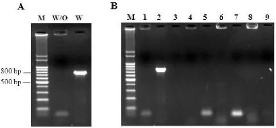
In order to confirm the specificity of the PCR test in the
presence of other bacteria two tests were performed. In the first
test, DNA from two species of Streptococcus (S. pyogenes and
S. uberis), Enterococcus faecalis, and E. coli, were mixed and
submitted to PCR amplification. No amplicons were detected
in this sample, but when S. agalactiae DNA was included in
the mixture the expected amplicon (779bp) was obtained
(Figure 1.A).
In a second experiment, DNA from clinical isolates of
eight bacterial species currently found in vaginal and rectal
samples (S. aureus, S. epidermidis, Acinetobacter sp., Serratia
sp., Salmonella sp., P. mirabilis, Citrobacter sp., and M.
morganii) were submitted to PCR amplification using the
atr-primers, showing PCR negative results (Figure 1.B).
These results indicate that the atr-primers designed are
highly specific for S. agalactiae, and that the presence of other
bacterial DNA did not interfere with atr-PCR amplification.
Figure 1. Results for PCR assay with the atr-primers to the mix of DNA with and without S. agalactiae (A) and for the cultures of clinical isolates (B). Lane M- 100-bp molecular-size standard. Lane W/O- mix of DNA without S. agalactiae. Lane W- mix of
DNA with S. agalactiae. Lane 1- Proteus mirabillis. Lane 2- S. agalactiae. Lane 3- Staphylococcus aureus. Lane 4-
Staphylococcus epidermidis. Lane 5- Acinetobacter sp. Lane 6- Serratia sp. Lane 7- Salmonella sp. Lane 8- Morganella morganii.
Performance of PCR for GBS detection
A total of 89 pregnant women were screened for GBS
colonization using the traditional enrichment/culture method,
and the combined enrichment-PCR based procedure (PCR)
proposed in this work. A total of 32 patients (35.9%) were
identified as carriers of GBS on the basis of PCR results, as
compared with just 20 patients (22.5%) on the basis of the
traditional enrichment/culture method.
All positive samples according to the culture methods
were also positive by the PCR technique, indicating that the
sensitivity of the PCR assay was 100%. The highest number of
positive samples detected by the PCR method can be attributed
to its high accuracy, as it is able to detect as low as 1-2 bacteria
per L, been particularly interesting for the diagnosis of
asymptomatic and lightly charged GBS patients.
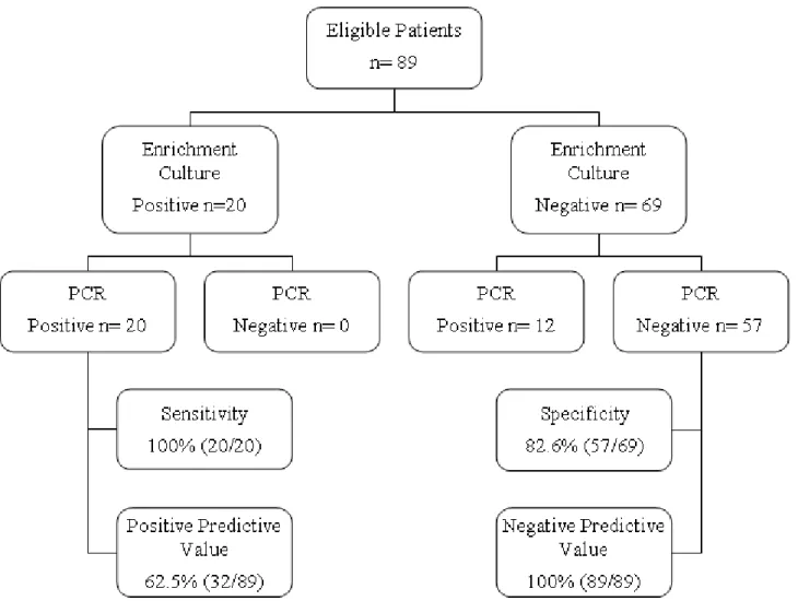
Among the 69 culture-negative samples for GBS, 12 were
positive by PCR and 57 were negative in both methods.
Considering the culture method as gold standard, the
enrichment/PCR method showed a specificity of 82.6% (Figure
2), and positive and negative predictive values of 62.5% and
100%, for the enrichment/culture and the PCR methods,
respectively. Moreover, the index of agreement (Kappa)
between the techniques was 0.68. This apparent lower
specificity is associated to the higher sensibility of PCR, and
not to a lower “specificity” of this technique.
DISCUSSION
The aim of this study was to develop a more accurate and
faster screening test for GBS than the standard culture method.
We compared two GBS identification screening methods in
pregnant women: enrichment culture (as recommended by
CDC) (10) and a combined enrichment/PCR method.
Nowadays, as recommended by CDC, the standard
method for the diagnosis of GBS in pregnant women consists
of vaginal and anal sampling, enrichment in selective broth,
and colony identification by routine microbiological methods
(10, 19). Alternatively, immunological tests have been
developed for GBS detection during labor in an effort to
optimize specific intrapartum antibiotic prophylaxis. However,
these antigen-based tests are neither sensitive nor specific
enough to substitute routine bacterial culture methods,
especially in lightly colonized patients (1, 3, 5, 10, 14). This is
a cause of concern as many infected neonates were born from
lightly colonized women, as showed in clinical studies (5).
In the last decade, molecular techniques emerged as
alternatives for the rapid and specific diagnostic of several
infectious agents including GBS (8, 12). PCR-based
approaches for GBS detection include direct amplification or
nested-PCR of 16S rRNA gene (18, 20, 40) and amplification
using primers specifically designed for the GBS CAMP factor
gene (cfb). Although efficient, these approaches are limited by
the increased risk of carryover of nested PCR and the absence
of CAMP factor in some GBS isolates. To overcome these
problems, in the present study, we used species specific
primers that amplified part of the housekeeping atr gene of
GBS.
Experimental data showed that the use of atr-primers for
GBS screening resulted in a high sensitivity (100%) when
compared with the values obtained by PCR amplification with
other primers (8, 12, 27, 30). The PCR approach showed a
specificity of just 82.6% in relation with the conventional
culture method. However, the low specificity should be regard
carefully as the formulae adopted considered the culture
method as gold standard, but experimental data have shown
that the conventional culture procedures are not sensitive
enough to detect GBS in low charge samples. In fact, the lower
specificity of PCR is due to its ability to detect GBS in samples
where the culture method failed. When amplifications are
performed after a short term culture of the specimens into
selective enrichment broth, the atr-primers showed high
analytical specificity for GBS. The prevalence of positive
results for GBS detection was considerable higher when
assessed by the combined enrichment/PCR method than with
the enrichment culture (35.9% versus 22.5%, respectively).
The prevalence of 35,9% found in this work for GBS
among pregnant women using the enrichment culture/PCR
method is higher than previous results obtained in Brazil which
varied between 15% and 26% (6, 7, 11, 22, 26, 35). This
difference can be attributed to the highest sensibility of the
method adopted in the present study. This enrichment/PCR
method integrates high specificity, sensibility and allows the
detection of GBS even in lightly colonized patients. Moreover,
the amplicons obtained with the selected primers can be also
used to perform epidemiologic studies by MLST approach.
Considering the newborn contamination risk imposed by
lightly colonized mothers, the high sensibility of PCR based
approaches, in particular the proposed enrichment/PCR
method, may contribute to the proper prophylaxis and
consequent reduction of the neonate infectious diseases caused
by GBS.
In addition to its accuracy, the enrichment/PCR method is
performed in a moderate time (24-36 hours) when compared to
standard culture-based methods (48-72 hours), allowing to
adopt the prophylactic protocols earlier and with better results.
Moreover, the swabs can be analyzed by the combined
enrichment/PCR method until 15 days after samples collection
(data not shown), without any reduction in the accuracy. This
fact may allow the screening of pregnant women from remote
In conclusion, the results demonstrated that the use of
selective enrichment broth followed by PCR targeting the atr
gene is an excellent test for GBS screening in pregnant women.
This PCR methodology may provide a diagnostic tool for GBS
detection, potentially allowing more accurate and effective
intrapartum antibiotic prophylaxis and lower newborn
mortality and morbidity.
ACKNOWLEDGEMENTS
This study was supported by grants from Conselho
Nacional de Desenvolvimento Científico e Tecnológico
(CNPq), Fundação de Amparo à Pesquisa do Estado do Rio
Grande do Sul (FAPERGS) and Hospital de Clínicas de Porto
Alegre (HCPA).
REFERENCES
1. Adler, A.; Block, C.; Engelstein, D.; Hochner-Celnikcier, D.; Drai-Hassid, R.; Moses, A.E. (2008). Culture-based methods for detection and identification of Streptococcus agalactiae in pregnant women-what are we missing? Eur J Clin Microbiol Infec Dis. 27 (3), 241-243.
2. Alsoub, H.; Najma, F.; Robida, A. (1997). Group B streptococcal endocarditis in children beyond the neonatal period.Pediatr Infect Dis J. 16 (4), 418-420.
3. American College of Obstetricians and Gynecologists. (2002) Committee Opinion: number 279, December 2002. Prevention of early-onset group B streptococcal disease in newborns. Obstet Gynecol. 100 (6), 1405-1412.
4. Baker, C.J.; Clark, D.J.; Barrett, F.F. (1973). Selective broth medium for isolation of group B streptococci.Appl Microbiol. 26 (6), 884-885. 5. Baker, C.J. (1973). Inadequacy of rapid immunoassays for intrapartum
detection of group B streptococcal carriers.Obstet Gynecol. 88 (1), 51-55.
6. Benchetrit, L.C.; Fracalanzza, S.E.L.; Peregrino, H.; Camelo, A.A.; Sanches, L.A.L.R. (1982). Carriage of Streptococcus agalactiae in women and neonates and distribution of serological types: a study in Brazil. J Clin Microbiol. 15 (5), 787-790.
7. Beraldo, C.; Brito, A.S.J.; Saridakis, H.O.; Matsuo, T. (2004). Prevalência da colonização vaginal e anorretal por estreptococo do grupo B em gestantes do terceiro trimestre. Rev Bras Ginecol Obstet. 26 (7), 543-549.
8. Bergeron, M.G.; Ke, D.; Menard, C.; Picard, F.J.; Gagnon, M.; Bernier, M.; Ouellette, M.; Roy, P.H.; Marcoux, S.; Fraser, W.D. (2000). Rapid detection of group B streptococci in pregnant women at delivery. N Engl J Med. 343 (3), 175-179.
9. Bossuyt, P.M.; Reitsma, J.B.; Bruns, D.E.; Gatsomis, C.A.; Glasziou, P.P.; Irwig, L.M.; Lijmer, J.G.; Moher, D.; Rennie, D.; de Vet, H.C.W. (2003). Towards complete and accurate reporting of studies of diagnostic accuracy: the STARD initiative. Clin Chem. 49 (1), 1-6
10. Centers for Disease Control and Prevention. (2002). Prevention of perinatal group B streptococcal disease. Revised guidelines from CDC.
MMWR CDC Surveill Summ. 51 (RR11), 1–22.
11. Costa, A.L.R.; Filho, F.L.; Chein, M.B.C.; Brito, L.M.O.; Lamy, Z.C.; Andrade, K.L. (2008). Prevalence of colonization by group B Streptococcus in pregnant women from a public maternity of Northwest region of Brazil. Rev Bras Ginecol Obstet. 30 (6), 274-280.
12. Davies, H.D.; Miller, M.; Faro, S.; Gregson, D.; Kehl, S.; Jordan, J. (2004). Multicenter study of a rapid molecular-based assay for the diagnosis of group B streptococcus colonization in pregnant women. Clin Infect Dis. 39 (8), 1129-1135.
13. Gardner, S.E.; Yow, M.D.; Leeds, L.J.; Thompson, P.K.; Mason, E.O.Jr.; Clark, D.J. (1979). Failure of penicillin to eradicate group B streptococcal colonization in the pregnant woman. A couple study.Am J
Obstet Gynecol. 135 (8), 1062-1065.
14. Hall, R.T.; Barnes, W.; Krishnan, L.; Harris, D.J.; Rhodes, P.G.; Fayez, J.; Miller, G.L. (1976). Antibiotic treatment of parturient women colonized with group B streptococci. Am J Obstet Gynecol. 124 (6), 630-634.
15. Heelan, J.S.; Struminsky, J.; Lauro, P.; Sung, C.J. (2005). Evaluation of a new selective enrichment broth for detection of group B streptococci in pregnant women.J Clin Microbiol. 43 (2), 896-897.
16. Jolley, K. A.; Chan, M.; Maiden, M. C. J. (2004). mlstdbNet – distributed multi-locus sequence typing (MLST) databases. BMC Bioinformatics, 5:86.
http://www.ncbi.nlm.nih.gov/pmc/articles/PMC459212/
17. Jones, N.; Bohnsack, J.F.; Takahashi, S.; Oliver, K.A.; Chan, M.S.; Kunst, F.; Glaser, P.; Rusniok, C.; Crook, D.W.; Harding, R.M.; Bisharat, N.; Spratt, B.G. (2003). Multilocus sequence typing system for group B streptococcus. J Clin Microbiol. 41 (6), 2530–2536.
18. Ke, D.; Ménard, C.; Picard, F.; Boissinot, M.; Ouellette, M.; Roy, P.; Bergeron, M. (2000). Development of conventional and real-time PCR assay for the rapid detection of group B streptococci. Clin Chem. 46 (3), 324-331.
19. Larsen, J.W.; Sever, J.L. (2008). Group B Streptococcus and pregnancy: a review. Am J Obstet Gynecol. 198 (4), 440-450.
21. Miura, E.; Martin, M.C. (2001). Group B streptococcal neonatal infections in Rio Grande do Sul, Brazil. Rev Inst Med Trop São Paulo. 43 (5), 243-246.
22. Mocelin, C.O.; Carvalho, D.A.F.; Brites, C.; Christofolli, D.; Mocelin, A.O.; Fracalanzza, S.E.L.; Longo, S.E.; Saridakis, H.O. (1995). Isolamento do Streptococcus agalactiae de gestantes na região de Londrina-PR. Rev Bras Ginecol Obstet. 17 (9), 915-918.
23. Nomura, M.L.; Passini Júnior, R.; Oliveira, U.M. (2005). Group B streptococcus colonization in preterm labor and preterm premature rupture of membranes. Int J Gynaecol Obstet. 91 (1), 69-70
24. Phares, C.R.; Lynfield, R.; Farley, M.M. (2008). Epidemiology of invasive group B streptococcal disease in the United States, 1999 – 2005.
JAMA. 299 (17), 2056-2065.
25. Pinheiro, R.S.; Ferreira, L.C.L.; Brun, I.R.; Guilherme, J.P.; Monte, R.L. (2007). Estudo dos fatores de risco maternos associados à sepse neonatal precoce em hospital terciário da Amazônia brasileira. Rev Bras Ginecol Obstet. 29 (8), 387-95.
26. Pogere, A.; Zoccoli, C.M.; Tobouti, N.R.; Freitas, P.F.; d’Acampora, J.; Zunino, J.N. (2005). Prevalência da colonização pelo estreptococo do grupo B em gestantes atendidas no ambulatório de pré-natal. Rev Bras
Ginecol Obstet. 27 (4), 174-180.
27. Rallu, F.; Barriga, P.; Scrivo, C.; Martel-Laferrière, V.; Laferrière, C. (2006). Sensitivities of antigen detection and PCR assays greatly increased compared to that of the standard culture method for screening for group B streptococcus carriage in pregnant women. J Clin Microbiol. 44 (3), 725–728.
28. Regan, J.A.; Klebanoff, M.A.; Nugent, R.P. (1991). The epidemiology of group B streptococcal colonization in pregnancy. Vaginal Infections and Prematurity Study Group.Obstet Gynecol. 77 (4), 604-610.
29. Schrag, S.J.; Zell, E.R.; Lynfield, R.; Roome, A.; Arnold, K.E.; Craig, A.S.; Harrison, L.H.; Reingold, A.; Stefonek, K.; Smith, G.; Gamble, M.; Schuchat, A. (2002). A population-based comparison of strategies to prevent early-onset group B streptococcal disease in neonates. NEJM.
347 (4), 233-239.
30. Schrag, S.J. (2004). The past and future of perinatal group B streptococcal disease prevention. Clin Infect Dis. 39 (8), 1136-1188. 31. Schuchat, A. (1998). Epidemiology of group B streptococcal disease in
the United States: shifting paradigms. Clin Microbiol Rev. 11 (3), 497-513.
32. Schwartz, B.; Schuchat, A.; Oxtoby, M.J.; Cochi, S.L.; Hightower, A.; Broome, C.V. (1991). Invasive group B streptococcal disease in adults. A population-based study in metropolitan Atlanta.JAMA. 266 (8), 1112-1114.
33. Sim, J.; Wright, C.C. (2005). The Kappa Statistic in Reliability Studies: Use, Interpretation, and Sample Size Requirements.Physical Therapy. 85 (3), 257-268.
34. Sledge, D.; Austin, E.; Sobczyk, W.; Rabalais, G. (1994). Group B streptococcal endocarditis involving the tricuspid valve in a 7-month-old infant. Clin Infect Dis. 19 (1), 166-168.
35. Smânia-Júnior, A.; Benchetrit, L.C.; Smânia, E.F.A.; Fracalanzza, S.E.L. (1986). Isolamento do estreptococos do grupo B de gestantes e neonatos em Florianópolis, Santa Catarina. Rev Bras Anal Clin. 18 (4), 103-108.
36. Streptococcus agalactiae Multi Locus Sequence Typing (MLST)
website. Available at: http://pubmlst.org/sagalactiae/. Accessed 26 June 2011.
37. Winn, H.N. (2007). Group B streptococcus infection in pregnancy.Clin Perinatol. 34 (3), 387-392.
38. Young, N.; Finn, A.; Powell, C. (1996). Group B Streptococcal epiglottitis.Pediatr Infect Dis J. 15 (1), 95-96.
39. Yu-Ping, J.; Dong-Shun, Z.; Hong-Kun, Z.; Ren-Zhong, W.; Wen-Qiang, L.; Jing-Dong, H. (2006). A nested PCR-based method for detection of Streptococcus agalactiae 16S rRNA in milk and its application. Chin J
Agric Biotechn. 3 (2), 115-118.
40. Zangwill, K.M.; Schuchat, A.; Wenger, J.D. (1992). Group B streptococcal disease in the United States, 1990, report from a multistate active surveillance system. MMWR CDC Surveill Summ. 41 (6), 25-32.