T
ABSTRACT
SEALING ABILITY OF GRAY MTA ANGELUS
TM, CPM
TMAND MBPC USED AS APICAL PLUGS
Fernando Accorsi OROSCO1, Clovis Monteiro BRAMANTE2, Roberto Brandão GARCIA3,
Norberti BERNARDINELI2, Ivaldo Gomes de MORAES3
1- MSc in Endodontics, Bauru School of Dentistry, University of São Paulo, Bauru, SP, Brazil.
2- PhD, Professor of Endodontics, Bauru School of Dentistry, University of São Paulo, Bauru, SP, Brazil.
3- PhD, Associate Professor of Endodontics, Bauru School of Dentistry, University of São Paulo, Bauru, SP, Brazil.
Corresponding address: Fernando Accorsi Orosco, Departamento de Odontologia Restauradora, Endodontia e Materiais Dentários, Faculdade de Odontologia de Bauru, Universidade de São Paulo - Alameda Dr. Octávio Pinheiro Brisolla 9-75 - 17012-901, Bauru, SP, Brasil - Phone: 55 14 3235-8344 - Fax: 55 14 3224-2788 - e-mail: [email protected]
Received: May 18, 2007 - Modification: August 8, 2007 - Accepted: October 23, 2007
his study evaluated the sealing ability of apical plugs fabricated with gray MTA AngelusTM sealer, CPM TM sealer and MBPc
sealer. The root canals of 98 extracted single-rooted human teeth were instrumented with #5 to #1 Gates Glidden drills according to the crown-down technique until the #1 drill could pass through the apical foramen. The specimens were then prepared with K-files, starting with an ISO 50 until an ISO 90 could be visualized 1 mm beyond the apex. After root canal preparation, the external surface of each root was rendered impermeable and roots were assigned to 3 experimental groups (n = 30), which received a 5-mm thick apical plug of gray MTA AngelusTM, CPM TM and MBPc, and two control groups (n=4). The remaining
portion of the canal in the experimental groups was filled by the lateral condensation technique. The teeth of each group, properly identified, were fixed on utility wax by their crowns and were placed in plastic flasks, leaving the apex free and facing upward. The flasks were filled with 0.2% Rhodamine B solution, pH 7.0, so as to completely cover the root apex of all teeth. The sealing ability was analyzed by measuring 0.2% Rhodamine B leakage after all groups had been maintained in this solution for 48 hours. Data were analyzed statistically by Kruskal-Wallis test and Dunn test with a=5%. The results showed that, among the tested materials used for fabrication of apical plugs, MBPc sealer had the least amount of leakage with statistically significant difference (p<0.05).
Uniterms:Apical plug. Sealing ability. Gray MTA AngelusTM. CPM TM. MBPc.
INTRODUCTION
When teeth with incomplete root formation undergo pulp necrosis, dentin formation is interrupted and root development ceases. Consequently, the root canal is large, with thin and fragile walls, and the apex remains open. These features impair root canal instrumentation and prevent the achievement of an adequate apical stop. In such cases, in order to allow condensation of the filling material and promote apical sealing, it is imperative to create an artificial apical barrier or induce closure of the apical foramen with calcified tissue (apexification)9. Calcium hydroxide apexification has been
successfully used for years, yet requires patient compliance and multiple sessions25. In an attempt to eliminate these
problems, especially the need of multiple sessions, some materials have been used as apical plugs.
In addition to calcium hydroxide, used in powder or paste forms25, other materials have been used as apical plugs,
especially dentin chips14. Recently, many studies and some
case reports have shown the use of MTA for fabrication of apical plugs2,3,4,8,10,11,12,17,18,24.
MTA was developed by Torabinejad in the early 1990’s; the first study on this material was published by Lee, et al.16
in 1993. The main components of MTA are tricalcium oxide, tricalcium silicate, bismuth oxide, tricalcium aluminate, tricalcium oxide, tetracalcium aluminoferrite and silicate oxide. In addition, there are a few other mineral oxides, which are responsible for the chemical and physical properties of MTA. The powder consists of fine hydrophilic particles that form a colloidal gel in the presence of water or moisture; this gel solidifies to form a hard sealer in less than four hours24. In
2001, the company Angelus Soluções Odontológicas introduced the MTA developed in Brazil, which is apparently identical to the MTA developed by Torabinejad7,13.
In 2004, CPMTM was developed in Argentina (Egeo S.R.L.,
moisture, solidifying to form a hard sealer in one hour. The main components are tricalcium silicate, tricalcium oxide, tricalcium aluminate and other oxides5.
In 1984, the investigators Ivaldo Gomes de Moraes, DDS, PhD, and Alceu Berbert, DDS, PhD, from the Department of Operative Dentistry, Dental Materials and Endodontics of the Dental School of Bauru, University of São Paulo, Brazil, developed a new epoxy resin sealer containing calcium hydroxide (MBPc), introduced as a retrograde filling material. MBPc is packed in glass vials as a hydrophobic paste/paste sealer, mixed in a 4:1 ratio (base paste/catalyst paste) with 4 hours of setting time6.
The purpose of this study was to evaluate the sealing ability of apical plugs fabricated with gray MTA AngelusTM
sealer, CPM TM sealer and MBPc sealer, evaluated by leakage
of 0.2% Rhodamine B dye.
MATERIAL AND METHODS
The study was approved by the Institutional Review Board of the Dental School of Bauru (133/2005). Ninety-eight extracted single-rooted human teeth were used for this study. The teeth were stored in 10% formalin and kept moist before the experiment. After coronal access, the canals were instrumented with #5 to #1 Gates Glidden drills (Dentsply-Maillefer Instruments SA, Ballaigues, Switzerland) by the crown-down technique until the #1 size drill could pass through the apical foramen. The specimens were then prepared with K-files (Dentsply-Maillefer Instruments SA, Ballaigues, Switzerland), starting with an ISO 50 until an ISO 90 could be visualized 1 mm beyond the root apex. The root canals were irrigated with 1 mL of 1% sodium hypochlorite (Biodinâmica Química e Farmacêutica Ltda, Ibiporã, PR, Brazil) throughout instrumentation. After root canal preparation, the roots were dried and one layer of epoxy adhesive (Araldite; Brascola Ltda, São Paulo, SP, Brazil) and two layers of nail polish (Niasi SA, São Paulo, SP, Brazil) were applied to the external surface of each root.
Then, root canal of each tooth as filled with 1mL of 17% EDTA (Biodinâmica Química e Farmacêutica Ltda) for 3 minutes. After this time, the root canals were irrigated with 5 mL of saline (Laboratório Tayuyna, Nova Odessa, SP, Brazil) and dried with paper points (Tanariman Industrial Ltda, Manacapuru, AM, Brazil). The teeth were then randomly assigned to 3 experimental groups with 30 teeth each, according to the sealer used as apical plug: group 1: gray MTA AngelusTM; group 2: CPM TM and group 3: MBPc; as
well as two control groups with 4 teeth each, which did not receive an apical plug. In the negative controls, the external surface of each root, including the apical foramen, was rendered impermeable; in the positive controls, the external surface of each root was rendered impermeable, except for the apical foramen. All apical plugs of the experimental groups had the same thickness (5 mm).
Gray MTA AngelusTM (Angelus Soluções Odontológicas,
Londrina, PR, Brazil) was prepared according to the manufacturer’s instructions, mixed at one portion of powder
and one drop of sterile water, and carried with a Lentulo spiral (Dentsply-Maillefer Instruments SA) at low speed up to 3 mm short of the apical foramen. The MTA was condensed up to the apical end with aid of an ISO 90 K-file wrapped in cotton. Another K-file involved with moistened cotton was used to remove the excess MTA from the dentinal walls. In case of overfilling, the excess material was also removed.
CPMTM (Egeo S.R.L., Buenos Aires, Argentina)) was also
prepared according to the manufacturer’s instructions, mixed at three portions of powder and one drop of saline solution, and carried with a Lentulo spiral in low speed as described for gray MTA AngelusTM. CPMTM condensation and excess
removal was performed as described for the MTA.
MBPc was mixed at a 4:1 ratio (base paste/catalyst paste). Before mixture, small cylindrical portions of the sealer were prepared, with smaller diameter than the root canal diameter. These cylinders were individually placed in the root canal using an ISO 70 K-file to the root canal end. The MBPc was condensed with pluggers and any overfilling material was removed with care avoid compressing the sealer against the apex. Radiographs were obtained from all teeth to check the thickness of the apical plug.
After fabrication of apical plugs, the remaining root canal portions were filled with a calcium hydroxide water-based paste (Odontopharma Indústria e Comércio Ltda, Porto Alegre, RS, Brazil) and placed in an oven at 37oC for 15 days. After
this period, the calcium hydroxide water-based paste was removed by saline irrigation aided with files. The root canals were dried with paper points (Tanariman Industrial Ltda) and filled by the lateral condensation technique with gutta-percha points (Tanariman Industrial Ltda) and Sealer 26 (Dentsply Indústria e Comércio Ltda, Petrópolis, RJ, Brazil).
Thereafter, the tooth crowns were rendered impermeable by immersion in sticky wax followed by application of two layers of nail polish. The teeth of each group, properly identified, were fixated on utility wax (Asfer Indústria Química Ltda, São Caetano do Sul, SP, Brazil) by their crowns and were placed in plastic flasks, leaving the apex free and facing upward. The flasks were filled with 0.2% Rhodamine B solution (Labsynth Produtos para Laboratórios Ltda, Araçatuba, SP, Brazil), pH 7.0, so as to completely cover the root apex of all teeth. The flasks were kept at 37oC for 48 hours. After this
period, the teeth were removed from the dye and washed in tap water for 24 hours. The impermeable layer was removed with a LeCron spatula, and the teeth were brushed and further washed for 12 hours. After drying, the teeth were submitted to longitudinal sectioning on the proximal surface using carborundum discs, in order to expose the apical plug and part of the root canal filling.
Then, sets of five specimens were placed on a sheet of utility wax and photographed with a digital camera (Canon EOS Rebel 300 D) on a tripod. The teeth were also photographed close to a millimeter plastic ruler.
considered the line with longer length of dye, on the apical plug-dentinal wall interface, from the most apical to the most cervical portion. Leakage was independently measured by three calibrated examiners.
RESULTS
Kappa statistics showed that agreement between the three examiners was higher than 90%. The results (mean of leakage values obtained by each examiner) obtained in millimeters were tabulated and analyzed by the Kruskal-Wallis test to investigate possible differences between materials, and Dunn’s test to confirm the significance of difference between groups. Significance level was set at 5%.
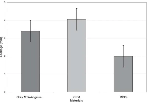
Figure 1 represents the mean leakage of 0.2% Rhodamine B, in millimeters, for the groups receiving the apical plug.
DISCUSSION
Several techniques have been used to analyze the sealing ability of root canal filling and retrograde filling, including evaluation of leakage of bacteria2,11,23, human saliva1, protein
complex24, fluid filtration15 and dye leakage16,18,20,22,27.
Among the dyes, use of methylene blue at different concentrations has been outstanding16,18,22. However, in 1998,
Wu, et al.26 conducted an interesting work and stated that
methylene blue suffers discoloration when in contact with some alkaline filling materials, which may cause unrealistic results of such materials in leakage studies. Methylene blue discoloration occurs because it is unstable when in contact
with alkaline materials. Such materials cause hydrolysis of methylene blue, resulting in formation of a clear compound named thionine. This would explain why methylene blue is discolored by calcium hydroxide. In relation to MTA, in the presence of water, the calcium oxide in the material could form calcium hydroxide, which would certainly cause discoloration of methylene blue26.
Moraes, et al.19 (2005) and Tanomaru Filho, et al.21(2005)
share the same opinion. Therefore, when performing the leakage test to assess the sealing ability of gray MTA-AngelusTM, CPMTM and MBPc, in this study, 0.2% Rhodamine
B was selected instead of methylene blue, based on the previously mentioned studies, since labeling by Rhodamine B is not influenced by alkaline materials19,21.
Investigation of gray MTA has been based on the frequency of utilization when apical plugs are necessary, with excellent results4,10,17. Gray MTA-AngelusTM was used instead
of gray ProRoot MTATM (Dentsply/Tulsa Dental, United
States of America) in order to use a national product, which is also easier to find in the market.
Concerning CPMTM, according to the manufacturer, this
material has similar or better physical, chemical and biological characteristics compared to MTA, with the same clinical indications5. As this material is also a mineral trioxide
aggregate, this study evaluated the possibility of using it as apical plug, as well as its sealing ability and marginal adaptation, since few studies are available on CPMTM.
MBPc was also used because its physical and chemical characteristics have been previously assessed, showing great results20; one study also assessed its biological properties6.
As the initial clinical indications of this material included only use for retrograde filling and in cases of root perforation18, it FIGURE 1- Mean leakage of 0.2% Rhodamine B, in millimeters, for the three groups receiving the apical plug. Data are mean
was deemed interesting to investigate the possibility of using this material for fabrication of apical plug.
The thickness of apical plug in different studies ranges from 1 mm 11,24 to 10 mm 3. Nevertheless, the best results have
more frequently been reported with apical plugs between 3 mm and 5 mm of thickness. Considering the studies of Matt, et al.18 (2004) and Al-Kahtani, et al.2 (2005), in which
5-mm-thick apical plugs showed the best results, this dimension was established for fabrication of the plugs in this study.
Data on Figure 1 show that, in general, the sealing ability of apical plugs with several types of material may be classified, in a descending numerical order, as follows: MBPc (1.99 ± 1.44mm), Gray MTA-AngelusTM(3.39 ± 1.39 mm) and CPMTM
(4.00 ± 1.00 mm). Dunn’s test showed that MBPc was significantly better than the other materials used as apical plugs (p<0.05).
The study of Bramante, et al.5 (2006) allows comparison
between the results obtained with CPMTM and those of the
present results. According to those authors, CPMTM has
dimensional adhesion stability through time, among other properties. However, the results observed for this material with regard to sealing ability were not so good, with a mean overall leakage of 4.00 ± 1.00 mm.
Regarding MTA, the excellent sealing ability of both Pro RootTM MTA and gray MTA-AngelusTM, used in this study,
have been highlighted by several authors16,18,22,27. Conversely,
in a recent study by Silva Neto and Moraes20 (2003), MTA
was not considered as a good sealer. When used as an apical plug, especially with 4- to 5-mm thickness, MTA has shown great sealing ability2,11,18,24. Consequently, the results observed
for gray MTA-AngelusTM in this study confirm those found
in the aforementioned studies.
Regarding MBPc, the study testing its sealing ability (Silva Neto and Moraes 20, 2003, using Rhodamine B in perforations)
has shown its ability as a good sealer. That fact was observed in this study, which revealed best outcomes for MBPc, with only 1.99 ± 1.44 mm of leakage, with statistically significant difference in relation to CPMTM and gray MTA-AngelusTM.
CONCLUSION
The results showed that, when analyzing the sealing ability of apical plugs of gray MTA-AngelusTM, CPMTM and
MBPc, it was observed that MBPc presented the best results, with statistically significant difference compared to the other two materials.
ACKNOWLEDGEMENTS
This research was supported by CNPq (Brazililian National Council for Technological and Scientific Development).
REFERENCES
1- Al-Hezaimi K, Naghshbandi J, Oglesby S, Simon JHS, Rotstein I. Human saliva penetration of root canals obturated with two types of mineral trioxide aggregate sealers. J Endod. 2005;31(6):453-6.
2- Al-Kahtani A, Shostad S, Schifferle R, Bhambhani S. In vitro evaluation of microleakage of an orthograde apical plug of mineral trioxide aggregate in permanent teeth with simulated immature apices. J Endod. 2005;31(2):117-9.
3- Aminoshariae A, Hartwell GR, Moon PC. Placement of mineral trioxide aggregate using two different techniques. J Endod. 2003;29(10):679-82.
4- Bramante CM, Bortoluzzi EA, Broon NJ. Agregado trióxido mineral (MTA) como plug apical para la obturación de conductos radiculares: descripción de la técnica y caso clínico. Endodoncia. 2004;22(3):155-161.
5- Bramante CM, Bramante AS, Moraes IG, Bernardineli N, Garcia RB. CPM es MTA: nuevos materiales de uso en endodoncia – experiencias clinicas en el manejo de los materiales. Rev Fac Odontol. 2006;17:7-10.
6- Cintra LTA, Moraes IG, Bernabé PFE, Gomes-Filho JE, Bramante CM, Garcia RB, et al. Evaluation of the tissue response to MTA and MBPc: microscopic analysis of implants in alveolar bone of rats. J Endod. 2006;32(6):556-9.
7- Duarte MAH, Demarchi ACCO, Yamashita JC, Kuga MC, Fraga SC. pH and calcium íon release of 2 root-end filling materials. Oral Surg Oral Med Oral Pathol Oral Radiol Endod. 2003;95:345-7.
8- El-Meligy OA, Avery DR. Comparison of apexification with mineral trioxide aggregate and calcium hydroxide. Pediatr Dent. 2006;28(3):248-53.
9- Felippe WT, Felippe MCS, Rocha JC. The effect of mineral trioxide aggregate on the apexification and periapical healing of teeth with incomplete root formation. Int Endod J. 2006;39(1):2-9.
10- Giuliani V, Baccetti T, Pace R, Pagavino G. The use of MTA in teeth with necrotic pulps and open apices. Dent Traumatol. 2002;18(4):217-21.
11- Hachmeister DR, Schindler WG, Walker WA, Thomas DD. The sealing ability and retention characteristics of mineral trioxide aggregate in a model of apexification. J Endod. 2002;28(5):386-90.
12- Hayashi M, Shimizu A, Ebisu S. MTA for obturation of mandibular central incisors with open apices: case report. J Endod. 2004;30(2):120-2.
13- Holland R, Souza V, Nery MJ, Fáraco Júnior IM, Bernabé PFE, Otoboni Filho JA et al. Reaction of rat connective tissue to implanted dentin tube filled with mineral trioxide aggregate, Portland cement or calcium hydroxide. Braz Dent J. 2001;12(1):3-8.
14- Jacobsen EL, Bery PF, BeGole EA. The effectiveness of apical dentin plugs in sealing endodontically treated teeth. J Endod. 1985;11(7):289-93.
15- Lamb EL, Loushine RJ, Weller RN, Kimbrough WF, Pashley DH. Effect of root resection on the apical sealing ability of mineral trioxide aggregate. Oral Surg Oral Med Oral Pathol Oral Radiol Endod. 2003;95(6):732-5.
17- Maroto M, Barbería E, Planells P, Vera V. Treatment of a non-vital immature incisor with mineral trioxide aggregate (MTA). Dent Traumatol. 2003;19(3):165-9.
18- Matt GD, Thorpe JR, Strother JM, McClanahan SB. Comparative study of white and gray mineral trioxide aggregate (MTA) simulating a one-or-two-step apical barrier technique. J Endod. 2004;30(12):876-9.
19- Moraes IG, Moraes FG, Mori GG, Gonçalves SB. Influence of calcium hydroxide on dyes for dentin labeling, analized by means of a new methodology. J Appl Oral Sci. 2005;13(3):218-21.
20- Silva Neto UX; Moraes IG. Sealing capacity by some materials when utilized under furcation perforations of human molars. J Appl Oral Sci. 2003;11(1):27-33.
21- Tanomaru M Filho, Figueiredo FA, Tanomaru JMG. Effect of different dye solutions on the evaluation of the sealing ability of mineral trioxide aggregate. Braz Oral Res. 2005;19(2):119-22.
22- Torabinejad M, Higa RK, McKendry DJ, Pitt Ford TR. Dye leakage of four root end filling materials: effects of blood contamination. J Endod. 1994;20(4):159-63.
23- Torabinejad M, Rastegar AF, Kettering JD, Pitt Ford TR. Bacterial leakage of mineral trioxide aggregate as a root-end filling material. J Endod. 1995; 21(3):109-12.
24- Valois CRA, Costa Jr ED. Influence of the thickness of mineral trioxide aggregate on sealing ability of root-end filling in vitro. Oral Surg Oral Med Oral Pathol Oral Radiol Endod. 2004;97(1):108-11.
25- Weisenseel JA, Lamar Hicks M, Pelleu GB. Calcium hydroxide as an apical barrier. J Endod. 1987;13(1):1-5.
26- Wu MK, Kontakiotis EG, Wesselink PR. Decoloration of 1% methylene blue solution in contact with dental filling materials. J Dent. 1998;26(7):585-9.