online | memorias.ioc.fiocruz.br
Acute gastroenteritis and enteric viruses in hospitalised children in
southern Brazil: aetiology, seasonality and clinical outcomes
Sonia Maria Raboni1,2/+, Guilherme Augusto Costa Damasio3,Carla EO Ferreira3, Luciane A Pereira1,
Meri B Nogueira1, Luine R Vidal1, Cristina R Cruz4, Sergio M Almeida1,5
1Laboratório de Virologia, Hospital de Clínicas 2Departamento de Doenças Infecciosas 3Programa de Pós-Graduação em Microbiologia, Parasitologia e Patologia 4Departamento de Pediatria, Universidade Federal do Paraná, Curitiba, PR, Brasil
5Faculdades e Instituto de Pesquisa Pelé Pequeno Príncipe, Complexo Pequeno Príncipe, Curitiba, PR, Brasil
Viral acute gastroenteritis (AG) is a significant cause of hospitalisation in children younger than five years. Group A rotavirus (RVA) is responsible for 30% of these cases. Following the introduction of RVA immunisation in Brazil in 2006, a decreased circulation of this virus has been observed. However, AG remains an important cause of hospitalisation of paediatric patients and only limited data are available regarding the role of other enteric viruses in these cases. We conducted a prospective study of paediatric patients hospitalised for AG. Stool samples were collected to investigate human adenovirus (HAdV), RVA, norovirus (NoV) and astrovirus (AstV). NoV typing was performed by nucleotidesequencing and phylogenetic analysis. From the 225 samples tested, 60 (26%) were posi-tive for at least one viral agent. HAdV, NoV, RVA and AstV were detected in 16%, 8%, 6% and 0% of the samples, respectively. Mixed infections were found in nine patients: HAdV/RVA (5), HAdV/NoV (3) and HAdV/NoV/RVA (1). The frequency of fever and lymphocytosis was significantly higher in virus-infected patients. Phylogenetic analysis of NoV indicated that all of these viruses belonged to genotype GII.4. The significant frequency of these pathogens in patients with AG highlights the need to routinely implement laboratory investigations.
Key words: gastroenteritis - children - rotavirus - norovirus - astrovirus - human adenovirus
enovirus (AdV)] (Santos et al. 2007). Other viruses, such as aichivirus, human parechovirus and human bocavi-rus have been described in faecal samples from patients with diarrhoea, but their association with AG has still not been established (Chhabra et al. 2013).
Since the mid-2000s, two RV vaccines became available, a monovalent RV vaccine (Rotarix®,
Glaxo-SmithKline Biologicals Inc) and a pentavalent RV vac-cine (RV5, RotaTeq®, Merck & Co, Inc). Both vaccines
are recommended by the World Health Organization, have been used in several countries and studies have demonstrated a significant reduction of hospitalisation and mortality due to RV gastroenteritis (Justino et al. 2012, Lopman et al. 2012, Soares-Weiser et al. 2012). Brazil was one of the first countries to introduce univer-sal vaccination against RVA, Rotarix®, which has been
provided free through the public health system since March of 2006. Before the introduction of the group A RV (RVA) vaccine in Brazil, the frequency for this virus in the population with AG in our institution was 30%. The number of RVA positive cases has decreased substantially since then (Pereira et al. 2011), while other pathogens are now reported more frequently.
NoVs have been recognised as the major causes of non-bacterial AG in all age groups in industrialised countries and are frequently associated with food and water-borne outbreaks. Human NoV cannot be grown in cell culture, they are non-enveloped RNA viruses with icosahedral symmetry and are divided into five geno-group (GI-GV), which are further divided into > 30 gen-otypes, with genogroups GI and GII being associated with human infections (Mead et al. 1999, Hardy 2005, Patel et al. 2009, Khamrin et al. 2010, Kroneman et al. 2013). Acute gastroenteritis (AG) is the most common
gas-trointestinal inflammatory condition affecting people in both developed and developing countries (Andreasi et al. 2008, Domínguez et al. 2009). Worldwide, diarrhoea remains the second leading cause of death in children younger than five years. Around 1.5 million of children die annually as a result of AG, which represents 15% of all deaths that occur in this population group (Boschi-Pinto et al. 2008, Black et al. 2010, Wardlaw et al. 2010 ). In the United States of America (USA), 200,000 children are hospitalised each year with this disease, resulting in 300-400 deaths, and thereby generating a high economic impact (McCollough & Sharieff 2006). In Brazil, AG represents a major cause of morbidity and mortality in the first year of life and, in 2006, about 120,000 hospi-talisations of children less than five years occurred due to AG (RIPSA 2008).
Over 20 different types of viruses have been iden-tified as etiologic agents of AG, but the major viruses associated with acute diarrhoea in children can be di-vided into four different families: Reoviridae [rotavirus (RV)], Caliciviridae [norovirus (NoV) and sapovirus], Astroviridae [astrovirus (AstVs)] and Adenoviridae
[ad-doi: 10.1590/0074-0276140066
+ Corresponding author: [email protected] Received 23 February 2014
Human AdV (HAdV) is a non-enveloped DNA virus with icosahedral symmetry. The AdV has been associ-ated with a wide spectrum of clinical manifestations, in-cluding respiratory, gastrointestinal, ocular, neurologi-cal and urinary tract infections. This family comprises 55 different serotypes (HAdV 1-55) grouped into seven subgenera (A-G), of which the genotypes 40, 41 and more rarely, 38 are related to acute diarrhoea (Wold & Horwitz 2007, Ramani & Kang 2009, Walsh et al. 2010).
Human AstVs (HAstVs) are non-enveloped RNA viruses with icosahedral symmetry. They are classi-fied in eight serotypes (HAstVs 1-8), which are further divided into four subtypes (1a, 1b, 1c and 1d). Studies have shown that colonisation or infection by HAstV may be associated with necrotising enterocolitis, es-pecially in preterm infants (Santos & Cardoso 2005, Marshall et al. 2007, Bagci et al. 2010).
To assess the impact of these enteric viruses in the aetiology of AG in hospitalised children in our institu-tion, this study aimed to identify (i) the frequency of RVA, HAdV, HAstV and NoV in stool samples, (ii) re-port the clinical findings of these infections, (iii) anal-yse the displacement of these viruses after the decrease in RVA cases during the period between September 2010-September 2011 and (iv) and genotypically char-acterise the NoV detected.
SUBJECTS, MATERIALS AND METHODS
Material - This study evaluated 225 stool samples from hospitalised paediatric patients, which were sent to the Virology Laboratory of the Faculty of Medicine Clinics Hospital, Federal University of Paraná (HC-UFPR) to test for RVA between the period of September 2010-September 2011. Medical records of the patients were reviewed to evaluate clinical, laboratory and de-mographic data. The HC-UFPR Institutional Review Board approved the study (IRB#0221.0.208.000-10).
The HC-UFPR is a tertiary academic care hospital where patients with severe infections are referred and AG is one of the most frequent causes of paediatric hos-pital admissions.
RV detection - RVA antigen detection was carried out using the enzymatic immunoassay Rotascreen II®
kit (Microgen Bioproducts, UK), according to the manu-facturer’s instructions.
Viral DNA/RNA extraction - DNA/RNA was extract-ed from 150 µL of clarifiextract-ed 10% faecal suspension with the buffer Tris-HCl (0.01 M)-CaCl2 (0.0015 M) by using a commercial kit (Intron Biotechnology Inc, South Ko-rea) according to the manufacturer’s instructions. Pseu-dotrabies viruses (PRV) were added to the lysis buffer at a concentration of 1.74 x 10⁻⁸ ng/µL for use as an internal
control for extraction.
AdV detection - Generic primers and amplification tests were carried out as described by Avellón et al. (2001) (Table I)with some modifications. Briefly, 2.5 µL of extraction product was added to 22.5 µL of a PCR mix (Bioron, Ludwigshafe, GE) containing 2.5 mM dNTPs, 1.25 U of Taq DNA Polymerase, 160 mM (NH4)2SO4, 670 mM Tris-HCl (pH 8.8), 25 mM MgCl2 and primary amplification primers for AdV and PRV at a concentra-tion of 10 pmol/µL. Amplificaconcentra-tion was carried out on a Mastercycler Personal (Eppendorf Inc, Hamburg, GE) thermocycler with the following cycling conditions: one hold of 94ºC/2 min, 40 cycles of 94ºC/60 s, 50ºC/60 s, 72ºC/60 s and one extension step of 72ºC/6 min. For the second reaction, 1 µL of primary amplification product was added to 24 µL of a new PCR mix similar to that for the primary amplification, but containing secondary amplification primers. PCR products were resolved on 1% agarose gel electrophoresis stained with ethidium
bromide (0.5 μg/mL).
TABLE I
Sequences of the primers used in polymerase chain reaction (PCR) and reverse transcription-PCR for the detection of adenovirus (AdV), pseudorabies viruses (PRV), norovirus (NoV) and astrovirus (AstV)
Primer Sequence 5’-3’ Gene target Nested-PCR Product Reference
ADHEX 2F CCCMTTYAACCACCACCG AdV
Hexon
Yes 169 bp Avellón et al. (2001)
ADHEX 1R ACATCCTTB CKGAAGTTCCA
ADHEX 2R KATGGGGTARAGCATGTT
ADHEX 1F AACACCTAYGASTACATGAAC
PRV 1+ CGCGTGGTCTACGGGGACACGGA PRV DNA
polymerase
Yes 140 bp Pozo and Tenorio (1999)
PRV 1- ATGACGCCGATGTACTTCTTCTT
PRV 2+ GGGACACGGACTCGGTCTCC
PRV 2- CCGGAAGGTCTTCTCGCACTC
JV12 ATACCACTATGATGCAGATTA NoV RNA
polymerase
No 430 bp Vinjé and Koopmans (1996)
JV13 TCATCATCACCATAGAAAGAG
Mon 270F CAACTCAGGAAACAGGGTGT AstV
ORF2
No 449 bp Noel et al. (1995)
Mon 269R TCAGATGCATTGTCATTGGT
AstV and NoV detection - Reverse transcription-polymerase chain reaction (RT-PCR) was performed for detection of HAstV and NoV. First-strand cDNA was synthesised using random primers and a RT system (Su-perScript III® Reverse Transcriptase, Invitrogen, USA). Briefly, cDNA was obtained by adding 1 μL random primer (3 µg/µL, Invitrogen™) and 1 μL of ultrapure
water in 7.5 mL of RNA, followed by incubation for 5 min at 65ºC. Then, it was added 10.5 µL of a RT-master
mix containing 4 μL of deoxynucleotide triphosphates (dNTPs) (2.5 mM each), 4 μL of cDNA buffer (5 x), 2 μL
of 0.1M dithiotreitol (DTT) (Invitrogen™) 0.25 µL of RNase inhibitor RNase OUT™ (40U/µL, Invitrogen™) and 0.25 µL of enzyme reverse transcriptase Super-Script® III (2.000U/µL, Invitrogen™), with subsequent
incubation for 5 min at 25ºC, 60 min at 50ºC and 15 min at 70°C. Generic primers and amplification tests for HAstV and NoV were carried out as reported by Noel et al. (1995) and Vinjé and Koopmans (1996), respectively (Table I). Briefly, 2.5 µL of extraction product were add-ed to 22.5 µL of a PCR mix (Bioron, GE) containing 2.5 mM dNTPs, 1.25 U of Taq DNA polymerase, 160 mM (NH4)2SO4, 670 mM Tris-HCl (pH 8.8), 25 mM MgCl2 and primary amplification primer for NoV or HAstV at a concentration of 10 pmol/µL. NoV amplification was carried out on a Mastercycler Personal (Eppendorf Inc) thermocycler programmed with the cycling condi-tions: 94ºC for 2 min followed by 30 cycles of 94ºC/60 s, 37ºC/30 s, 72ºC/30 s and an elongation step of 72ºC/6 min. HAstV amplification was performed using the same cycling protocol described for HAdV. Both PCR products were resolved on 1% agarose gel
electrophore-sis stained with ethidium bromide (0.5 μg/mL). The gels
were photographed under ultraviolet light and the bands were analysed using the E-Capt program.
Nucleotide sequencing - Nucleotide sequencing of positive samples was performed to genotype the NoV detected. The initial amplification was performed us-ing three sets of primers described by Vinjé et al. (2004) (Table II), with one being GI specific, one being GII
spe-cific and the third exhibiting no genogroup spespe-cificity. Briefly, 2.5 µL of cDNA was added to 22.5 µL of a PCR mix (Bioron) containing 2.5 mM dNTPs, 1.25 U Taq DNA polymerase, 160 mM (NH4)2SO4, 670 mM Tris-HCl (pH 8.8), 25 mM MgCl2 and primary genotyping primers for NoV at a concentration of 10 pmol/µL. The amplification was carried out on a Mastercycler Per-sonal (Eppendorf Inc) thermocycler programmed with the cycling conditions: 94ºC for 1 min, followed by 40 cycles of 94ºC/60 s, 44ºC/60 s, 72ºC/60 s and an elonga-tion step of 72ºC/10 min. Afterwards, the PCR prod-ucts were resolved on 1.5% agarose gel electrophoresis
stained with ethidium bromide (0.5 μg/mL).
The amplicons obtained from one of the three sets of primers were subjected to sequencing reactions with the same primers. PCR products were purified using the Invisorb® Fragment Clean up (Invitek GmbH,
Ber-lin, GE) kit, according to the manufacturer’s instruc-tions. Sequencing reactions were carried out using a Big Dye® Terminator v.3.1 kit and the ABI3130 automated
sequencer (Applied Biosystems, CA, USA).
Multiple sequence alignments were carried out using CLUSTALW software from BioEdit v.7.0.9 package (Ibis Biosciences, CA, USA) and phylogenetic analysis was performed using MEGA 5.1 (Tamura et al. 2011). The se-quences (VP1 protein and RNA polymerase genes) were compared to a panel of reference sequences obtained from GenBank. Phylogenetic trees were generated using the neighbour-joining algorithm with bootstrap of 1,000 replicates and evolutionary distances were calculated us-ing the Kimura two-parameter method.
Statistical analysis - Demographic and clinical data were compiled using JMP software v.5.2.1 and ana-lysed using GraphPad Prism® v.5.03. Fisher exact or χ2
tests were used to assess differences between groups and the Mann Whitney test was used for continuous variables, as appropriate. Results for continuous data have been expressed as median ± interquartile ranges. All p-values are two-tailed and a value of < 0.05 was considered as significant.
TABLE II
Sequences of primers used for genome amplification and nucleotide sequencing of norovirusa
Primer Sequence 5’-3’ Product Gene target Genogroup
Cap A GGCWGTTCCCACAGGCTT 177 bp VP1 GI
Cap B1 TATGTTGACCCTGATAC
Cap B2 TATGTIGAYCCWGACAC
Cap C CCTTYCCAKWTCCCAYGG 253 bp VP1 GII
Cap D1 TGTCTRSTCCCCCAGGAATG
Cap D3 TGYCTYITICCHCARGAATGG
MJV12 TAYCAYTATGATGCHGAYTA 327 bp RNA
polymerase
GI and GII
Reg A CTCRTCATCICCATARAAIGA
RESULTS
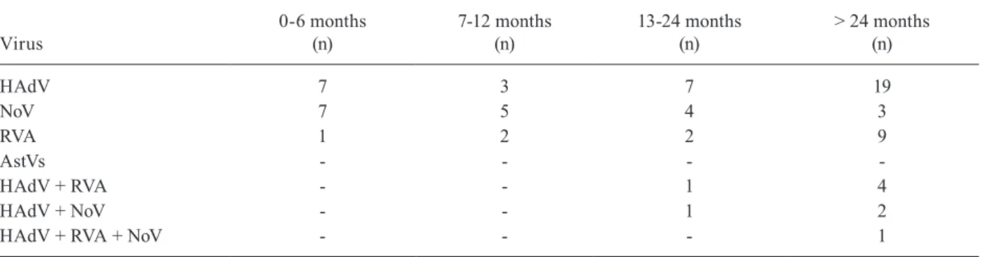
From 225 samples sent to the virology laboratory between September 2010-September 2011, 60 (26%) were positive for at least one viral agent. HAdV was detected in 36 (16%) samples, NoV in 19 samples (8%) and RVA in 14 samples (6%). Additionally, we ob-served five cases of mixed infection involving HAdV and RVA, three cases of HAdV combined with NoV and one case of triple infection involving RVA, HAdV and NoV. No cases of AstV infection were detected. Most positive samples were from patients older than two years. There were five case of RVA infection in a patient aged less than two years, who had not received the full vaccination or had underlying diseases with immunological impairment (Table III).
The mean number of stool samples sent to the lab-oratory was approximately 17 samples/month (± 4.8), with an average of five positive samples/month (± 2.6). The distribution of positive samples and its rela-tion to the average monthly temperature (ºC) and pre-cipitation (cm3) between September 2010-September
2011 revealed the absence of a seasonal pattern for the studied viruses, as the pathogen detections occurred throughout the year (Fig. 1).
The group of patients that presented infection by one or more viral agents (infected group) was compared with the group of individuals who tested negative for any of the searched viral agents (not infected group). Signifi-cant differences were observed for the presence of fever (p = 0.0051) and the number of lymphocytes in the blood count (p = 0.0224), which were higher in the infected group. Results were also obtained for faecal occult blood, parasites, reducing substances and stool culture, but the majority of the samples tested produced negative results, with no significant differences between the groups.
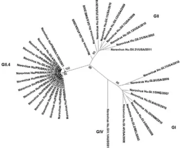
Nineteen samples were positive for NoV and from these we obtained 14 sequences for VP1 gene and 10 sequences for RNA polymerase gene to perform the phylogenetic analysis. These sequences were compared to VP1 genes, RNA polymerase genes and entire genome sequences of different NoV genotypes collected from GenBank. The final sequence alignment revealed 208 informative posi-tions for analysis of VP1 gene and a phylogenetic tree was
constructed (Fig. 2). Phylogenetic analysis of RNA poly-merase gene was carried out, the results were the same as that performed with VP1 region (data not shown) and all sequences belonged to the genotype GII.4.
DISCUSSION
Analysis conducted previously in this hospital to as-sess the impact of vaccination against RVA indicated a reduction of 54.2% and 39.4% in medical consulta-tions for children less than 12 months old and between 12-months, respectively, and a reduction of 44% in the number of hospitalisations for gastroenteritis in children less than 12 months. However, diarrhoea still represents a frequent cause of hospitalisation and, except for RVA, the role of other enteric viruses was not determined in our patients and their incidence was likely underesti-mated. Previously, we have carried out RV genotypic characterisation in all positive samples and G2 P[4] was the most prevalent genotype after the vaccine implemen-tation in 2006 (Pereira et al. 2013).
Overall, 26% of samples were positive for enteric viruses, with HAdV the most frequent virus detected, corresponding to a frequency of 16%. The prevalence of HAdV in prior studies has been variable, ranging from 0.7% to > 30% (Domínguez et al. 2009, Stroparo et al. 2010). In central Brazil, the state of Mato Grosso do Sul, AdVs were found in 3.6% of cases of AG (Andreasi et al. 2008), while other studies, such as those by Simpson et
TABLE III
Enteric virus detected according to stratified age group
Virus
0-6 months (n)
7-12 months (n)
13-24 months (n)
> 24 months (n)
HAdV 7 3 7 19
NoV 7 5 4 3
RVA 1 2 2 9
AstVs - - -
-HAdV + RVA - - 1 4
HAdV + NoV - - 1 2
HAdV + RVA + NoV - - - 1
AstVs: astrovirus; HAdV: human adenovirus; NoV: norovirus; RVA: group A rotavirus.
al. (2003) and Cunliffe et al. (2010), have reported higher frequencies of HAdV (7.9% and 15%, respectively). Pre-viously, Pereira Filho et al. (2007) also reported a fre-quency of 2% of AdV antigen detection in hospitalised and community children with diarrhoea in Rio de Janei-ro and Salvador, respectively. These lower frequencies might have been underestimated due to the technique used, that is, antigen detection. Subsequent introduction of molecular methods for studying this virus has led to a significant increase in the frequency of AdV detection in stools (Rohayem et al. 2004)
It is important to note that this study was carried out using a combination of generic primer for HAdVs detection, whose molecular sequencing of the products did not allow genotyping of detected virus. Then, the identified virus in the sample should not necessarily be considered the causative agent of the AG, as infections caused by some HAdV genotypes can result in an in-termittent viral excretion in the stools after a previous infection. However, analysis of patient medical records did not reveal any further diseases that could be asso-ciated with other HAdV serotype infections. NoV was the second most common virus detected in the studied patients, responsible for 8% (19/225) of AG cases, with
RVA cases accounting for 6% (14/225). Andreasi et al. (2008), in Brazil, reported similar findings, with NoV identified in 7.6% of the cases. Unlike the results of this study, Ferreira et al. (2012) in the states of Rio de Ja-neiro and Ribeiro et al. (2008) Espírito Santo identified this pathogen as the major viral agent in cases of gastro-intestinal infections. Additionally, a study by Nakagomi et al. (2008) identified NoV in 15% of samples from the municipality of Recife, with clinical severity similar to that for RV, while in the municipality of São Paulo, Castilho et al. (2006) reported 33% positive cases of NoV and Siqueira et al. (2013a) found 36.5% of positive samples in the municipality of Belém.
HAstV was not observed in this study. Some studies from other regions of Brazil and other countries (e.g., Japan, Greece and India) have also reported minimal circulation of this pathogen compared to other enteric viruses (Santos et al. 2007, Domínguez et al. 2009, Le-vidiotou et al. 2009, Chan-it et al. 2010, Verma et al. 2010). Usually, HAstV infections are not associated with severe AG, consequently, its identification in hospital-ised patients might be uncommon. On contrary of our findings, a study carried out in Rio de Janeiro in 2004 shown a prevalence of 14% of AstV in the samples col-lected from hospitalised children with AG (Victoria et al. 2007). Interestingly, the protocol used in the present study was very similar to that reported, including the use of random primer to obtain cDNA and the sequences of specific primers employed in the PCR. Indeed, the prev-alence of this virus in several studies is quite variable, epidemiological and seasonal factors should be consid-ered in such analyses and studies that include laboratory surveillance for a longer time will be able to demonstrate the real impact of this pathogen on the children health.
HAdV and the NoV were detected during most of the months for which data were collected and although higher frequencies were observed in the fall and winter, no seasonal pattern or association with relative humid-ity was identified for these two viruses. Levidiotou et al. (2009) also failed to identify any seasonal pattern to the HAdV detected in Greece. However, Ozdemir et al.
(2010) reported the presence of HAdV in the fall and winter months in Turkey, Kitajima et al. (2010)observed a higher frequency of NoV in winter and spring in Ja-pan and Zeng et al. (2012) observed that the frequency of NoV peaks only during the cold months in China. Interestingly, similar to that reported by Siqueira et al. (2013b), we observed that the distribution of RVA and NoV positive cases throughout the year presented a dis-tinct seasonal profile and a “seesaw effect”.
Demographic data revealed no significant dif-ferences for age and gender between patients with or without viral infection. However, a trend was observed with infection occurring in younger children (median 30 months of age). Fever and vomiting were the most common symptoms presented and these were more frequent in the infected group than in the not infected group. Furthermore, significant lymphocytosis was observed in the infected group, although this param-eter should be evaluated carefully since the quantity of lymphocytes in the blood can vary according to a child’s age. Investigation of other pathogens associated
to diarrhoea, as enteric bacteria and parasites, as well as other evidence of infection, such as the presence of faecal occult blood resulted as negative.
Phylogenetic analysis identified only genotype GII.4 at the study site, including the NoV detected in a sample collected in 2001. This result confirms the dominance of the genotype GII.4 circulation, already observed in other regions of Brazil, such as São Paulo, Vitória, Rio de Ja-neiro, Salvador and Belém, and other countries such as Venezuela, Mexico and Thailand (Castilho et al. 2006, Ferreira et al. 2008, Xavier et al. 2009, Aragão et al. 2010, Barreira et al. 2010, Khamrin et al. 2010, González et al. 2011, Gómez-Santiago et al. 2012).
Since 1995, NoV has caused five pandemics of AG and despite it has over 30 genotypes circulating, it is only a single genotype - GII.4 - that cause mass outbreaks and pandemics (Lindesmith et al. 2008). Factors intrinsic to the host and virus are associated with the higher prevalence of this genotype, among these are cited (i) GII.4 viruses bind to all blood group antigens and the ability to use blood groups antigens may affect the viral infectivity, differently of other genotypes (Tan & Jiang 2005, Bull et al. 2010), (ii) the high capacity of the virus to alter its carbohydrate-binding targets over time, allowing to escape from pro-tective immunity (Lindesmith et al. 2008), (iii) the high-er mutation rate and rate of evolution of GII.4 genotype compared to the less frequently detected NoV (GII.b, GII.3 and GII.7 strains) and (iv) GII.4 lineage had on average a 1.7-fold higher rate of evolution within the capsid sequence allowing the viral persistence (Bull et al. 2010).
Recently, Kroneman et al. (2013) proposed for a new NoV nomenclature: the identification of new genotypes should be based on the sequences of both genomic re-gions, open reading frame (ORF)1 and VP1. In the pres-ent study it was performed partial sequencing of both VP1 and ORF1 regions and in the phylogenetic analysis all vi-ruses found were identified as GII.4, without any finding suggestive of recombinant forms between these samples.
Prior to implementation of the RVA vaccine, a reduc-tion in the number of RVA positive samples in our hospi-tal had already been observed, likely due to a number of factors including: improved sanitary conditions, greater access to basic healthcare and conversion of the institu-tion from primary to tertiary care, where only patients with severe disease are referred. With the introduction of the vaccine, the reduction was even more impressive. However, the reduction in RVA circulation has been fol-lowed by an increased occurrence of HAdV and NoV and it is likely that we can expect an escalation in the fre-quency of occurrence of other enteric viruses, implying the need for more accurate laboratory investigation and improved epidemiological surveillance to guide preven-tion, therapeutic measures and control of outbreaks.
To our knowledge, this work represents the first re-port detailing the observed frequency of enteric viruses, other than RVs, which are associated with severe condi-tions responsible for hospitalisation of paediatric patients in southern Brazil. It is important to note the limitations of this study. First, genetic characterisation of the AdV detected should be performed to confirm the presence of the most frequent strains associated with AG, par-ticularly genotypes 40 and 41. Second, the investigation
should be extended over a longer period to better assess the population distribution of these viruses and better evaluate seasonal patterns. Furthermore, prospective case-control studies, including the investigation AdVs in hospitalised children without diarrhoea should be imple-mented aiming to define the causal relation between the detection of these viruses and the presence of disease.
In conclusion, this study demonstrated that 26% of the AG identified in hospitalised children was associated with enteric viruses and that the introduction of RVA immunisation was associated with reduced frequency of this virus. However, reduction of RVA was followed by an increasing presence of other pathogens associated with severe clinical manifestations of AG in this popula-tion, highlighting the need to implement methods to rou-tinely investigate these cases in a virology laboratory.
REFERENCES
Andreasi MS, Cardoso DD, Fernandes SM, Tozetti IA, Borges AM, Fiaccadori FS, Santos RA, Souza M 2008. Adenovirus, calicivi-rus and astrovicalicivi-rus detection in fecal samples of hospitalized chil-dren with acute gastroenteritis from Campo Grande, MS, Brazil.
Mem Inst Osvaldo Cruz 103: 741-744.
Aragão GC, Oliveira DS, Santos MC, Mascarenhas JD, Oliveira CS, Linhares AC, Gabbay YB 2010. Caracterização molecular de no-rovírus, sapovírus e astrovírus em crianças com gastroenterite aguda em Belém, Pará, Brasil. Rev Pan-Amaz Saude 1: 149-158.
Avellón A, Pérez P, Aguilar JC, Lejarazu R, Echevarría JE 2001. Rap-id and sensitive diagnosis of human adenovirus infections by a generic polymerase chain reaction. J Virol Methods 92: 113-120.
Bagci S, Eis-Hübinger AM, Yassin AF, Simon A, Bartmann P, Franz AR, Mueller A 2010. Clinical characteristics of viral intestinal infection in preterm and term neonates. Eur J Clin Microbiol In-fect Dis 29: 1079-1084.
Barreira DM, Ferreira MS, Fumian TM, Checon R, de Sadovsky AD, Leite JP, Miagostovich MP, Spano LC 2010. Viral load and geno-types of noroviruses in symptomatic and asymptomatic children in southeastern Brazil. J Clin Virol 47: 60-64.
Black RE, Cousens S, Johnson HL, Lawn JE, Rudan I, Bassani DG, Jha P, Campbell H, Walker CF, Cibulskis R, Eisele T, Liu L, Mathers C, for the Child Health Epidemiology Reference Group of WHO, UNICEF 2010. Global, regional and national causes of child mor-tality in 2008: a systematic analysis. Lancet 375: 1969-1987.
Boschi-Pinto C, Velebit L, Shibuya K 2008. Estimating child mortal-ity due to diarrhoea in developing countries. Bull World Health Organ 86: 710-717.
Bull RA, Eden JS, Rawlinson WD, White PA 2010. Rapid evo-lution of pandemic noroviruses of the GII.4 lineage. PLoS Pathog 6: e1000831.
Castilho JG, Munford V, Resque HR, Fagundes-Neto U, Vinjé J, Rácz ML 2006. Genetic diversity of norovirus among chil-dren with gastroenteritis in São Paulo state, Brazil. J Clin Microbiol 44: 3947-3953.
Chan-it W, Thongprachum A, Okitsu S, Mizuguchi M, Ushijima H 2010. Epidemiology and molecular characterization of sapovirus and astrovirus in Japan, 2008-2009. Jpn J Infect Dis 63: 302-303.
Cunliffe NA, Booth JA, Elliot C, Lowe SJ, Sopwith W, Kitchin N, Nakagomi O, Nakagomi T, Hart CA, Regan M 2010. Healthcare-associated viral gastroenteritis among children in a large pediat-ric hospital, United Kingdom. Emerg Infect Dis 16: 55-62.
Domínguez A, Godoy P, Torner N, Cardeñosa N, Martínez A 2009. Las gastroenteritis víricas: un problema de salud pública. Rev Esp Salud Publica 83: 679-687.
Ferreira MS, Xavier MP, Fumian TM, Victoria M, Oliveira SA, Pena LH, Leite JP, Miagostovich MP 2008. Acute gastroenteritis cases associated with noroviruses infection in the state of Rio de Ja-neiro. J Med Virol 80: 338-344.
Ferreira MS, Xavier MP, Tinga AC, Rose TL, Fumian TM, Fialho AM, de Assis RM, Costa FAC, de Oliveira SA, Leite JP, Miagos-tovich MP 2012. Assessment of gastroenteric viruses frequency in a children’s day care center in Rio de Janeiro, Brazil: a fifteen year study (1994-2008). PLoS ONE 7: 1-7.
Gómez-Santiago F, Ribas-Aparicio RM, García-Lozano H 2012. Molecular characterization of human calicivirus associated with acute diarrheal disease in Mexican children. Virol J 9: 1-9.
González GG, Liprandi F, Ludert JE 2011. Molecular epidemiology of enteric viruses in children with sporadic gastroenteritis in Valen-cia, Venezuela. J Med Virol 83: 1972-1982.
Hardy ME 2005. Norovirus protein structure and function. FEMS Microbiol Lett 253: 1-8.
Justino MCA, Araújo EC, van Doorn L-J, Oliveira CS, Gabbay YB, Mascarenhas JDP, Miranda YS, Guerra SFS, da Silva VB, Linhares AC 2012. Oral live attenuated human rotavirus vac-cine (RotarixTM) offers sustained high protection against severe
G9P[8] rotavirus gastroenteritis during the first two years of life in Brazilian children. Mem Inst Oswaldo Cruz107: 846-853.
Khamrin P, Maneekarn N, Thongprachum A, Chaimongkol N, Okitsu S, Ushijima H 2010. Emergence of new norovirus vari-ants and genetic heterogeneity of noroviruses and sapoviruses in children admitted to hospital with diarrhea in Thailand.
J Med Virol 82: 289-296.
Kitajima M, Oka T, Haramoto E, Takeda N, Katayama K, Katayama H 2010. Seasonal distribution and genetic diversity of genogroups I, II and IV noroviruses in the Tamagawa River, Japan. Environ Sci Technol 44: 7116-7122.
Kroneman A, Vega E, Vennema H, Vinje J, White PA, Hansman G, Green K, Martella V, Katayama KK, Koopmans M 2013. Pro-posal for a unified norovirus nomenclature and genotyping. Arch Virol 158:2059-2068.
Levidiotou S, Gartzonika C, Papaventsis D, Christaki C, Pri-avali E, Zotos N, Kapsali E, Vrioni G 2009. Viral agents of acute gastroenteritis in hospitalized children in Greece. Clin Microbiol Infect 15: 596-598.
Lindesmith LC, Donaldson EF, LoBue AD, Cannon JL, Zheng DP, Vinje J, Baric RS 2008. Mechanisms of GII.4 norovirus persis-tence in human populations. PLoS Med 5: e31.
Lopman BA, Payne DC, Tate JE, Patel MM, Cortese MM, Parashar UD 2012. Post-licensure experience with rotavirus vaccina-tion in highand middle income countries: 2006 to 2011. Curr Opin Virol2:434-442.
Marshall JA, Bruggink LD, Sturge K, Subasinghe N, Tan A, Hogg GG 2007. Molecular features of astrovirus associated with a gastroenteritis outbreak in an aged-care centre. Eur J Clin Microbiol Infect Dis 26: 67-71.
McCollough M, Sharieff GQ 2006. Abdominal pain in children.
Pediatr Clin N Am 53: 107-137.
Mead PS, Slutsker L, Dietz V, McCaig LF, Bresee JS, Shapiro C, Griffin PM, Tauxe RV 1999. Food-related illness and death in the United States. Emerg Infect Dis 5: 607-625.
Nakagomi T, Correia JB, Nakagomi O, Montenegro FM, Cuevas LE, Cunliffe NA, Hart CA 2008. Norovirus infection among children with acute gastroenteritis in Recife, Brazil: disease severity is comparable to rotavirus gastroenteritis. Arch Virol 153: 957-960.
Noel JS, Lee TW, Kurtz JB, Glass RI, Monroe SS 1995. Typing of hu-man astroviruses from clinical isolates by enzyme immunoassay and nucleotide sequencing. J Clin Microbiol 33: 797-801.
Ozdemir S, Delialioğlu N, Emekdaş G 2010. Investigation of rota -virus, adenovirus and astrovirus frequencies in children with acute gastroenteritis and evaluation of epidemiological features.
Mikrobiyol Bul 44: 571-578.
Patel MM, Hall AJ, Vinjé J, Parashar UD 2009. Noroviruses: a com-prehensive review. J Clin Virol 44: 1-8.
Pereira Filho E, Faria NR, Fialho AM, Assis RS, Almeida MM, Ro-cha M, Galvão M, Santos FB, Barreto ML, Leite JPG 2007. Ad-enoviruses associated with acute gastroenteritis in hospitalized and community children up to 5 years old in Rio de Janeiro and Salvador, Brazil. J Med Microbiol 56: 313-319.
Pereira LA, Ferreira CE, Turchetto GD, Nogueira MB, Vidal LR, Cruz CR, Debur MC, Almeida SM, Raboni SM 2013. Molecular charac-terization of rotavirus genotypes in immunosuppressed and non-im-munosuppressed pediatric patients. J Pediatr (Rio J) 89: 278-285.
Pereira LA, Raboni SM, Nogueira MB, Vidal LR, Almeida SM, De-bur MC, Cruz C 2011. Rotavirus infection in a tertiary hospital: laboratory diagnosis and impact of immunization on pediatric hospitalization. Braz J Infect Dis 15: 215-219.
Pozo F, Tenorio A 1999. Detection and typing of lymphotropic herpesvi-ruses by multiple polymerase chain reaction. J Virol Meth 79: 9-19.
Ramani S, Kang G 2009. Viruses causing childhood diarrhoea in the developing world. Curr Opin Infect Dis 22: 477-482.
Ribeiro LR, Giuberti RSO, Barreira DMPG, Saick KW, Leite JPG, Miagostovich MP, Spano LC 2008. Hospitalization due to noro-virus and genotypes of rotanoro-virus in pediatric patients, state of Espírito Santo. Mem Inst Oswaldo Cruz 103: 201-206.
RIPSA - Rede Interagencial de Informacão para a Saúde Brasil 2008. Indicadores básicos para a saúde no Brasil: conceitos e aplicações. Available from: tabnet.datasus.gov.br/tabdata/livroidb/2ed/indi-cadores.pdf.
Rohayem J, Berger S, Juretzek T, Herchenröder O, Mogel M, Poppe M, Henker J, Rethwilm A 2004. A simple and rapid single-stpe multiplex PCR to detect norovirus, astrovirus and adenovirus in clinical stool samples. J Virol Methods 118: 49-59.
Santos RA, Cardoso DD 2005. Astrovírus. Rev Patol Trop 34: 161-174.
Santos RAT, Borges AMT, da Costa PSS, Teixeira JMS, Giugliano LG, Leite JPG, Cardoso DDP 2007. Astrovirus infection in children living in the Central West Region of Brazil. Mem Inst Oswaldo Cruz 102: 209-213.
Simpson R, Aliyu S, Iturriza-Gómara M, Desselberger U, Gray J 2003. Infantile viral gastroenteritis: on the way to closing the di-agnostic gap. J Med Virol 70: 258-262.
Siqueira JAM, Linhares AC, Carvalho TCN, Aragão GC, Oliveira DS, Santos MC, Sousa MS, Justino MCA, Mascarenhas JDP, Gabbay YB 2013a. Norovirus infection in children admitted to hospital for acute gastroenteritis in Belém, Pará, northern Brazil.
J Med Virol 85: 737-744.
rota-virus and nororota-virus display sharply distinct seasonal profiles in Belém, northern Brazil. Mem Inst Oswaldo Cruz108: 661-664.
Soares-Weiser K, MacLehose H, Bergman H, Ben-Aharon I, Nagpal S, Goldberg E, Pitan F, Cunliffe N 2012. Vaccines for prevent-ing rotavirus diarrhoea: vaccines in use. Cochrane Database Syst Rev 12: CD008521.
Stroparo E, Cruz CR, Debur MC, Vidal LR, Nogueira MB, Almeida SM, Pereira LA, Rotta I, Raboni SM 2010. Adenovirus respira-tory infection: significant increase in diagnosis using PCR com-paring with antigen detection and culture methods. Rev Inst Med Trop Sao Paulo 52: 317-321.
Tamura K, Peterson D, Peterson N, Stecher G, Nei M, Kumar S 2011. MEGA5: molecular evolutionary genetics analysis using maxi-mum likelihood, evolutionary distance and maximaxi-mum parsimony methods. Mol Biol Evol 28: 2731-2739.
Tan M, Jiang X 2005. Norovirus and its histo-blood group antigen recep-tors: an answer to a historical puzzle. Trends Microbiol 13: 285-293.
Verma H, Chitambar SD, Gopalkrishna V 2010. Astrovirus associ-ated acute gastroenteritis in western India: predominance of dual serotype strains. Infect Genet Evol 10: 575-579.
Victoria M, Carvalho-Costa FA, Heinemann MB, Leite JPG, Mia-gostovich MP 2007. Genotypes and molecular epidemiology of human astroviruses in hospitalized children with acute gastroen-teritis in Rio de Janeiro, Brazil. J Med Virol 79: 939-944.
Vinjé J, Hamidjaja RA, Sobsey MD 2004. Development and appli-cation of a capsid VP1 (region D) based reverse transcription-PCR assay for genotyping of genogroup I and II noroviruses.
J Virol Methods 116: 109-117.
Vinjé J, Koopmans MP 1996. Molecular detection and epidemiology of small round-structured viruses in outbreaks of gastroenteritis in the Netherlands. J Infect Dis 174: 610-615.
Walsh MP, Seto J, Jones MS, Chodosh J, Xu W, Seto D 2010. Computational analysis identifies human adenovirus type 55 as a re-emergent acute respiratory disease pathogen. J Clin Microbiol 48: 991-993.
Wardlaw T, Salama P, Brocklehurst C, Chopra M, Mason E 2010. Diarrhoea: why children are still dying and what can be done.
Lancet 375:870-872.
Wold WS, Horwitz MS 2007. Adenoviruses. In DM Knipe, PM Howley, Fields virology, Lippincott Williams & Wilkins, Philadelphia, p. 2395-2436.
Xavier MP, Oliveira SA, Ferreira MS, Victoria M, Miranda V, Silva MF, Strina A, Barreto ML, Miagostovich MP, Leite JP 2009. De-tection of caliciviruses associated with acute infantile gastroen-teritis in Salvador, an urban center in Northeast Brazil. Braz J Med Biol Res 42: 438-444.