Human Papillomavirus Infection and Cervical Cancer in
Brazil: a Retrospective Study
Sílvia MB Cavalcanti/
+, Flávia CC Deus, Lucília G Zardo
*,
Izabel CPP Frugulhetti, Ledy HS Oliveira
Departamento de Microbiologia e Parasitologia, Instituto Biomédico, Universidade Federal Fluminense, Rua Ernani Melo 101, 24210-030 Niterói, RJ, Brasil *Setor de Anatomia Patológica e Citopatologia, Hospital Luíza
Gomes de Lemos, Instituto Nacional de Câncer, Rua Visconde de Santa Isabel 274, 20560-120 Rio de Janeiro, RJ, Brasil
Two hundred and thirty paraffin-embedded biopsies obtained from female cervical lesions were tested for the presence of human papillomavirus (HPV) types 6/11,16/18 and 31/33/35 DNA using non-isotopic in situ hybridization. Specimens were classified according to the Bethesda System in low grade squamous intraepithelial lesion (LSIL), high grade SIL (HSIL) and squamous cell carcinoma (SCC). HPV prevalence ranged from 92.5% in LSIL to 68.5% in SCC. Benign types were prevalent in LSILs while oncogenic types infected predominantly HSILs and SCC. HPV infection showed to be age-depen-dent, but no significant relation to race has been detected. Patients were analyzed through a five-year period: 20.7% of the lesions spontaneously regressed while 48.9% persisted and 30.4% progressed to carcinoma. Patients submitted to treatment showed a 19.4% recurrence rate. High risk types were present in 78.6% (CrudeOR 13.8, P=0.0003) of the progressive lesions, and in 73.7% of the recurrent SILs (COR 19.3, P=0.0000001). Possible co-factors have also been evaluated: history of other sexually trans-mitted diseases showed to be positively related either to progression (Adjusted OR 13.0, P=0.0002) or to recurrence (AOR 17.2, P=0.0002) while oral contraceptive use and tobacco smoking were not sig-nificantly related to them (P>0.1). Association of two or more co-factors also proved to be related to both progression and recurrence, indicating that they may interact with HPV infection in order to in-crease the risk of developing malignant lesions.
Key words: human papillomavirus - progression - risk factors - cervical cancer
Work supported by CNPq.
+Corresponding author. Fax: 55-21-719.2588
Received 2 October 1995 Accepted 11 April 1996
Experimental and epidemiological data have been accumulated to support a central role for hu-man papillomavirus (HPV) in the aetiology of anogenital cancer, specially cervical carcinoma (Schiffman 1994). Nearly 70 different types of HPV have now been described, and 28 have been isolated from benign and malignant genital lesions (De Villiers 1989). These viruses have been fur-ther classified according to their malignant poten-tial. High risk types (e.g. HPV16,18 and to a lesser extent 31,33 and 35) have been linked to high grade intraepithelial lesions (HSILs) and invasive carci-noma, whereas low risk types (e.g. HPV6 and 11) have been associated to benign or low grade SILs (LSILs). Differences in oncogenic potential of low and high risk types were also demonstrated by transformation experiments (Woodworth et al. 1989).
Cervical carcinogenesis is a multi-step process in which certain factors are thought to be required for the HPV-induced lesions to progress (Roman & Fife 1989). Cigarette smoking, oral contracep-tive use and concurrent sexually transmitted dis-eases (STD) may be such co-factors. The elucida-tion of their role in this malignant process leads to the possibility of distinguishing the HPV infected women at increased risk for progression to cancer from those eventually regressing without treatment, what may affect the assessment of the prognosis, the choice of treatment and the follow-up modali-ties.
Cervical cancer is the second leading cause of cancer deaths in women world-wide. In many de-veloping countries, it is still the main cause of death from cancer in women (Lowy et al. 1994). In Bra-zil, high annual cancer incidences have been re-ported. Nevertheless, there have been few pub-lished epidemiological studies concerning the prevalence of HPV DNA in the female genital tract and the natural history of the infection.
women and to search for specific risk factors asso-ciated with progression to malignancy and with post-treatment recurrence.
MATERIALS AND METHODS
Specimens - The material of the present study includes a series of 230 cases of normal, benign, premalignant and malignant cervical biopsies of women attended at Hospital Luíza Gomes de Lemos, State of Rio de Janeiro, from 1989 to 1994. Patients were selected within a group referred to histopathologic examination after an altered cytol-ogy (Pap smear). The follow-up period was six years, with intervals of six months, in average. The medical records of all investigated patients were reviewed for possible risk factors and previously diagnosed STDs, including HIV infection, genital herpes, gonorrhoea, chlamydial infection, bacte-rial pelvic inflammatory disease, thrichomoniasis and syphilis. Forty-two percent of the patients were treated: partial ablation with diathermy and electrocauterization were used for LSILs and conization was the treatment of choice for HSILs. Patients left untreated were those who did not come back for treatment and/or for preventive exam, so their follow-up intervals were longer than for treated patients. Formalin-fixed, paraffin-embed-ded biopsy specimens were available for studies in the files of the Department of Pathology. Five µm sections were cut onto Aminopropyl triethoxysilane (Sigma) coated slides, heated at 60°C for 2 hr and stored at room temperature (RT).
Histological diagnosis - Slides were stained with haematoxylin and eosin (HE) for grading of the cervical lesions according to the Bethesda Sys-tem (1993) in LSIL, including condylomata and cervical intraepithelial neoplasia grade I (CIN I), in HSIL, including CIN II and CIN III/ in situ car-cinoma, and in squamous cell carcinoma (SCC). Histologically normal tissues presenting only re-active/reparative changes were classified as Reac-tive Changes (RC).
Probes - HPV 6b, 11, 16 and 18 DNAs were kindly provided by Dr De Villiers (Heidelberg). They were prepared and purified through caesium-chloride gradients. Plasmids were labelled using a nick translation kit (BRL) and biotin-11-d UTP (Sigma). HPV 31, 33 and 35 DNAs were available in Digene In situ Hybridization Kit.
In situ hybridization (ISH)- ISH procedure has been described in detail elsewhere (Cavalcanti et al. 1994). Briefly, sections have been deparaffinized in two changes of xylol and two changes of 100% ethanol. Nucleic acids were un-masked by digestion with 0.5 mg/ml proteinase K (Sigma). The hybridization mixture contained 5xSSC, 5% polyethileneglycol, 0.1 mg/ml
dena-tured carrier DNA (herring sperm), biotinylated probes (1.0µg/ml) and 50% deionized formamide. Each section was layered with 20 µl of hybridiza-tion mixture under a coverslip, denatured by heat-ing at 92°C for 10 min on a heatheat-ing block, and hybridized at 37°C for 2 hr. Coverslips have been removed by soaking the slides in 4xSSC at RT for 10 min. Further the slides have been washed in 0.1xSSC/50% formamide, 4xSSC and phosphate buffered saline pH7.2 (PBS) at RT for 10 min each. The DNA-DNA hybrids were visualized by using streptavidin-alkaline phosphatase complex at RT for 30 min. Unbound conjugate has been removed by two washes in Buffer 1 (0.1 M Tris HCl, 0.15 M NaCl, pH 7.5) for 10 min and once in Buffer 3 (0.1 M Tris HCl, 0.1 M NaCl, 50 mM MgCl2, pH 9.5) for 5 min at RT. The slides have then been incubated in NBT (nitroblue tetrazolium) and BCIP (5-bromo-4-chloro-3-indolylphosphate) dissolved in Buffer 3 at RT for 30 min in the dark. Slides have been rinsed in distilled water to stop reaction, air dried and mounted in glycerine jelly without counterstaining. This method is meant to be capable of detecting approximately 50 to 100 viral genome copies per cell. Positive cells are de-tected by strong nuclear signal in the upper epithe-lial layers especially when associated with koilocytic features. Occasional intermediate and parabasal cells with or without koilocytosis also show a purple precipitate in the nuclei under light microscopy (Syrjanen 1990).
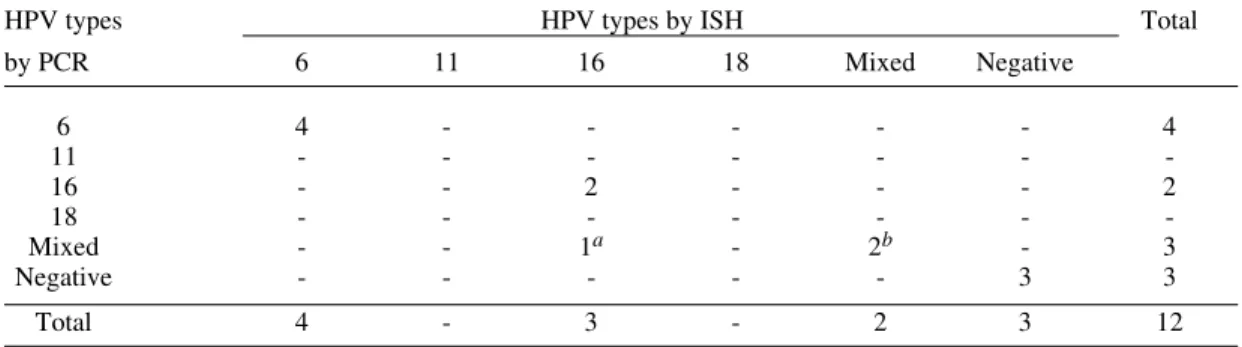
HPV-positive and negative cervical biopsies have been used as controls in every experiment. They were composed by 3 RC, 4 LSIL, 4 HSIL and 1 SCC and have been previously tested by Southern blot followed by hybridization onto nytrocellulose membrane (data not shown). These controls have also been submitted to HPV detection by using type specific primers in PCR (Table III).
Polymerase Chain Reaction (PCR) - Briefly, 10µm sections were cut from the paraffin blocks, deparaffinized in octane and added to 150µl of di-gestion buffer (50mM Tris-HCl pH 8.5, 10mM EDTA, 200µg/ml proteinase K - final concentra-tion of 100µg/ml) for 18 hr at 37oC. 100µl PCR reaction mixture containing 2.5 units of Taq poly-merase have been prepared and subjected to 40 cycles of amplification. Cycles were carried out using the following method: denaturation at 92°C for 1min, annealing at 55°C for 2min, extension at 72°C for 2 min, ending at 72°C for 10 min.
been identified by size determination in 12% poly-acrylamide gel stained with ethidium bromide. Various concentrations of purified cloned DNA of HPV 6 and 16, DNA from CaSki and HeLa cells, containing HPV 18 and 16 genomes, respectively, were used as positive controls. To analyze the qual-ity of target DNA, samples have been subjected to PCR using β-globin gene specific primers (Vandenvelde et al. 1990).
Statistical analysis- The statistical significance of the results has been analyzed by using the chi-square test for heterogeneity with Yates continu-ity correction, Fisher’s exact test and a Mantel-Haentszel procedure for trend, when appropriate. Multivariate logistic regression analysis was per-formed to examine the associations of progression/ regression and recurrence/cure and various risk factors simultaneously. Odds ratios were used as a measure of association. All analyses were done using Epi Info (version 5) microcomputer software (USD, Stone Mountain, GA).
RESULTS
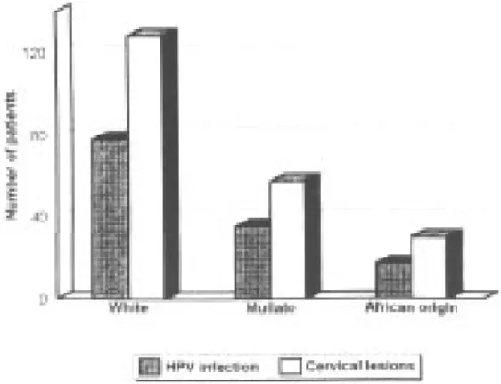
Two hundred and thirty biopsies from female cervical lesions have been investigated to detect the presence of HPV DNA. The average age of participants was 38.7 years, ranging from 16 to 75 years old. The studied population was composed of 128 (59.5%) white, 57 (26.5%) mulatto and 30 (14.0%) African origin women.
No significant correlation has been found for HPV infection and race (P> 0.1). Although a higher number of white women presenting cervical lesions were studied, HPV infection rates were similar within the three studied groups (Fig.). Different age distri-bution patterns have been shown according to the histological grade of the HPV infected lesions: the LSIL group (Xage= 31.5) exhibit statistically signifi-cant differences when compared to the malignant SCC (Xage= 46.9, P<0.001) (Table I).
As shown in Table II, the overall prevalence of HPV DNA in the studied group was 73.9% (170/ 230) ranging from 92.5% (49/53) in LSIL to 68.5%
TABLE II
Prevalence of different human papillomavirus types according to the histological diagnosis
Diagnosis Number of HPV types HPV prevalence
patients 6/11 16/18 31/33/35 mixed (%)
Reactive changes 20 2 - 1 - 3 (15.0)%
LSIL 53 30 4 2 13a 49 (92.5%)
HSIL 103 11 33 16 21b 81 (78.6%)
SCC 54 1 21 11 4c 37 (68.5%)
Total 230 44 58 30 38 170 (73.9%)
a: 10 HPV 6/11, 16/18; 3 HV 6/11, 31/33/35; b: 12 HPV 6/11, 16/18; 9 HPV 16/18, 31/33/35; c: 2 HPV 6/11, 16/ 18; 1 HPV 16/18, 31/33/35; 1 HPV 6/11,16/18, 31/33/35.
TABLE I
Average age of HPVa infected women according to the histological grade of lesions
Histological diagnosis of patients
Reactive Changes LSIL HSIL SCC (n=3) (n=49) (n=81) (n=37)
Χ age 34.6 31.5 36.7 46.9 a: HPV 6, 11, 16, 18, 31, 33 and 35.
HPV infection in cervical lesions of women of different races.
(37/54) in SCC. The control group (RC) presented 15.0% (3/20) of HPV positivity. Statistically sig-nificant differences have been detected among the cervical lesions and the control (RC) (P<0.0001) for HPV infection of the cervix.
and 1.4, respectively, with P>0.1) for both. Data are presented in Table IV.
From the 230 studied women, 98 were treated. Partial ablation with diathermy and electrocauter-ization were the treatments of choice for LSILs, while conization was the procedure applied for HSILs. After treatment, 19 patients (19.4%) showed recurrence, but none of them presented lesion progression in the follow-up period. As shown in Table V, 14 of the recurrent lesions (73.7%) presented oncogenic HPV DNA, with sig-nificant association of recurrence and HPV types 16/18 (COR19.3, P=0.000001). Analysis of recur-rence rates in treated women pointed out that 31.6% (6/19) of the patients showing recurrence presented other STDs, comparing to the cured cases (2.5%, 2/79) and statistically significant difference was detected (AOR 17.2, P= 0.0002). No other factor, such as cigarette smoking or OC use, presented significant role in differentiating recurrent from cured lesions (P>0.5).
DISCUSSION
In our study, 170 out of 230 (73.9%) cervical biopsies were infected by HPV. Similar prevalence rates have been described previously, even when diverse detection systems were used (Syrjanen 1989, Kiviat et al. 1989): 44 biopsies (25.9%) con-tained HPV6/11, 58 (34.1%) concon-tained HPV16/18 and 30 (17.6%) contained HPV31/33/35. Oncogenic HPV 16 and 18 were the most com-mon types in malignant lesions, showing an up-ward trend with high statistical significance from LSIL to SCC (Table II, P<0.0002). Low risk HPV 6 and 11 do not seem to play an important role in carcinogenesis, since their prevalences tended to decrease significantly from LSIL to SCC (P<0.00001). It is also worth noting that HPV6 and 11 have not been found in the premalignant and malignant lesions, except when in mixed infec-PCR results for the 12 tested controls were
nearly the same obtained by in situ hybridization (Table III), except for one sample that PCR re-vealed to be a double infection by HPV16 and 33, and ISH had only indicated the presence HPV16 DNA. No other differences in sensibility or speci-ficity have been detected.
The retrospective analysis showed that from 1989 to 1994, in 20.7% (19/92) of the untreated patients HPV-induced lesions regressed spontane-ously, in 48.9% (45/92) the lesion persisted and in 30.4% (28/92) the lesion showed clinical progres-sion to in situ carcinoma or to invasive cancer. Spontaneously regressing cases presented low risk HPVs 6/11 in 68.4% of the lesions, showing to be inversely related to progression (Crude OR 0.02, P=0.000009), what means directly related to re-gression (X2 = 19.7, P=0.000009) while persisting and progressive cases showed high risk types in 63.3% and 78.6% of the samples, respectively. Oncogenic HPV types16/18 were strongly associ-ated with progression (COR 13.8, P=0.0003), while HPVs types 31/33/35 did not show significant cor-relation (COR 1.8, P=0.7).
Co-factors currently related to cancer progres-sion in HPV infected women have been investi-gated. From patients with progressive lesions, 42.9% (12/28) were tobacco smokers, 75% showed other STD (21/28) and 35.7% (10/28) used oral contraceptive regularly. From women showing spontaneous regression, 26.3% (5/19) were smok-ers, 15.8% (3/19) had other STD and 26.3% (5/ 19) used oral contraceptives. After adjustment for age, race, and HPV types, the presence of concur-rent STDs showed to be significantly related to malignant evolution (Adjusted OR 13.0, P=0.0002). Cigarette smoking and oral contracep-tive (OC) use presented approximately the same rates for progressive and regressing lesions, and thus were not associated to progression (AOR 1.8
TABLE III
Comparison of human papillomavirus typing by in situ hybridization (ISH) and the polymerase chain reaction (PCR)
HPV types HPV types by ISH Total
by PCR 6 11 16 18 Mixed Negative
6 4 - - - 4
11 - - -
-16 - - 2 - - - 2
18 - - -
-Mixed - - 1a - 2b - 3
Negative - - - 3 3
Total 4 - 3 - 2 3 12
TABLE IV
Risk factors associated to progression of Low Squamous Intraepithelial Lesions to cancer
Risk factors Number of Number of Crude Odds Adjusted Odds Ratio P values
regressive progressive Ratio (95%CI)
cases cases (95%CI)
HPV types 6/11
negative 6 27 1.0 1.0 0.000009
positive 13a 1 0.02 (0.0-0.2) 0.02 (0.0-0.2)
HPV types 16/18
negative 15 6 1.0 1.0 0.0003
positive 4b 22c 13.8 (2.7-76.8) 13.2 (1.9-58.6)
HPV types 31/33/35
negative 17 23 1.0 1.0 0.7
positive 2 5d 1.8 (0.3-15.8) 1.2 (0.2-7.3)
Concurrent STD
negative 16 7 1.0 1.0 0.0002
positive 3 21 16.0 (3.0-98.6) 13.0 (1.9-56.3)
Tobacco smoking
negative 14 16 1.0 1.0 0.4
positive 5 12 2.1 (0.5-9.1) 1.8 (0.5-7.2)
Oral contraceptive use
negative 14 18 1.0 1.0 0.7
positive 5 10 1.6 (0.4-6.8) 1.4 (0.4-5.6)
Associated risk factors
(tobacco+oral contraceptive) 0.08
negative 15 14 1.0 1.0
positive 4 14 3.8 (0.0-17.8) 4.6 (1.1-18.4)
Associated risk factors
(STD+tobacco+oral contraceptive) 0.0002
negative 9 0 U U
positive 10 28
U Fisher exact test (P=0.00007); a/b: 4 and 1 HPV mixed infections, respectively; c/d: 5 and 2 HPV mixed infec-tions, respectively.
tions, associated with high or intermediate risk HPVs. Our results reinforce the role for HPV16 and 18 in the pathogenesis of cervical cancer and indicates a relevant proportion of HPV 31, 33 and 35 associated to malignancy (29.7%).
According to Yun and Sherwood (1992), non-isotopic in situ hybridization proved to be a useful tool for distinguishing between high and low risk cervical lesions as well as a safe and rapid method for routine HPV diagnosis. Besides, it allows the simultaneous assessment of the morphological al-terations associated to the studied lesion. PCR us-ing type-specific primers confirmed the results obtained by using non-isotopic in situ hybridiza-tion (Table III), except for one sample that showed to be infected by HPV 16 byISH and PCR revealed to be a mixed infection containing both HPV 16 and 33. No other differences were detected, indi-cating the satisfactory sensibility of the in situ hy-bridization when using biotinylated probes. Clearly, however, ISH is less sensitive than the more recently developed PCR assay, which has demonstrated even higher rates of HPV DNA (Young et al. 1989). The enhanced sensitivity of
PCR is due to both its ability to detect lower copy numbers of HPV DNA as well as to confer the possibility of evaluating a broader spectrum of HPV types, potentially all the 20 recognized geni-tal types as opposed to the seven types usually de-tected by ISH (Schiffman 1994).
Multiple HPV infections were found in 22.4% (38/170) of all positive biopsies. As described by Reid et al. (1987), high rates of such infections are often detected and may be due to promiscuous sexual behaviour. But its occurrence is still not clearly associated to progression to malignancy. In our study, multiple HPV infection were not re-lated to cancer establishment since nearly the same rates of mixed infection were detected for regres-sive (26.3%, 5/19) and progresregres-sive lesions (25.0%, 7/28) (X2=0.9, P>0.1).
TABLE V
Risk factors associated to recurrence of Squamous Intraepithelial Lesions caused by human papillomavirus
Risk factors Number of Number of Crude Odds Adjusted Odds P values
cured recurrent Ratio Ratio
cases cases (95%CI) (95%CI)
HPV types 6/11
negative 37 16 1.0 1.0 0.007
positive 42 3 0.01 (0.04-0.7) 0.01 (0.1-0.2)
HPV types 16/18
negative 69 5 1.0 1.0 0.0000001
positive 10a 14b 19.3 (5.0-79.9) 27.4 (7.2-101.2)
HPV types 31/33/35
negative 72 17 1.0 1.0 0.8
positive 7c 2 1.2 (0.16-7.3) 1.2 (0.6-2.2)
Concurrent STD
negative 77 13 1.0 1.0 0.0002
positive 2 6 17.8 (2.8-145.1) 17.2 (7.2-32.9)
Tobacco smoking
negative 68 15 1.0 1.0 0.7
positive 11 4 1.7 (0.4-6.8) 2.0 (0.1-0.4)
Oral contraceptive use
negative 60 16 1.0 1.0 0.6
positive 19 3 0.6 (0.12-2.5) 0.4 (0.2-1.1)
Associated risk factors (tobacco+oral contraceptive)
negative 73 12 1.0 1.0 0.002
positive 6 7 34.9 (6.1-262.9) 7.0 (3.0-15.2)
Associated risk factors (STD+tobacco+oral contraceptive)
negative 41 2 1.0 1.0 0.0000004
positive 10 17 9.2 (3.6-25.3) 9.0 (3.4-24.2)
a/c: 7 and 5 HPV mixed infections, respectively; b: 6 HPV mixed infection.
number of white women showing SILs. Similar results have been previously described (Cavalcanti et al. 1994) and have to be further investigated.
Evaluation of the natural history of HPV in-fection indicated high rates of progression (30.4%) and persistence (48.9%). Regressing lesions have been demonstrated in 20.7% of the cases. Syrjanen (1989) described similar rates, although progres-sion rates were lower (20%) than those here de-scribed. Kataja et al. (1992), during a follow-up of eight years, showed that nearly 65% of the HPV infections have regressed spontaneously while 14% of the patients progressed to in situ carcinoma or SCC. These results are considerably different from ours. In fact, HPV types distribution in Europe are also quite different from Brazil, where oncogenic HPV 18 are commonly detected in HSILs and SCCs (Cavalcanti et al. 1994). This could explain such poor prognosis of LSILs in our country. Other authors (Kristiansen et al. 1994) also related an association of HPV 18 and poor prognosis, for which there is some support, associated to efficient and fast neoplastic transformation promoted by HPV 18 in vivo and in vitro (Walker et al. 1989, Barnes et al. 1990).
required to analyze their real oncogenic potential in cervical lesions.
As described in Table IV, concurrent STD were significantly related to progressive lesions and even after adjustment it has showed to be significantly associated to progression (Adjusted OR 13.0, P=0.0002). STDs probably contributed to progres-sion of the HPV leprogres-sions by depressing local im-munity of the genital tract. Although poorly un-derstood, host cellular defence mechanisms seem to play an important role in controlling HPV in-fection. Patients with cellular immune deficiencies (such as HIV-positive ones) have higher rates of HPV infection and are more susceptible to develop high grade SILs (Ho et al. 1994). In accordance to our findings, Meekin et al. (1992) described a link between previous or current STDs and progression to HPV-induced high grade lesions, proposing that these infections might act as markers for develop-ing SILs. Other studies suggested that other cervi-cal infections may interact with HPV increasing the risk of developing HSILs (Koutsky et al. 1992). The effect of OC use on cervical carcinogenesis has been widely investigated. The incidence of cervical carcinoma was found to be higher among women who had used OC compared to non-users. Stern et al. (1977) have suggested that the progres-sion rate might be higher in OC users than in other women. Nevertheless, we have not found any sig-nificant correlation when statistical significance were analyzed (AOR1.4, P=0.7). These results are in agreement with those from other authors (Davidson et al. 1994).
Regarding the relationship between smoking and cervical cancer, studies indicated a slight in-crease in the risk of the disease among smokers, attributed to a local impairment of cell-mediated immunity, but the effect was small (Vessey 1986). In our study no correlation could be demonstrated. In fact, our preliminary results pointed out that cigarette smoking and OC use were not related to cancer progression, in accordance to results from Van Doornum et al. (1993) and Eluf-Neto et al. (1994). But when these factors were associated, differences were observed in a great number of smokers+OC users showing progressive lesions, when compared to the other groups (only OC us-ers, only smokus-ers, non-OC user/non-smoker) (AOR 4.6, P=0.08). Association of three study fac-tors (STDs+cigarette+OC) has shown that 100% of the progressive cases were related to at least one of the analyzed risk factors, while only 47.3% of the regressing lesions presented them (P=0.0002). These results are similar to those described by Vandenvelde and Van Beers (1993) who found that well-known risk factors for cervical cancer seem to co-operate with HPV DNA in order to increase
the risk of malignant progression. Molecular mechanisms could explain the fact: HPV infection with high risk types and consequent synthesis of oncoproteins E6 and E7 would not be sufficient for malignant transformation. When other factors increasing the risk of mutational events and trans-formation are associated, the probability of cancer development increases.
From the 98 treated patients, 19 (19.4%) showed recurrence (Table V). Twenty out of the 98 treated and cured women were HPV negative (20.4%). Hence, from the 78 HPV-positive lesions submitted to treatment, 19 recurred (24.4%). In these recurrent lesions, 73.7% (14/19) of the cases presented oncogenic HPV types (OR 27.4, P=0.0000001). In relation to risk factors, the asso-ciation was the same seen in progressive cases: concurrent STDs (AOR 17.2, P=0.0002) but nei-ther smoking nor OC use were associated to recur-rence (P>0.1). Association of the three parameters were related to recurrence (AOR 9.0, P=0.0000004), reinforcing the idea of co-opera-tion between factors increasing the risk of devel-oping CINs and cancer. Some authors described that 90% of the patients achieved a two-year cure by using treatment procedures similar to ours: cau-terization and conization (Yliskoski et al. 1991), contrasting to the 80% of cure showed by us in a five-year study. These observations could be at-tributed to the different periods of analysis, or to the higher frequency of oncogenic HPVs detected in our patients what could be responsible for these lower efficiency in achieving cure.
In our study, we present the results of a retro-spective five-year analysis. Despite the adjustment for possible confounding factors, oncogenic HPV types 16 and 18 and history of STD showed to be significantly associated to progression to cancer and to unsuccessful treatment of HPV-induced le-sions. Smoking and the use of OC did not show relation to these events but when they were associ-ated, a significant co-operative role in both pro-gression and recurrence were demonstrated and thus a poor prognosis for women presenting SILs were described in Brazil.
ACKNOWLEDGEMENTS
To Dr Luiza L Villa for kindly providing the HPV primers for PCR. To Edson Alves de Souza and Vera Lúcia S Peixoto for their technical assistance.
REFERENCES
Barnes W, Woodworth C, Wagooner S 1990. Rapid dysplastic transformation of human genital cells by human papillomavirus type 18. Gynecol Oncol 38: 343-346.
Cavalcanti SMB, Frugulhetti ICP, Passos MRL, Fonseca MEF, Oliveira LHS 1994. Prevalence of human papillomavirus DNA in female cervical lesions from Rio de Janeiro, Brazil. Mem Inst Oswaldo Cruz89: 575-580.
Davidson M, Schnitzer PG, Bulkow LR, Schloss ML, Fitzgerald MA, Knight JA, Kiviat NB, Toomey KE, Reeves WC, Schmidt DS, Stamm WE 1994. The prevalence of cervical infection with human papillomaviruses and cervical dysplasia in Alaska native women. J Infect Dis 169: 792-800.
De Villiers EM 1989. Heterogeneity of human papillomavirus group. J Virol63: 4898-4903. Eluf-Neto J, Booth M, Muñoz N, Bosch F, Meijer CLM,
Walboomers JM 1994. Human papillomavirus and invasive cervical cancer in Brazil. Brit J Cancer 69: 114-119.
Ho GYF, Burk RD, Fleming I, Klein RS 1994. Risk of genital human papillomavirus infection in women with human immunodeficiency virus-induced immu-nosuppression. Int J Cancer 56: 788-792.
Kataja V, Syrjanen S, Mantijarvi R 1992. Prognostic factors in cervical human papillomavirus infections. Sex Transm Dis 19: 154-160.
Kataja V, Syrjanen S, Yliskoski M 1993. Risk factors associated with cervical human papillomavirus in-fections: a case-control study. Am J Epidem 138: 735-745.
Kiviat NB, Koutsky LA, Paavonen JA 1989. Prevalence of genital papillomavirus infection among women attending a college student clinic or a sexually trans-mitted disease clinic. J Infec Dis 159: 293-302. Koutsky LA, Holmes KK, Crichtlow CW, Stevens CE,
Paavonen J, Beckman AM, DeRouen TA, Galloway DA, Vernon CT, Kiviat NB 1992. A cohort study of the risk of cervical intraepithelial neoplasia grade 2 or 3 in relation to papillomavirus infection. N Engl J Med18: 1272-1278.
Kristiansen E, Jenkins A, Kristiansen G, Ask E, Kaern J, Abeler J, Lindqvist BH, Tropé C, Kristiansen BE 1994. Human papillomavirus infection in Norwe-gian women with cervical cancer. APMIS 102: 122-128.
Lowy DR, Kirnbauer R, Schiller JT 1994. Genital hu-man papillomavirus infection. Proc Nat Acad Sci USA 91: 2436-2440.
Meekin GE, Sparrow MJ, Fernwicke RJ, Tobias M 1992. Prevalence of genital human papillomavirus infec-tion in Wellington women. Genitorin Med68: 228-232.
Reid R, Greenberg M, Jenson AB, Stanhope R, Hershman VR, Smith JP, Lancaster WD 1987. Sexu-ally transmitted papillomavirus infections. I. The anatomic distribution and pathologic grade of
neo-plastic lesions associated with different viral tyes. Am J Obstet Gynecol 156: 212-222.
Roman A, Fife KH 1989. Human papillomaviruses: are we ready to type? Clin Microbiol Rev 2: 166-190. Schiffman MH 1994. Epidemiology of cervical human
papillomavirus infections. Cur Microbiol Immunol 186: 56-78.
Stern E, Forsythe AB, Youkles L, Coffelt CF 1977. Ste-roid contraceptive use and cervical cancer: increased risk of progression. Science 196: 1460-1463. Syrjanen KJ 1989. Epidemiology of human
papillomavirus (HPV) infections and their associa-tion with genital squamous cell cancer. APMIS 97: 957-970.
Syrjanen SM 1990. Basic concepts and practical appli-cations of recombinant DNA techniques in the de-tection of human papillomavirus (HPV) infections. APMIS 98: 95-110.
Vandenvelde C, Van Beers D 1993. High risk genital papillomaviruses and degree of dysplastic changes in the cervix. J Med Virol 39: 273-277.
Vandenvelde C, Vestraeten M, Van Beers 1990. Fast multiplex polymerase chain reaction on boiled clini-cal samples for rapid viral diagnosis. J Virol Meth-ods 30: 215-227.
Van Doornum GJJ, Van den Hoek JAR, Van Ameijden EJC, Van Haastrecht HJA, Roos MTL, Henquet CJM, Quint WGV, Coutinho RA 1993. Cervical HPV infection among HIV-infected prostitutes ad-dicted to hard drugs. J Med Virol 41: 185-190. Vessey MP 1986. Epidemiology of cervical cancer: role
of hormonal factors, cigarette smoking and occupa-tion. Banbury Report Series 21: 29-43.
Yliskoski M, Saarikoski S, Syrjanen K 1991. Conization for CIN associated to human papillomavirus infec-tion. Arch Gynecol Obstet 249: 59-65.
Young LS, Bevan IS, Jonhson MA, Blomfield PI, Bromigde T, Maitland NJ, Woodman CBJ 1989. The polymerase chain reaction: a new epidemiological tool for investigating cervical human papillomavirus infection. Brit Med J 298: 14-18.
Yun K, Sherwood MJ 1992. In situ hybridization at light and electron microscopic levels: identification of human papillomavirus nucleic acids. Pathol 24: 91-98.
Walker J, Bloss JD, Liao SY, Berman M, Berhen S, Wilczynski SP 1989. Human papillomavirus geno-type as a prognostic indicator in carcinoma of the uterine cervix. Obstet Gynecol 74: 781-785. Woodworth CD, Doniger J, Di Paolo JA 1989.