ORIGINAL
ARTICLE
Chlamydia trachomatis
as a probable cofactor in
human papillomavirus infection in aboriginal women
from northeastern Argentina
Authors
Gerardo Daniel Deluca1 Jorge Basiletti2 Eduardo Schelover3 Nicolás Díaz Vásquez4 José Mario Alonso5 Héctor Marcelo Marín6 Raúl Horacio Lucero7 María Alejandra Picconi8
1PhD, MSc, Professor, Faculty of Medicine, Northeast National University, Corrientes, Argentina 2Biochemist; Researcher, Oncogenic Virus Service, National Reference Laboratory on Human Papillomavirus, National Institute of Infectious Diseases “Carlos G. Malbrán”, Buenos Aires, Argentina 3MD, Pathologist, Institute of Regional Medicine, Northeast National University, Chaco, Argentina
4MD, Gynecologist, Institute of Regional Medicine, Northeast National University, Chaco, Argentina
5PhD, Head of the Immunology Service, Institute of Regional Medicine, Northeast National University, Chaco, Argentina 6Biochemist, Researcher, Molecular Biology Laboratory, Institute of Regional Medicine, Northeast National University, Chaco, Argentina
7Biochemist, Head of the Molecular Biology Laboratory, Institute of Regional Medicine, Northeast National University, Chaco, Argentina
8PhD, Head of the Oncogenic Virus Service, National Reference Laboratory on Human Papillomavirus, National Institute of Infectious Diseases “Carlos G. Malbrán”, Buenos Aires, Argentina
Submitted on: 05/14/2011 Approved on: 07/08/2011
Correspondence to: Gerardo Daniel Deluca Roldán 1307 (CP:3500) Resistencia, Chaco, Argentina [email protected]
Financial Support: This work was partially funded by the General Secretariat of Science and Technology of National Northeast University, Argentina.
We declare no conflict of interest.
©2011 Elsevier Editora Ltda. All rights reserved. ABSTRACT
Objectives: High-risk types of human papillomavirus (HPV) are strongly associated with cervical cancer (CC), and Chlamydia trachomatis (CT), the most frequent sexually transmitted bacterial infection (STBI) worldwide, seems to be a risk factor for HPV infection and for CC. It is also known that both agents are more prevalent in vulnerable communities where lack of adequate primary health care is a cause for concern. The aim of this work was to determine the impact of CT and HPV infections in women belonging to an isolated aboriginal population (Pilaga community) from a poor region in Northern Argentina (province of Formosa). For this purpose, a cross-sectional study was performed in all sexually active Pilaga women, who attended a local community-based gynecological health screening project. The polymerase chain reaction (PCR) method on a cervi-cal brush specimen was used to detect both agents. Results: A total of 227 women (20% of the total female population of the Pilaga community) were studied and the overall prevalence was 26.4% for CT, 46.7% for HPV and 16.3% for concurrent infection. CT infection was higher in HPV DNA positive (34.2%) than in HPV DNA negative women (19%; OR: 2.22/95% CI = 1.16-4.28 / p = 0.009) and the most prevalent HPV types were HPV-16 (19.4%), 6 and 18 (5.3%), 58 (3.5%) and 33 (3.1%). Conclusions: The prevalence of CT and HPV observed in Pilaga women are among the worst regis-tered in Latin America. Also, data collected suggest that chlamydial infection may play an important role in the natural history of HPV infection. On this respect, we propose that the association between these two agents seems to be more related to a mutual potentiation than to the fact that they share a common route of transmission.
Keywords: Chlamydia trachomatis; papillomavirus infections; indigenous population; uterine cervi-cal diseases.
INTRODUCTION
Chlamydia trachomatis (CT) is the most fre-quent sexually transmitted bacterial infection (STBI) worldwide, with an estimated 89 million new cases per year.1,2 Up to 70% of such
infec-tions are asymptomatic and the detection of cases has an important epidemiological implica-tion, because the main consequences (infertility, ectopic pregnancy, pelvic inflammatory disease and perinatal infections) of symptomatic and asymptomatic infections are often identical.2,3
Although many authors refer to an association between CT infection and dysplastic or neoplas-tic cervical atypia, epidemiological data have not yet provided consistent evidence about its real implication in cervical cancer (CC) devel-opment.4-6 However, the asymptomatic nature
of CT infection, its long-term persistence in the absence of treatment, and its capability of
inducing metaplasia and chronic inflamma-tion are well-defined characteristics to strongly consider this bacteria as a potential cofactor for high-risk persistent HPV infection (the main proven cause of CC).7-9 HPV genital infection
is highly frequent worldwide and its prevalence in the general population ranges from 2-44% depending on the region of the world.10
How-ever, it is known that most HPV infections are transient and only a small proportion of women with high-risk persistent HPV infection will develop CC.11-13 Therefore, numerous cofactors
that might promote virus persistence or enhance its oncogenicity have been studied in recent dec-ades to better understand the oncogenic pro-cess.14,15 CT is one of these cofactors.4,8,9
of the country,16 where a wide spectrum of Indian and
iso-lated white communities live in unfavorable socioeconomic and health conditions.
The aim of this study was to determine the prevalence of CT and HPV infections among vulnerable populations of native Indians belonging to the Pilaga community in north-ern Argentina.
METHODS
Study population
This study was nested in a community-based gyneco-logical health screening project for the Pilaga aboriginal people of the central region of Formosa province (north-ern Argentina). Two hundred and twenty-seven women aged 13-68 years (mean age 30 years), corresponding to 20% of all sexually active women of the Pilaga Indians in Argentina, were recruited from different aboriginal settlements between March 2007 and March 2009. A standardized questionnaire was used to interview the subjects regarding their clinical history, sexual behavior, cultural habits and socio-economic and living conditions. After providing informed consent, all women were exam-ined by a gynecologist.
The study was reviewed and approved by the Ethics Committee of the Institute of Regional Medicine of North-east National University, Argentina.
Sample collection and DNA extraction
Cervical exfoliated cells were collected from all women by sampling the ecto- and endocervix using a wooden spatula for PAP smears and a cytobrush for HPV and CT DNA de-tection. Cervical cells from the latter were eluted in sterile phosphate-buffered saline (PBS) contained in a tube, and then transported on ice to the processing laboratory. Upon arrival to the lab and after checking the sample container’s integrity, the cells of each sample were pelleted by centrifu-gation and kept frozen at -70°C until processing.
DNA was obtained by treating pellets with 400-700 µL of homogenization solution (2% cetyltrimethylammonium bromide-CTAB, 1.4 M NaCl, 0.2% β-mercaptoethanol, 20 mM EDTA, 100 mM Tris-HCl pH 5.0), extracted with chloroform isoamyl alcohol method, then precipitated with absolute ethanol and resuspended in 50-100 μL of ster-ile double-distilled water. All samples were checked to assess DNA quality and integrity by amplifying a known region of the human β-globin gene (268 bp). The samples negative for the β-globin test were discarded.
Cytological analysis
PAP smears were processed and analyzed according to the 2001 Bethesda Classification System. The final diagnosis was based on the worst morphological picture.
CT and HPV DNA amplification
CT DNA was detected in cervical samples by polymerase chain reaction (PCR) using the KL1/KL2 primers, as pre-viously described.17 This primer pair amplifies a 241 bp
fragment of the chlamydial multicopy, and a genetically conserved, cryptic plasmid. PCR was performed in a final volume of 20 µL using 3 µL of DNA sample; 10 mM Tris-HCl pH = 8.3, 50 mM KCl, 3.5 mM MgCl2, 5 nmol of each
deoxyribonucleoside triphosphate (dNTP), 20 pmol of each primer, 1.25 U Taq DNA polymerase (Go-Taq® DNA polymerase, Promega, USA). Each amplification reaction was carried out as follows: an initial cycle at 93°C for 3 min; 35 cycles of 1 min at 93°C, 1 min at 64°C and 1 min at 72°C and a final extension at 72°C for 5 min. Finally, the PCR products were electrophoresed on a 2% agarose gel, stained with ethidium bromide (EB) and photographed under UV light.
HPV DNA amplification and typing were conduct-ed using the widely known MY09/11 PCR (450 bp PCR product) followed by the restriction fragment-length poly-morphism (RFLP) technique, as previously described.18,19
Briefly, the products of MY09/11 PCR were first electro-phoresed on a 2% agarose gel, stained with EB and visualized under UV light. Then, the amplicons of positive samples were digested by seven restriction enzymes (BamHI, DdeI, HaeIII, HinfI, PstI, RsaI and Sau3AI) in individual microtubes. After that, the RFLP product of each sample was electrophoresed on a 3% agarose gel, stained with EB, and photographed under UV light. Each restriction pattern was compared with published data to identify the genotype(s) involved.
Statistical analysis
Continuous variables were categorized, and the risk associated with CT positivity was calculated for each category of a variable, adjusted by age. Univariate analysis was performed for all variables, and multivariate logistic regression analysis was performed in order to identify the independent variables that influence the relative risk of CT positivity (95% CI). The association between CT and HPV infections was evaluated in all women regardless of their cytological diagnosis. Data were analyzed using the Epi-InfoTM 3.5.1 Statistical Program (Centers for
Dis-ease Control and Prevention, Atlanta, GA) and statistical significance was determined by Fisher’s exact test, and p-values ≤ 0.05 were considered significant.
RESULTS
was more notorious for HPV infection and particularly for high-risk HPV types. The HPV prevalence estimates were 27.8% in women with normal pap smear, 46.2% in wom-en with reactive cellular changes (RCC), 87.5% in patiwom-ents with low-grade squamous intraepithelial lesion (L-SIL), and 100% in patients with high-grade squamous intraepithelial lesion (H-SIL) and CC (p < 0.05) (Table 1).
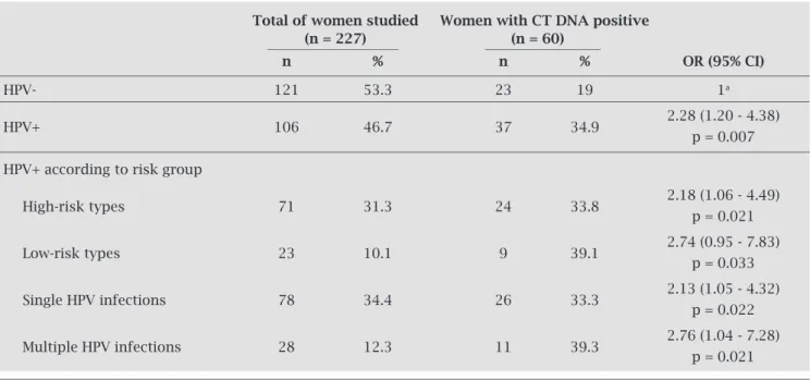
The prevalence of CT infection was significantly higher among women with positive HPV DNA (37/106 = 34.9%) than among those with negative HPV DNA (23/121 = 19%; OR: 2.28, 95% CI = 1.20 - 4.38, p = 0.007). This association re-mained significant when high- and low-risk HPV types were considered separately, as well as infections with one or
multi-Table 1. Chlamydia trachomatis and human papillomavirus infections stratified by grade of cervical status among sexually active women of the Pilaga community
Cytology n (%) HPV p High-risk HPV p CT p
positive (%) positive (%) positive (%)
Negative for SIL
36 (15.8) 10 (27.8) 7 (19.4) 8 (22.2) or malignancy
RCC 173 (76.2) 80 (46.2) 50 (28.9) 47 (27.2)
L-SIL 16 (7) 14 (87.5) 0.015 12 (75.0) 0.010 5 (31.3) 0.968 H-SIL 1 (0.4) 1 (100) 1 (100) 0 (0)
CC 1 (0.4) 1 (100) 1 (100) 0 (0) Total 227 (100) 106 (46.7) 71 (31.3) 60 (26.4)
SIL, squamous intraepithelial lesion; RCC, reactive cellular changes; L-SIL, low-grade squamous intraepithelial lesion; H-SIL, high-grade squamous intraepithelial lesion; ISC, in situ carcinoma.
Table 2. Association between Chlamydia trachomatis and human papillomavirus infections in cervical samples
Total of women studied Women with CT DNA positive (n = 227) (n = 60)
n % n % OR (95% CI)
HPV- 121 53.3 23 19 1a
HPV+ 106 46.7 37 34.9 2.28 (1.20 - 4.38) p = 0.007 HPV+ according to risk group
High-risk types 71 31.3 24 33.8 2.18 (1.06 - 4.49) p = 0.021
Low-risk types 23 10.1 9 39.1 2.74 (0.95 - 7.83) p = 0.033
Single HPV infections 78 34.4 26 33.3 2.13 (1.05 - 4.32) p = 0.022
Multiple HPV infections 28 12.3 11 39.3 2.76 (1.04 - 7.28) p = 0.021
a Reference group.
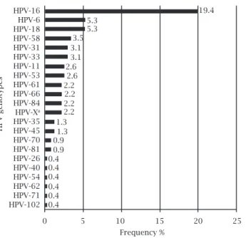
The individual prevalence for the 21 different de-tected HPV types is presented in Figure 1. Among the high-risk types, HPV-16 was clearly the most prevalent (44/227; 19.4%), followed by HPV-18 (12/227; 5.3%), -58 (8/227; 3.5%), and -31 and -33 (7/227; 3.1%); while among low-risk type, HPV-6 was the most prevalent (12/227; 5.3%), followed by HPV-11 (6/227; 2.3%), and -61 and -84 (5/227; 2.2%). Taking into account all the types found in this series, the most frequent were, in order, HPV-16, 18, 6, 58 and 33. In addition, 28 infections by multiple HPV types were ob-served, corresponding to 12.3% of the total women studied. According to the data obtained, the only independ-ent variable associated with CT-DNA positivity was concurrent HPV infection.
DISCUSSION
Despite the numerous logistical and cultural barriers that hindered this work in the Pilaga population, we were able to recruit and study 20% of the sexually active women of the community.
At large, developing countries seem to have higher prev-alence of CT infections.20 These nations have values that
range from 5% to 22% in the general population and can reach peaks as high as 41% among female adolescents.1,21-24
On the other hand, in developed countries, the prevalence levels range from less than 2% to 8-10% in general popula-tion and from less than 5% to 20% among female
adoles-cents.1,25 In Argentina, many prevalence studies of CT
infec-tion have been conducted among women, but these studies have two important shortcomings: (I) most of them used non-standardized populations, different analytical methods to detect CT or only considered symptomatic cases; and (II) there is scarce epidemiological information about popula-tions from northern Argentina, the region with the highest reported clinical diagnosis of STBI of the country. The avail-able data show that prevalence ranges from 1.8% to 13% in the general population and from 20% to 47% in women with gynecological alterations.20,26-28 The overall prevalence of CT
found in our study (26.4%) is clearly the highest reported in Argentina for the general population and is very close to the worst prevalence rates worldwide.
For HPV, the worldwide infection prevalence ranges from 2-44% in the general population. Once again, devel-oping countries usually have the highest prevalence rates.10
A recent worldwide meta-analysis on global HPV preva-lence demonstrated an infection rate of 10.4% for the world and 12.9% for Latin America,29 and HPV-16 was the most
common type in both normal and pathological populations. In our study, the overall prevalence of HPV was very high (46.7%), similar to the values found in developing countries with serious problems with implementing and maintaining a sustainable gynecological health policy. Also, it is com-parable with the results obtained by other authors in other indigenous populations of northern Argentina (prevalence ranging from 52% to 60%), but clearly exceeds the mean frequency for populations from the central and southern regions of the country and for Latin American women.10,29,30
In relation to the genotypes found, HPV-16 clearly stands out among the 21 different types identified in this work, which coincides with worldwide data and with previous studies on different populations in Argentina.31-34 In addition, the four
most prevalent HPV types among Pilaga women (HPV-16, 18, 6 and -1) represent 70% of all infections detected. These are very important baseline data to take into account for fu-ture post-vaccination epidemiological surveillance.
It is well-known that CT infection may play a major role in the etiology of cervical intraepithelial neoplasia by facilitating high-risk HPV entrance and persistence. Prob-ably, this is due to the chronic inflammation induced by the bacteria, and to the resistance to cell apoptosis that persis-tent CT infections appear to confer. In this respect, our data highlight a moderate but significant association between concurrent CT and HPV infection. However, due to the cross-sectional design of this study, it was not possible to determine the primary infection in patients with concur-rent CT and HPV infections. CT and HPV DNA detection by PCR-based assay only indicates an acute or chronic in-fection in the first case and a transient or persistent HPV infection. Nonetheless, the association between these two agents seems to be more related to a mutual potentiation
Figure 1: Prevalence of individual HPV types in cervical scrapes among Pilaga women detected by broad-spectrum polymerase chain reaction (MY 09/11 PCR) and a restriction fragment-length polymorphism assay (RFLP).
aHPV genotypes that could not be defined by the RFLP
technique.
HPV-16 HPV-6 HPV-18 HPV-58 HPV-31 HPV-33 HPV-11 HPV-53 HPV-61 HPV-66 HPV-84
HPV-Xa
HPV-35 HPV-45 HPV-70 HPV-81 HPV-26 HPV-40 HPV-54 HPV-62 HPV-71 HPV-102
HPV genotypes
Frequency %
0 5 10 15 20 25
19.4 5.3
5.3 3.5 3.1 3.1 2.6
than to the fact that they share a common route of trans-mission.35 In fact, controversial and discordant information
exists on this topic, and since the role of CT in the natural history of HPV infection is not sufficiently clear, this par-ticular issue merits further study.
CONCLUSIONS
The overall prevalence of CT and HPV infections found in this study is very high, even greater than other Latin American prevalences. The epidemiological data ob-tained in this work referred to a specific population, but numerous small communities exist in a similar situa-tion in northern Argentina. One could then suppose that similar infection rates prevail requiring effective and sus-tainable health policies to be implemented. In the case of HPV, this is very important in order to prevent CC de-velopment. For CT, its control is essential for preventing pelvic inflammatory disease and infertility. Our data may help physicians to prevent diseases in their gynecologi-cal practices, particularly when dealing with vulnerable populations.
ACKNOWLEDGEMENTS
The authors gratefully acknowledge the Pilaga aborigi-nal community, the health care volunteers of Las Lomitas (Province of Formosa) and Dr. Horacio Lucero for his as-sistance in HPV-DNA testing.
REFERENCES
1. World Health Organization. Global prevalence and incidence of selected curable sexually transmitted infections-Overview and estimates [Internet]. Geneva; 2001. Available from: http://www.who.int/hiv/pub/sti/who_hiv_aids_2001.02.pdf 2. Stamm WE. Chlamydia trachomatis infections of the adult. In:
Holmes KK, Sparling PF, Stamm WE et al., editors. Sexually transmitted diseases. New York: McGraw-Hill Medical;2008. pp. 575-94.
3. Stamm WE. Chlamydia trachomatis – the persistent
path-ogen: Thomas Parran Award Lecture. Sex Transm Dis. 2001;28(12):684-9.
4. Smith JS, Bosetti C, Muñoz N, et al. Chlamydia
trachoma-tis and invasive cervical cancer: a pooled analysis of
the IARC multicentric case-control study. Int J Cancer. 2004;111(3):431-9.
5. Lehtinen M, Ault KA, Lyytikainen E, et al. Chlamydia
tra-chomatis infection and risk of cervical intraepithelial neo-plasia. Sex Transm Infect [Internet]. 2011; Available from: http://www.ncbi.nlm.nih.gov/pubmed/21471141
6. Oliveira M L, Amorim MMR, Souza PRE, et al. Chlamydia in-fection in patients with and without cervical intra-epithelial le-sions tested by real-time PCR vs. direct immunofluorescence. Braz J Infect Dis. 2008;12(4):324-8.
7. Silins I, Ryd W, Strand A, et al. Chlamydia trachomatis infec-tion and persistence of human papillomavirus. Int J Cancer. 2005;116(1):110-5.
8. Smith JS, Muñoz N, Herrero R, et al. Evidence for Chlamydia trachomatis as a human papillomavirus cofactor in the eti-ology of invasive cervical cancer in Brazil and the Philip-pines. J Infect Dis. 2002;185(3):324-31.
9. Simonetti AC, Melo JH de L, de Souza PRE, et al. Im-munological’s host profile for HPV and Chlamydia tra-chomatis, a cervical cancer cofactor. Microbes Infect. 2009;11(4):435-42.
10. Bosch FX, Burchell AN, Schiffman M, et al. Epidemiology and natural history of human papillomavirus infections and type-specific implications in cervical neoplasia. Vaccine. 2008;26:K1-16.
11. Schiffman M, Castle PE, Jeronimo J, et al. Human papilloma-virus and cervical cancer. Lancet. 2007;370(9590):890-907. 12. Kjaer SK, van den Brule AJC, Paull G, et al. Type specific
persistence of high risk human papillomavirus (HPV) as in-dicator of high grade cervical squamous intraepithelial le-sions in young women: population based prospective follow up study. BMJ. 2002;325(7364):572.
13. Ho GY, Burk RD, Klein S, et al. Persistent genital human papillomavirus infection as a risk factor for persistent cer-vical dysplasia. J Natl Cancer Inst. 1995;87(18):1365-71. 14. Almonte M, Albero G, Molano M, et al. Risk factors for
human papillomavirus exposure and co-factors for cervi-cal cancer in Latin America and the Caribbean. Vaccine. 2008;26 Suppl 11:L16-36.
15. Drain PK, Holmes KK, Hughes JP, et al. Determinants of cervical cancer rates in developing countries. Int J Cancer. 2002;100(2):199-205.
16. Arrossi S. Proyecto para el mejoramiento del Programa Nacional de Prevención de Cáncer de Cuello Uterino en Argentina. Informe final: Diagnóstico de situación del Pro-grama Nacional y ProPro-gramas Provinciales [Internet]. 1st ed. Buenos Aires: Organización Panamericana de la Salud, OPS; 2008. Available from: http://www.msal.gov.ar/cancer-cervico-uterino/pdf/pub64_ops.pdf
17. Mahony JB, Luinstra KE, Sellors JW, et al. Confirmatory poly-merase chain reaction testing for Chlamydia trachomatis in first-void urine from asymptomatic and symptomatic men. J Clin Microbiol. 1992; 30(9):2241-5.
18. Bernard HU, Chan SY, Manos MM, et al. Identification and assessment of known and novel human papillomaviruses by polymerase chain reaction amplification, restriction fragment length polymorphisms, nucleotide sequence, and phylogenetic algorithms. J Infect Dis. 1994; 170(5):1077-85.
19. Manos MM, Ting Y, Wright DK, et al. The use of polymerase chain reaction amplification for the detection of genital human papillomavirus. Cancer Cells. 1989; (7):209-14.
20. Franceschi S, Smith JS, van den Brule A, et al. Cervical infec-tion with Chlamydia trachomatis and Neisseria gonorrhoeae in women from ten areas in four continents. A cross-sectional study. Sex Transm Dis. 2007; 34(8):563-9.
21. León SR, Konda KA, Klausner JD, et al. Chlamydia trachomatis infection and associated risk factors in a low-income marginal-ized urban population in coastal Peru. Rev Panam Salud Pub-lica. 2009; 26(1):39-45.
22. Martínez TMA, Reid SI, Arias C, et al. Prevalence of cervical infec-tion by Chlamydia trachomatis among Chilean women living in the Metropolitan Region. Rev Med Chil. 2008; 136(10):1294-300. 23. Rodrigues MM, Fernandes PÁ, Haddad JP, et al.
Frequen-cy of Chlamydia trachomatis, Neisseria gonorrhoeae,
My-coplasma genitalium, MyMy-coplasma hominis and
Ureaplas-ma species in cervical samples. J Obstet Gynaecol. 2011;
24. Santos C, Teixeira F, Vicente A, et al. Detection of Chlamydia trachomatis in endocervical smears of sexually active wom-en in Manaus-AM, Brazil, by PCR. Braz J Infect Dis. 2003; 7(2):91-5.
25. Wilson JS, Honey E, Templeton A, et al. A systematic review of the prevalence of Chlamydia trachomatis among European women. Hum Reprod Update. 2002; 8(4):385-94.
26. de Cristófano MA, Livellara B, Galli MA, et al. Extent of
en-demic Chlamydia trachomatis in the metropolitan area of
Buenos Aires (Argentina). Enferm Infecc Microbiol Clin. 1997;15(3):134-9.
27. Di Bartolomeo S, Rodríguez M, Sauka D, et al. Microbiologic profile in symptomatic pregnant women’s genital secretions in Gran Buenos Aires, Argentina. Enferm Infecc Microbiol Clin. 2001 19(3):99-102.
28. Golijow CD, Abba MC, Mourón SA, et al. Chlamydia
tra-chomatis and human papillomavirus infections in cervical dis-ease in Argentine women. Gynecol Oncol. 2005;96(1):181-6. 29. de Sanjosé S, Diaz M, Castellsagué X, et al. Worldwide
preva-lence and genotype distribution of cervical human papilloma-virus DNA in women with normal cytology: a meta-analysis. Lancet Infect Dis. 2007;7(7):453-9.
30. Clifford G, Gallus S, Herrero R, et al. Worldwide distribution of human papillomavirus types in cytologically normal women in the International Agency for Research on Cancer HPV preva-lence surveys: a pooled analysis. Lancet. 2005;366(9490):991-8. 31. Tonon SA, Picconi MA, Zinovich JB, et al. Prevalence of cervi-cal infection by human papilloma virus (HPV) in the Caucasian and Guaraní populations residing in the province of Misiones, Argentina. Rev Argent Microbiol. 2003;35(4):205-13.
32. Picconi MA, Gronda J, Alonio LV, et al. Human papilloma virus in Quechua women from Jujuy with high frequency of cervi-cal cancer: viral types and HPV-16 variants. Medicina (B Aires). 2002;62(3):209-20.
33. Matos E, Loria D, Amestoy GM, et al. Prevalence of human pap-illomavirus infection among women in Concordia, Argentina: a population-based study. Sex Transm Dis. 2003;30(8):593-9. 34. Sijvarger CC, González JV, Prieto A, et al. Cervical infection
epi-demiology of human papillomavirus in Ushuaia, Argentina. Rev Argent Microbiol. 2006;38(1):19-24.