Paul Richard Julian Ames
ATHEROGENESIS AND ATHEROSCLEROSIS IN
PRIMARY ANTIPHOSPHOLIPID SYNDROME
Faculdade de Ciência Médicas
Universidade Nova de Lisboa, Portugal, 2013
Dissertation presented to obtain the PhD degree in
“
Medicina
-Especialidade Medicina Interna” at the Faculdade de Ciências
iii
THIS WORK ORIGINATED THE FOLLOWING PUBLICATIONS:
CHAPTER III
Ames PRJ. Antiphospholipid antibodies, thrombosis and atherosclerosis in systemic lupus
erythematosus: a unifying 'membrane stress syndrome' hypothesis. Lupus 1994; 3: 371-377
CHAPTER IV
Ames PRJ, Pyke S, Iannaccone L, Brancaccio V. Antiphospholipid antibodies, haemostatic
variables and thrombosis-a survey of 144 patients. Thromb Haemost. 1995; 73: 768-773
Ames PRJ, Tommasino C, Iannaccone L, Brillante M, Cimino R, Brancaccio V. Coagulation
activation and fibrinolytic imbalance in subjects with idiopathic antiphospholipid
antibodies--a cruciantibodies--al role for antibodies--acquired free protein S deficiency. Thromb Hantibodies--aemost 1996; 76: 190-194
Ames PRJ, Iannaccone L, Alves JD, Margarita A, Lopez LR, Brancaccio V. Factor XIII in
primary antiphospholipid syndrome. J Rheumatol 2005; 32: 1058-1062
CHAPTER V
Ames PRJ, Matsuura E, Batuca JR, Ciampa A, Lopez LL, Ferrara F, Iannaccone L, Alves
JD. High-density lipoprotein inversely relates to its specific autoantibody favoring oxidation
in thrombotic primary antiphospholipid syndrome. Lupus 2010; 19: 711-716
Ames PRJ, Tommasino C, Alves J, Morrow JD, Iannaccone L, Fossati G, Caruso S, Caccavo
F, Brancaccio V. Antioxidant susceptibility of pathogenic pathways in subjects with
antiphospholipid antibodies: a pilot study. Lupus 2000; 9: 688-695
CHAPTER VI
Ames PRJ, Batuca JR, Ciampa A, Iannaccone L, Delgado Alves J. Clinical relevance of
nitric oxide metabolites and nitrative stress in thrombotic primary antiphospholipid syndrome.
J Rheumatol 2010; 37: 2523-2530
CHAPTER VII
Ames PRJ, Antinolfi I, Ciampa A, Batuca J, Scenna G, Lopez LR, Delgado Alves J,
Iannaccone L, Matsuura E. Primary antiphospholipid syndrome: a low-grade
iv
CHAPTER VIII
Ames PRJ, Margarita A, Delgado Alves J, Tommasino C, Iannaccone L, Brancaccio V.
Anticardiolipin antibody titre and plasma homocysteine level independently predict intima
media thickness of carotid arteries in subjects with idiopathic antiphospholipid antibodies.
Lupus 2002; 11: 208-214
Ames PRJ, Delgado Alves J, Lopez LR, Gentile F, Margarita A, Pizzella L, Batuca J, Scenna
G, Brancaccio V, Matsuura E. Antibodies against beta2-glycoprotein I complexed with an
oxidised lipoprotein relate to intima thickening of carotid arteries in primary antiphospholipid
syndrome. Clin Dev Immunol 2006; 13: 1-9
Ames PRJ, Antinolfi I, Scenna G, Gaeta G, Margaglione M, Margarita A. Atherosclerosis in
thrombotic primary antiphospholipid syndrome. J Thromb Haemost 2009; 7: 537-542
CHAPTER IX
Ames PRJ, Scenna G, Antinolfi I, Lopez L, Iannaccone L, Matsuura E, Margarita A.
Atherosclerosis in primary antiphospholipid syndrome. Expert Rev Clin Immunol 2008; 4:
53-60
v
DEDICATION
This thesis is dedicated to my dear friend Giulio with whom I shared the happiness of
youth, the goliardy of our school days, the enthusiasm for our life projects and the emotions
of his first rugby matches. It is during one of these that tragedy befell and he passed away
without ever reaching the try-line. His friendship lived on within me and I would like to
vi
ACKNOWLEDGEMENTS
No part of this thesis would have been written without the friendship and support of my
colleague Doctor Vincenzo Brancaccio (Haemostasis Unit of the Cardarelli Hospital, Naples,
Italy), of his Senior MLSO Mr Luigi Iannaccone, both of whom I collaborated with for almost
two decades and of Doctor Antonio Ciampa from the Haemostasis Unit, Moscati Hospital,
Avellino, Italy. When I moved to London to pursue a career in Rheumatology, I walked into
the large bookstore close to University College with the intention of securing the best
available Rheumatology textbook, but I came out with a book on free radicals instead. The
encounter with Jaffar Nourooz Zadeh, at the time Senior Scientist in lipid biochemistry at
University College London, and now Professor of Biochemistry (Urmia University of
Medical Sciences, Iran) widened my perspectives on free radicals and on lipid peroxidation to
the extent that we published a seminal article on oxidative stress in systemic lupus
erythematosus and primary antiphospholipid syndrome. The ideas on oxidative stress fostered
further collaboration with Doctor Luis Lopez (Corgenix Inc., Colorado, USA) and Professor
Eiji Matsuura (Department of Cell Chemistry, Okayama University, Okayama, Japan) who
helped me with several of the novel immune assays. But most of all I am grateful to all the
patients whom I had come across over the twenty years of collaboration with my Neapolitan
colleagues, so much so that when Doctor Brancaccio retired I set up an association for
patients with antiphospholipid syndrome and inherited thrombophilia and subsequently I set
vii
ABSTRACT
Background
In the late seventies the term “Haematological Stress Syndrome” defined some haematological abnormalities appearing in the course of acute and chronic disorders, such as
raised plasma levels of fibrinogen (FNG) and factor VIII, reduced fibrinolytic activity and
hyperviscosity. In the early nineties the “Membrane stress syndrome hypothesis” proposed the unification of the concepts of haematological stress syndrome with those of oxidation,
inflammation and immune activation to explain the pathogenesis of the antiphospholipid
syndrome (APS)
Antiphospholipid antibodies, coagulation, fibrinolysis and thrombosis
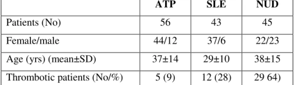
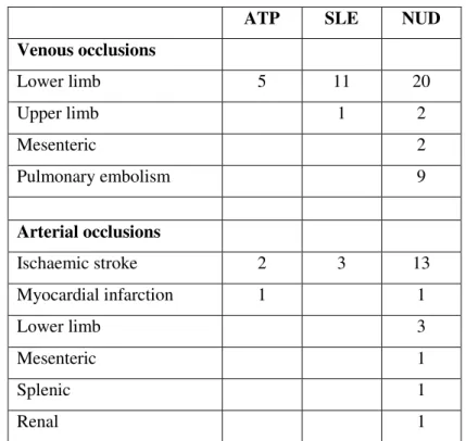
This chapter investigated the occurrence of the “Haematological Stress Syndrome” and thrombosis in 144 participants positive for aPL detected by clotting and immune tests. Among
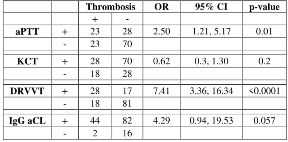
the clotting assays for the detection of lupus anticoagulant, dilute Russell's viper venom time
better correlated with a history of venous thrombosis than activated partial thromboplastin
time (p<0.0002 vs p<0.009) and was the only test correlated with a history of arterial
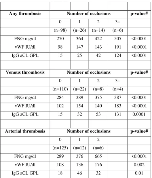
thrombosis (p<0.01). By regression analysis, serum levels of IgG anticardiolipin antibodies
(aCL) associated with the number of venous occlusions (p<0.001). With regards to FNG and
von Willebrand factor (vWF), the former rose by 36% (95% CI; 21%, 53%) and the latter by
50% (95% CI; 29%, 75%) at the first venous occlusion and remained unchanged after
subsequent occlusions. At variance FNG rose by 45% (95% CI; 31%, 60%) per arterial
occlusion and vWF by 27% (95% CI; 10%, 47%) per arterial occlusion throughout.
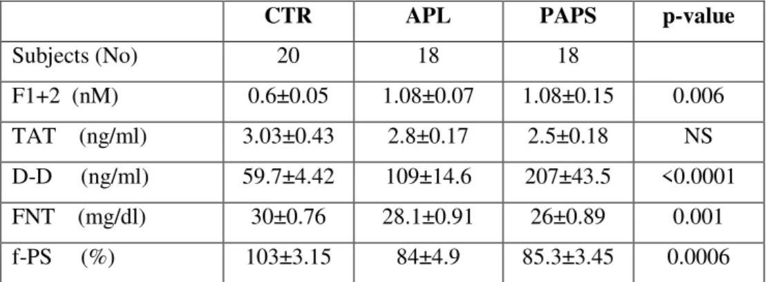
The coagulation/fibrinolytic balance was cross-sectionally evaluated on 18 thrombotic
PAPS patients, 18 subjects with persistence of idiopathic aPL and in healthy controls.
Markers of thrombin generation prothrombin fragment 1+2 (F1+2), thrombin-antithrombin
viii
and non-thrombotic subjects (p=0.0001) than in controls as were those of D-D (p<0.0001 and
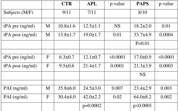
p=0.003 respectively). TAT levels did not differ. Gender analysed data revealed blunted tPA
release (hence a negative venous occlusion test) in thrombotic females but neither in
thrombotic males (p=0.01) nor in asymptomatic subjects of either sex. Also, in both patient
groups females had higher mean PAI than males (p<0.0002) and control females (p<0.02).
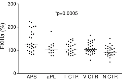
The activity of factor XIII (FXIIIa) was evaluated was evaluated in 29 patients with
PAPS, 14 persistent carriers of aPL without thrombosis, 24 thrombotic patients with inherited
thrombophilia, 28 healthy controls and 32 patients with mitral and aortic valve prosthesis as
controls for FXIII only. FXIIIa was highest in PAPS (p=0.001), particularly in patients with
multiple (n=12) than single occlusion (p=0.02) and in correlation with PAI (p=0.003) and
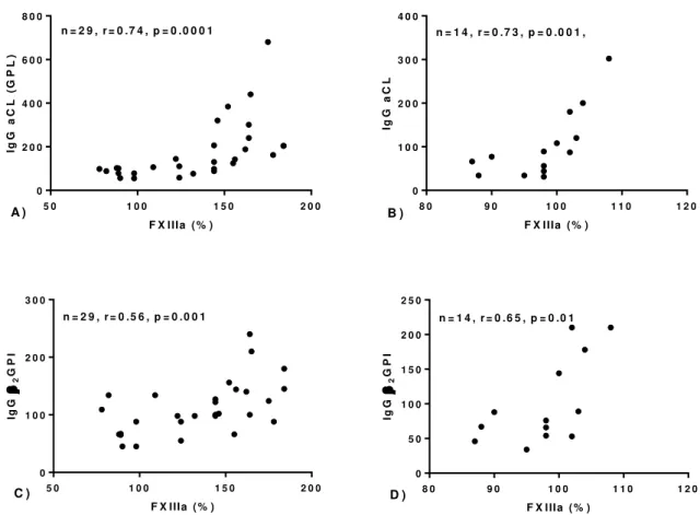
FNG (p=0.005). Moreover FXIIIa was strongly associated with IgG aCL and IgG anti-2GPI
(p=0.005 for both) in the PAPS group and to a lesser degree in the aPL group (FXIIIa with
IgG aCL, p=0.02, with IgG anti-2GPI, p=0.04). Altogether these results indicate: 1) a
differential relationship of aPL, vWF and FNG with venous and arterial thrombosis; 2)
heightened thrombin generation, accelerated fibrin turnover and fibrinolysis abnormalities
also in asymptomatic carriers of aPLs; 3) enhanced FXIIIa that may contribute to
atherothrombosis via increased fibrin/fibrinogen cross-linking.
Lipid profile, lipid peroxidation and anti-lipoprotein antibodies in thrombotic primary antiphospholipid syndrome.
Given the atherogenic lipid profile of SLE, the same possibility was explored in PAPS by
comparing high-density lipoprotein (HDL), low-density lipoprotein (LDL), total cholesterol
(CHO), apolipoprotein AI (ApoAI), apolipoprotein B (ApoB), triglycerides (TG),
anti-lipoprotein antibodies, beta-2-glycoprotein I complexed to oxidized low-density anti-lipoprotein
(oxLDL-2GPI) and C-reactive protein (CRP) in 34 thrombotic PAPS patients compared to
ix
for antiphospholipid antibodies (aPL) with no underlying autoimmune or non-autoimmune
disorders and to 28 healthy controls. Average concentrations of HDL (p<0.0001), LDL
(p<0.0001), CHO (p=0.0002), ApoAI (p=0.002) were lower in PAPS whereas average TRY
was higher (p=0.01) than other groups. Moreover PAPS showed higher IgG
anti-HDL (p=0.01) and IgG anti-ApoAI (p<0.0001) as well as greater average oxLDL-2GPI
(p=0.001) and CRP (p=0.003). Within PAPS, IgG anti-HDL correlated negatively to HDL
(p=0.004) and was an independent predictor of oxLDL-2GPI (p=0.009). HDL and ApoAI
correlated negatively with CRP (p=0.001 and p=0.007, respectively). IgG anti-HDL may
hamper the antioxidant and anti-inflammatory effect of HDL favouring low-grade
inflammation and enhanced oxidation in thrombotic PAPS. Indeed plasma
8-epi-prostaglandin F2α (a very specific marker of lipid peroxidation) was significantly higher in 10 patients with PAPS than 10 age and sex matched healthy subjects (p=0.0002) and strongly
related to the titre of plasma IgG aCL (r=0.89, p=0.0004). Hence oxidative stress, a major
player in atherogenesis, also characterises PAPS.
Nitric oxide and nitrative stress in thrombotic primary antiphosholipid syndrome.
Oxidative stress goes hand in hand with nitrative stress and to address the latter plasma
nitrotyrosine (NT, marker of nitrative stress), nitrite (NO2-) and nitrate (NO3-) were measured
in 46 thrombotic PAPS patients, 21 asymptomatic but persistent carriers of antiphospholipid
antibodies (PCaPL), 38 patients with inherited thrombophilia (IT), 33 patients with systemic
lupus erythematosus (SLE) and 29 healthy controls (CTR). Average crude NT was higher in
PAPS and SLE (p=0.01) whereas average plasma NO2- was lower in PAPS and average NO3
-highest in SLE (p<0.0001). In PAPS, IgG aCL titer and number of vascular occlusions
negatively predicted NO2-, (p=0.03 and p=0.001, respectively) whereas arterial occlusions and
smoking positively predicted NO3- (p=0.05 and p=0.005). Moreover CRP (an inflammatory
x
number of vascular occlusions and to aPL titers, whereas nitrative stress relates to low grade
inflammation and both phenomena may have implications for thrombosis and atherosclerosis
in PAPS
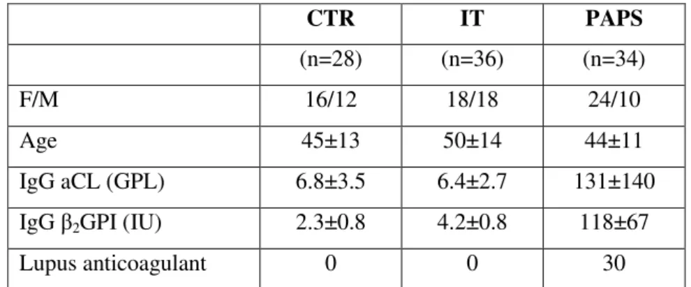
Inflammation and immune activation in thrombotic primary antiphospholipid syndrome.
To investigate inflammation and immune activation in thrombotic PAPS high-sensitivity
CRP (hs-CRP), serum amyloid A (SAA), oxLDL-2GPI, CRP bound to oxLDL-2GPI
(CRP-oxLDL-2GPI) (as inflammatory markers) neopterin (NPT) and soluble CD14 (sCD14)
(as immune activation markers) were measured by ELISA in 41 PAPS patients, in 44 patients
with inherited thrombophilia (IT) and 39 controls (CTR). Compared to other groups, PAPS
presented with higher plasma concentrations of inflammatory, hs-CRP (p=0.0004), SAA
(p<0.01), CRP-oxLDL-2GPI (p=0.0004) and immune activation markers, NPT (p<0.0001)
and sCD14 (p=0.007). By regression analysis SAA independently predicted thrombosis
number (p=0.003) and NPT independently predicted thrombosis type (arterial, p=0.03) and
number (p=0.04). These data confirm that low-grade inflammation and immune activation
occur and relate to vascular features of PAPS.
Antiphosholipid antibodies, haemostatic variables and atherosclerosis in thrombotic primary antiphospholipid syndrome
To evaluate whether IgG aCL titre, haemostatic variables and the lipid profile bore any
relationship to the intima media thickness (IMT) of carotid arteries high-resolution
sonography was applied to the common carotid (CC), carotid bifurcation (CB) and internal
carotid (IC) of 42 aPL subjects, 29 with primary thrombotic antiphospholipid syndrome and
13 with persistence of aPL in the absence of any underlying disorder. The following were
measured: plasma FNG, vWF, PAI, homocysteine (HC), CHO, TG, HDL, LDL, platelet
numbers and aCL of IgG and IgM isotype. By multiple regression analysis, IgG aCL titre
xi
FNG and HC independently predicted IMT at the CB (p=0.001 and p<0.0001, respectively)
and IC (p=0.03 and p<0.0001, respectively). These data strongly support an atherogenic role
for IgG aCL in patients with aPL in addition to traditional risk factors. The atherosclerosis
hypothesis was investigated in an age and sex-matched case-double-control study including
49 thrombotic PAPS patients (18 M, 31 F, mean age 37 ± 11), 49 thrombotic patients for IT
and 49 healthy subjects. Average IMT was always greater in PAPS than control patients (CC:
p=0.004, CB: p=0.013, IC: p=0.001). By dividing participants into age tertiles the IMT was
greater in the second (CC: p=0.003, CB: p=0.023, IC: p=0.003) and third tertiles (CC: p=0.03,
CB: p=0.004, IC: p=0.007).
Conclusion
Coagulation activation, fibrinolysis depression, hightened fibrin turnover, oxidative and
nitrative stress in parallel with low grade inflammation and immune activation characterise
thrombotic PAPS: all these are early atherogenic processes and contribute to the demonstrated
xii
RESUMO
Introdução
No final dos anos setenta algumas alterações hematológicas como níveis elevados de
fibrinogénio (FNG) e factor VIII, diminuição da actividade fibrinolítica e hiperviscosidade
foram agrupadas na definição “Síndrome de Stress Hematológico”.
Posteriormente, a hipótese da “Síndrome de Stress Membranar” veio propor a unificação dos conceitos de síndrome de stress hematológico, oxidação, inflamação e activação
imunológica, numa tentativa de explicação da patogénese da síndrome de anticorpos
antifosfolípidos (APS).
Anticorpos antifosfolípidos, coagulação, fibrinólise e trombose
Este capítulo aborda os conceitos da “Síndrome de stress hematológico” e trombose em 144 doentes com anticorpos antifosfolípidos (aPL), detectados por testes imunológicos e de
coagulação. Entre os testes para detecção do anticoagulante lúpico (LA), o “tempo de veneno de cobra” (DRVVT) apresentou uma melhor correlação com a ocorrência de tromboses venosas que o tempo de tromboplastina parcial activado (p < 0.0002 vs p < 0.009) e foi o
único teste em que se verificou uma correlação com a presença de tromboses arteriais
(p < 0.01). A análise de regressão, após correcção para potenciais factores de confundimento,
verificou uma associação entre os níveis séricos de anticorpos anti-cardiolipina (aCL) IgG e o
número de tromboses venosas (p < 0.001). No que se refere ao FNG e factor de Von
Willebrand (vWF), verificou-se uma elevação de 36% do primeiro (95% CI; 21%, 53%) e de
50% do segundo (95% CI; 29%, 75%) na primeira trombose venosa. Estes valores
mantiveram-se inalterados nos eventos subsequentes. Em contraste, verificou-se uma subida
de 45% do FNG (95% CI, 31%-60%) e de 27% do vWF (95% CI; 10%-47%) por cada evento
trombótico arterial. O equilíbrio entre coagulação e fibrinólise foi avaliado de forma
xiii
controlos saudáveis. Marcadores de geração de trombina como o fragmento 1+2 da
protrombina (F1+2) e complexos trombina-anti-trombina (TAT) e da renovação da fibrina
como os dímeros D (DD) estavam mais elevados em doentes com tromboses (p=0.006) e
mesmo em doentes sem tromboses (p=0.0001) do que no grupo controlo, tal como os DD
(p<0.0001 e p=0.003 respectivamente). Os valores de TAT não foram significativamente
diferentes.
Níveis médios de proteína S livre mais baixos foram encontrados no grupo com PAPS
(p=0.0006) e nos indivíduos sem tromboses (p=0.002) em comparação com os controlos
saudáveis. Em ambos os grupos de estudo, os níveis de F1+2 eram mais elevados em doentes
com níveis baixos de proteína S quando comparados com os indivíduos que apresentavam
valores de proteína S normais (p=0.01). No que diz respeito ao género, observou-se uma
redução do activador tissular do plasminogénio (rPA) (avaliado através do teste de oclusão
venosa), em mulheres com PAPS (de 16.80 ± 0.79 para 21.3 ± 3.9 ng/ml, NS), mas não em
homens com PAPS (de 18.2 ± 2.0 para 33.7 ± 4.9 ng/ml, p=0.01), nem nos doentes com
anticorpos mas sem eventos trombóticos, independentemente do género.
Da mesma forma, em ambos os grupos de doentes, as mulheres apresentaram níveis de
inibidor do activador de plasminogénio (PAI) mais elevados que os homens (p<0.0002) e que
os controlos femininos (p<0.02).
A actividade do factor XIII (FXIIIa) foi avaliada em 29 doentes com PAPS, 14 portadores
de anticorpos antifosfolípidos mas sem eventos trombóticos, 24 doentes com trombofilias
hereditárias e tromboses, 28 controlos saudáveis e 32 doentes com próteses mitrais ou aórticas
(utilizados apenas para controlo dos níveis de FXIII).
A FXIIIa estava mais elevada em doentes com PAPS (p=0.001), particularmente em
doentes com mais de um evento trombótico (158 ± 45% vs 118 ± 38%; p=0.02, n=12) e
xiv
A FXIIIa estava ainda fortemente associada aos títulos de aCL IgG e anti-2GPI IgG
(p=0.005 para ambos) no grupo com PAPS e, embora de forma menos significativa, no grupo
portador de anticorpos (aCL IgG, p=0.02; anti-2GPI IgG, p=0.04).
Em conjunto estes resultados mostram: 1) uma relação diferencial de aPL, vWF e FNG
com as tromboses arteriais e venosas; 2) o aumento da formação de trombina, o aumento da
renovação da fibrina e a diminuição da actividade fibrinolítica ocorrem também nos
portadores assintomáticos de aPL; 3) um papel central da deficiência de proteína S livre
“adquirida” no risco trombótico dos doentes com APS; 4) um aumento da actividade do FXIII que poderá contribuir para aterotrombose através de um aumento das ligações cruzadas entre
fibrina e fibrinogénio.
Perfil lipidico, peroxidação lipidica e anticorpos anti-lipoproteinas na síndrome de anticorpos antifosfolípidos
Estes aspectos foram estudados comparando os níveis de HDL, LDL, colesterol total
(CHO), apolipoproteína AI (ApoAI), apolipoproteína B (ApoB), triglicéridos (TG),
anticorpos anti-lipoproteínas, complexos beta-2-glicoproteína-I e LDL oxidada
(oxLDL-2GPI) e proteína C reactiva (CRP) em 34 doentes com PAPS, 36 doentes com trombofilias
hereditárias e tromboses, 18 portadores persistentes de anticorpos antifosfolípidos e 28
controlos saudáveis.
Os níveis de HDL (p<0.0001), LDL (p<0.0001), CHO (p=0.0002) e ApoAI (p=0.002)
foram mais baixos no grupo com PAPS, enquanto os TG foram mais elevados (p=0.01) que
nos outros grupos. Doentes com PAPS mostraram ainda ter títulos mais elevados de anti-HDL
IgG (p=0.01) e anti-ApoAI IgG (p<0.0001), e níveis superiores de oxLDL-2GPI (p=0.001) e
CRP (p=0.003).
Dentro do grupo com PAPS, os níveis de anti-HDL IgG correlacionaram-se
xv
valores de oxLDL-2GPI (p=0.009). HDL e ApoAI também se correlacionaram
negativamente com a CRP (p=0.001 e p=0.007, respectivamente).
Anticorpos anti-HDL IgG podem bloquear os efeitos anti-oxidante e anti-inflamatório das
HDL, favorecendo um estado de inflamação persistente e um aumento da oxidação no PAPS.
De facto, a 8-epi-prostaglandina F2α (um marcador específico de peroxidação lipídica) estava significativamente mais elevado em 10 doentes com PAPS quando comparados com um
grupo igual controlado para sexo e idade (234 ± 56 pg/ml vs 72 ± 14 pg/ml, p=0.0002) e
correlacionou-se directamente com os títulos de aCL IgG (r=0.89, p=0.0004). Estes dados
confirmam o facto de o stress oxidativo ser um factor importante no PAPS.
Óxido nítrico (NO), stress nitrativo e PAPS
O stress oxidativo associa-se ao stress nitrativo e para estudar o segundo, avaliaram-se os
níveis plasmáticos de nitrotirosina (NT), nitrito (NO2-) e nitrato (NO3-) em 46 doentes com
PAPS, 21 portadores assintomáticos de anticorpos aPL, 38 doentes com trombofilias
hereditárias (IT), 33 doentes com lupus sistémico (SLE) e 29 controlos saudáveis. Os níveis
de NT foram mais elevados nos grupos com PAPS e SLE (p=0.01), enquanto que os níveis de
NO2- foram mais baixos no PAPS e os de NO3- mais elevados no SLE (p<0.0001).
No grupo com PAPS, os títulos de aCL IgG e o número de oclusões vasculares foram
predictores negativos dos níveis de NO2-, (p=0.03 e p=0.001, respectivamente), enquanto as
oclusões arteriais e consumo de tabaco foram predictores positivos de NO3- (p=0.05 e
p=0.005). Para além destes dados, os valores de CRP foram predictores positivos dos níveis
de NT (p=0.004). Os metabolitos do NO estão associados ao tipo e número de tromboses
vasculares e aos títulos de aPL, enquanto que o stress nitrativo se associa à inflamação de
xvi
Inflamação e activação imunológica no PAPS
Para avaliar estes mecanismos em doentes com PAPS, foram medidos os níveis de CRP de
alta sensibilidade (hs-CRP), amilóide sérico A (SAA), oxLDL-2GPI e CRP ligada a
oxLDL-2GPI (CRP-oxLDL-2GPI) (como marcadores inflamatórios), e neopterina (NPT) e CD14
solúvel (sCD14) (como marcadores de activação imunológica) em 41 doentes com PAPS, 44
doentes com IT e 39 controlos saudáveis. Comparado com os outros grupos, os doentes com
PAPS apresentaram valores mais elevados de hs-CRP (p=0.0004), SAA (p<0.01),
CRP-oxLDL-2GPI (p=0.0004), NPT (p<0.0001) e sCD14 (p=0.007). A análise de regressão
mostrou que o SAA é predictor independente do número de tromboses (p=0.003) e a NPT do
tipo de trombose (arterial, p=0.03) e do número de eventos (p=0.04).
Anticorpos antifosfolípidos, variáveis hemostáticas e aterosclerose no PAPS
Para avaliar uma possível relação entre os títulos de aCL IgG, variáveis hemostáticas e
perfil lipídico, e a espessura da íntima e da média da parede arterial (IMT), realizaram-se
eco-dopplers da carótida comum (CC), bifurcação (B) e carótida interna (IC) em 42 doentes com
aPL (29 com PAPS e 13 portadores de aPL sem tromboses). Foram avaliados: FNG, vWF,
PAI, homocisteína (HC), CHO, TG, HDL, LDL, número de plaquetas e aCL (IgG e IgM). A
análise de regressão mostrou que os níveis de aCL IgG são predictores independentes da IMT
em todos os segmentos (p<0.005 para todos). FNG e HC foram predictores independentes da
IMT na bifurcação (p=0.001 e p<0.0001, respectivamente) e na IC (p=0.03 e p<0.0001,
respectivamente).
Estes dados suportam o papel pró-aterogénico dos aCL IgG em associação aos factores de
risco tradicionais. Estes dados foram complementados com o estudo de 49 doentes com
PAPS, 49 doentes com IT e 49 controlos saudáveis. A IMT média foi sempre maior no grupo
do PAPS (CC: p=0.004; B: p=0.013; IC: p=0.001). Ao dividir os indivíduos estudados em
xvii
p=0.023; IC: p=0.003) e (CC: p=0.03; B: p=0.004; IC: p=0.007), respectivamente. Desta
forma confirma-se a existência de aterosclerose como manifestação integrante do síndrome de
xviii
TABLE OF CONTENTS
DEDICATION ... v
ACKNOWLEDGEMENTS ... vi
ABSTRACT ... vii
RESUMO ... xii
TABLE OF CONTENTS ... xviii
LIST OF ABBREVIATIONS ... xxi
CHAPTER I THE ANTIPHOSPHOLIPID SYNDROME: DIAGNOSIS, CLASSIFICATION AND CLINICAL MANIFESTATIONS ... 1
HISTORICAL MILESTONES ... 1
BETA2-GLYCOPROTEIN I ... 3
DIAGNOSIS AND CLASSIFICATION OF THE ANTIPHOSPHOLIPID SYNDROME ... 4
CLINICAL MANIFESTATIONS ... 7
CHAPTER II ANTIPHOSPHOLIPID ANTIBODIES AND ATHEROTHROMBOSIS IN SYSTEMIC LUPUS ERYTHEMATOSUS: A UNIFYING 'MEMBRANE STRESS SYNDROME' HYPOTHESIS ... 11
CHAPTER III METHODS ... 20
CHAPTER IV ANTIPHOSPHOLIPID ANTIBODIES, COAGULATION, FIBRINOLYSIS AND THROMBOSIS ... 26
INTRODUCTION ... 26
EXPERIMENTAL DATA ... 28
1) RESULTS ANTIPHOSPHOLIPID ANTIBODIES, FIBRINOGEN, VON WILLEBRAND FACTOR AND THROMBOSIS ... 28
2) ANTIPHOSPHOLIPID ANTIBODIES, THROMBIN GENERATION AND FIBRINOLYSIS ... 35
3) ANTIPHOSPHOLIPID ANTIBODIES, FACTOR XIII AND FIBRINOLYSIS ... 39
DISCUSSION ... 43
xix
CHAPTER V
LIPID PROFILE, LIPID PEROXIDATION AND ANTI-LIPOPROTEIN ANTIBODIES IN
THROMBOTIC PRIMARY ANTIPHOSPHOLIPID SYNDROME ... 49
INTRODUCTION ... 49
EXPERIMENTAL DATA ... 52
1) THE LIPID PROFILE, ANTI LIPOPROTEIN ANTIBODIES AND C-REACTIVE PROTEIN ... 53
2) OXIDATIVE STRESS AS A RESULT OF LDL OXIDATION ... 57
DISCUSSION ... 58
CHAPTER VI NITRIC OXIDE AND NITRATIVE STRESS IN THROMBOTIC PRIMARY ANTIPHOSHOLIPID SYNDROME ... 60
INTRODUCTION ... 60
EXPERIMENTAL DATA ... 63
1) NITRIC OXIDE AND NITRATIVE STRESS IN PRIMARY THROMBOTIC ANTIPHOSPHOLIPID SYNDROME ... 63
MATERIALS AND METHODS ... 64
RESULTS ... 67
DISCUSSION ... 73
CHAPTER VII INFLAMMATION AND IMMUNE ACTIVATION IN THROMBOTIC PRIMARY ANTIPHOSPHOLIPID SYNDROME. ... 76
INTRODUCTION ... 76
EXPERIMENTAL DATA ... 81
RESULTS ... 84
DISCUSSION ... 88
CHAPTER VIII ANTIPHOSHOLIPID ANTIBODIES, HAEMOSTATIC VARIABLES AND ATHEROSCLEROSIS IN THROMBOTIC PRIMARY ANTIPHOSPHOLIPID SYNDROME .... 92
INTRODUCTION ... 92
EXPERIMENTAL DATA ... 95
xx
2) ANTI β2GPI/oxLDL, ANTI β2GPI-oxLig-1 AND INTIMA MEDIA THICKNESS OF
CAROTID ARTERIES ... 99
3) DEFINITE EVIDENCE FOR ATHEROSCLEROSIS IN PAPS ... 106
DISCUSSION ... 110
CHAPTER IX CONCLUSION ... 116
REFERENCES REFERENCES TO CHAPTER I ... 120
REFERENCES TO CHAPTER II ... 123
REFERENCES TO CHAPTER III ... 134
REFERENCES TO CHAPTER IV ... 135
REFERENCES TO CHAPTER V ... 139
REFERENCES TO CHAPTER VI ... 143
REFERENCES TO CHAPTER VII ... 149
REFERENCES TO CHAPTER VIII ... 158
xxi
LIST OF ABBREVIATIONS
2GPI: beta-2-glycoprotein-I
aCL: anticardiolipin aPL: antiphospholipid ApoA-I: apolipoprotein A-I APS: antiphospholipid syndrome
aPTT: activated partial thrombplastin time CHD: coronoary artery disease
CL: cardiolipin
CRP: C-reactive protein CVD: cardiovascular disease D-D: dimer D
DRVVT: dilute Russel’s viper venom time
DVT: deep vein thrombosis
eNOS: endothelial nitric oxide synthase ET: endothelin
F1+2: prothrombin fragment 1+2 FNG: fibrinogen
FNT: fibronectin f-PS: free protein S FXIIIa: factor XIII activity HC: homocysteine
xxii IMT: intima media thickness
iNOS: inducible nitric oxide synthase IS: ischaemic stroke
IT: inherited thrombophilia KCT: kaolin clotting time LA: lupus anticoagulant LDL: low density lipoprotein MI: myocardial infarction NO•: nitric oxide
NO2
-: nitrite NO3-: nitrate NPT: neopterin NT: nitrotyrosine OxLDL: oxidated LDL
PAI: plasminogen activator inhibitor PAPS: primary antiphospholipid syndrome PON: paraoxonase
ROS: reactive oxygen species SAA: serum amyloid A
xxiii TNF-:Tumor necrosis factor-alpha
tPA: tissue plasminogen activator TxB2: thromboxane B2
1
CHAPTER I
THE
ANTIPHOSPHOLIPID
SYNDROME:
DIAGNOSIS,
CLASSIFICATION AND CLINICAL MANIFESTATIONS
HISTORICAL MILESTONES
In the early ‘80s a syndrome characterized by thrombosis, thrombocytopenia and recurrent miscarriages was described in patients with systemic lupus erythematosus (SLE) who were
found positive for antibodies to cardiolipin (CL). Anti CL antibodies (aCL) belong to the
wider family of “antiphospholipid” antibodies (aPL) the specificity of which was initially attributed to negatively charged phospholipids. It became evident this was not the case and
that the major aPL specificity was towards proteins bound to negatively charged
phospholipids. The major antigenic target has been identified in beta-2-glycoprotein-I
(2GPI), a plasma protein of liver origin whose functions are well beyond those of
coagulation regulation. The immunisation of different strains of mice with 2GPI induce the
appearance of specific antibodies that have thrombogenic properties in vitro and in vivo, and
more interestingly 2GPI immunisation of low density lipoprotein knockout mice induce the
appearance of premature atherosclerosis.
Although the antiphospholipid syndrome (APS) was described in 1983, the existence of
aCL and lupus anticoagulant (LA) was already known. In 1906 Wasserman (1) described a
complement fixation test to detect “reagin” in the sera of syphilitic patients and in 1941 Pangborn (2) demonstrated that reagin bound to an antigen extracted from ox heart muscle.
This antigenic extract was later named cardiolipin. In the following years Moore and Mohr
(3) recognized that the “reagin test” was also positive in some patients who had never contracted a treponemal infection. By analysing these patients they identified a first group in
2
infection, and a second group in which the false positive reaction was a persistent
phenomenon. In this latter group there was a high prevalence of patients with SLE.
The first description of a circulating anticoagulant in patients with SLE was given by
Conley and Hartmann (4) in 1952. Their patients showed a bleeding tendency, contrary to the
increased thrombotic risk recognized by Bowie in 1963 (5). A decade later Feinstein and
Rappaport (6) named the inhibitor “lupus anticoagulant”. This was also found in association with placental infarctions and recurrent miscarriages. In 1983 Harris and colleagues (7) first
detected aPLs as aCL by a radioimmunoassay and then by an enzyme linked immunoassay
(Elisa) (8). In 1990, three independent groups of scientists (9-11) discovered that the aCL
detected by Elisa was not directed towards CL alone. Purified IgG from aCL positive patients
failed to bind CL, unless a “cofactor” was present. The “cofactor” was identified as 2GPI, a
plasma apolipoprotein with phospholipid binding properties involved in several steps of the
coagulation pathway.
From the clinical perspective the independent existence of APS outside SLE had been
suspected in the mid ’80 (12, 13) and in the late ‘80s a multicenter study documented the major clinical and serologic characteristics of a primary APS. Essentially these patients
suffered deep vein thromboses, often accompanied by pulmonary embolism, eventually
complicated by thromboembolic pulmonary hypertension, arterial occlusions (ischaemic
strokes and myocardial infarctions) or fetal loss; thrombocytopaenia, haemolytic anaemias
and isolated Coombs positivity were also described. The negativity of antinuclear antibodies,
antibodies against dsDNA and exctractable nuclear antigens ENA was the main serologic
features that discriminated them from SLE (14). These findings were confirmed in a later
Eurpean survey (15). A survey performed ten years later revealed that patients with primary
3
BETA2-GLYCOPROTEIN I
2GPI is a highly glycosylated protein synthesized in hepatocytes and circulates in the
plasma at a concentration range between 50–500 mg/mL though approximately 40% associates with lipoproteins; this single chain protein consists of 326 amino acids and is
arranged in five mutually homologous domains consisting of approximately 60 amino acids
bridged by disulphide bonds to form five short consensus repeats called Sushi domains (17).
2GPI circulates in blood in a circular conformation that shields the epitope for aPL binding
within domain I, but after interaction of domain V with anionic surfaces in vivo or with
anionic phospholipids in vitro, the circular structure opens up, the cryptic epitope is exposed
and aPL can bind to it (18).
Beta-2-glycoprotein I, antiphospholipid antibodies and thrombogenesis
2GPI regulates several steps of the coagulation pathway, exerting both procoagulant and
anticoagulant activities. β2GPI binds factor XI in vitro preventing activation of FXI to FXIa
by thrombin and FXIIa. Proteolytic cleavage of the phospholipid binding domain of β2GPI abolishes its inhibition of FXI activation. β2GPI protects thrombin from heparin inactivation
(19), modulates protein C activation, inhibits von Willebrand factor dependent platelet
adhesion and aggregation (20) and stimulates fibrinolysis (21). Whenever aPL binds β2GPI
the activity of the natural anticoagulant functions is lost and this effect, combined with the
aPL induced over-expression of tissue factor on monocytes and endothelial cells lead to
excessive thrombin generation. Thrombin promotes fibrinogen polimerisation, platelet
activation and further endothelial cell activation with a shift from an adhesive and
anti-thrombotic phenotype to a pro-adhesive and pro-anti-thrombotic phenotype. Finally enhanced
thrombin generation coupled with aPL impairment of fibrinolysis causes increased fibrin
4
Beta-2-glycoprotein I, antiphospholipid antibodies and atherogenesis
Free radicals generated by endothelial cells and circulating neutrophils, monocytes and
platelets may induce oxidative modifications within LDL (oxLDL) the uptake of which by
mononuclear cells is mediated by their scavenger receptors and is followed by monocyte
migration under the endothelial layer and then by their transformation in foam cells or lipid
laden macrophages. During this process monocytes/macrophages release a number of
inflammatory and chemotactic cytokines that contribute locally to the development of
atherosclerosis. Immune-staining of human atherosclerotic lesions co-localized oxLDL with
β2GPI. Indeed 2GPI may interact with oxLDL in an antioxidant fashion initially via
electrostatic interactions that over time become covalent: this process takes place in the
arterial intima of atherosclerotic lesions and produces stable and non-dissociable
oxLDLβ2GPI complexes (reviewed in 22). From the cellular point of view, after
co-incubation of IgG anti-2GPI antibodies with oxLDL and 2GPI, the monocyte/macrophage
uptake and intracellular accumulation of oxLDL accelerates and leads to up-regulation and
enhanced surface expression of scavenger (CD36) and FcγRI receptors that perpetuates this cycle. Therefore, while antibodies again 2GPI favor the development of thrombosis,
antibodies against oxLDL2GPI may favor the development of atherosclerosis (22).
DIAGNOSIS AND CLASSIFICATION OF THE ANTIPHOSPHOLIPID SYNDROME
Clinical diagnosis of antiphospholipid syndrome
The diagnosis of the APS relies on clinical and laboratory criteria. The clinical criteria
include one or more clinical episodes of arterial, venous, or small-vessel thrombosis in any
organ or tissue confirmed by adequate imaging or histopathology findings. Occlusions may
5
Investigations should be instigated if any of these occur in younger individuals (males < 55 y;
females < 65 y) or in the absence of other risk factors. With regards to pregnancy morbidity,
the criteria include one or more late-term (>10 weeks' gestation) spontaneous abortions, one
or more premature births of a morphologically healthy neonate at or before 34 weeks of
gestation for severe preeclampsia or eclampsia or severe placental insufficiency; three or
more unexplained, consecutive, spontaneous abortions before 10 weeks of gestation (23).
Clinical classification of antiphospholipid syndrome
The APS may appear in different settings: 1) as primary APS (PAPS) in patients with no
underlying disorder but for the presence and persistence of aPLs and clinical manifestations,
2) in patients with definite SLE, 3) as secondary APS in a variety of other autoimmune and
non-autoimmune conditions and as a “catastrophic” syndrome in patients with rapidly progressive multi-organ failure (23).
Laboratory diagnosis of antiphospholipid syndrome
The laboratory criteria include (1) medium to high levels of immunoglobulin (Ig) G or
IgM aCL, (2) anti-2GPI, or (3) lupus anticoagulant on at least 2 occasions at least 12 weeks
apart. Although the detection principles of these assays are different, the common
denominator of these three assays is that a positive result depends on the presence of 2GPI
(24).
Immunological detection of antiphospholipid antibodies
Two different but established enzyme linked immune assays are available for the detection
of aPL: in the first, the microplate is coated with CL as the target antigen (though there is a
minute amount of 2GPI) in the second the microplate is coated with 2GPI as the target
antigen. The former assay is more sensitive but less specific, whereas the latter is highly
6
Lupus anticoagulant testing
Clotting assays for the identification of a LA rely on the ability of the immunoglobulin to
interfere with phospholipid dependent stages of coagulation. Minimal criteria for the
diagnosis of a LA are 1) prolongation of PL dependent coagulation test 2) discrimination
between a clotting factor deficiency and circulating anticoagulant 3) confirmation of the lupus
inhibitor activity of the anticoagulant. Common screening tests include kaolin clotting time
(KCT), activated partial thromboplastin time (aPTT) and dilute Russell viper venom time
(dRVVT). The KCT is highly sensitive, but lacks specificity and may be affected by residual
platelets in plasma samples after centrifugation. The aPTT is widely used as a screening
procedure, although its ability to detect LA varies according to the sensitivity of the reagent
employed for the assay. The dRVVT is highly specific being associated with arterial
thrombosis. Once a screening test shows prolongation of the clotting time, mixing studies
using patient and normal plasma become mandatory. Correction of the prolonged clotting
time suggests a clotting factor deficiency whereas failure of correction is diagnostic of an
inhibitor. In this case a thrombin and a reptilase time must exclude the presence of heparin or
an acquired dysfibrinogenaemia respectively. The antiphospholipid nature of the
anticoagulant can be evaluated by several approaches. The inhibitor effect may be potentiated
by decreasing the phospholipid concentration in the test systems, as in the dRVVT and the
dilute aPTT. The inhibitor effect may be overcome by increasing the phospholipid
concentration. This applies to the platelet or phospholipids neutralisation procedures in aPTT
or dRVVT systems and to the KCT performed with low and high rabbit brain cephalin
concentrations (25). Phospholipids with an altered configuration have been used to bypass the
inhibitor effect of LA and a comparison between sensitive and insensitive reagents to LA has
7
Laboratory classification of antiphospholipid syndrome
The previous immunological and coagulation assays allow a classification of the
laboratory criteria into 4 categories: category IIa as lupus anticoagulant alone, IIb as aCL
alone, IIc as anti-2GPI alone and category I as any combination of the previous (23).
CLINICAL MANIFESTATIONS
aPL and venous thrombosis
In the setting of SLE, an early study from the ‘90s revealed LA as the strongest risk factor for venous thrombosis with an odds ratio of 6.6 (27) whereas a later meta-analysis from the
same decade found an odds ration of 5.6 for LA and 2.2 for aCL (28). The more recent
LUMINA Study showed that 9% of SLE patients had at least one episode of venous
thrombosis independently predicted by LA (29). Outside the autoimmune setting, the
association of venous thrombosis with aPLs has been assessed in patients with venous
thrombosis as well as in population-based prospective studies. A meta-analysis from the mid
‘90s found an overall odds ratio of 11.1 for LA whereas the odds ratio for aCL of any titer was approximately 1.6, and for high titer aCL was 3.2 (30). A later systematic review data
from 25 primary studies including more than 7000 patients and controls, revealed LA as a
strong risk factor for venous thrombosis with an odds ratios ranging from 5 to 16; a weaker
association was found for aCL that did not reach statistical significance in about half of the
studies reviewed (31).
aPL and ischemic stroke
Amongst arterial vessels the cerebral circulation is most often occluded in APS patients
leading to ischemic stroke or transient ischemic attacks; the middle cerebral artery is
8
presentation in 29.9% of adults with APS (33) and a lower 18% presentation prevalence was
derived from APS patients of Latin American origin (34). Another study detected a very high
odds ratio of 43.1 for LA tests with regards to ischemic strokes in a population with a low
positivity rate for LA (35). Ischaemic stroke is also the most frequent recurrent occlusive
event and responsible for elevated mortality. aPL-associated ischaemic strokes accounted for
11.8% of these events in a predominantly young female cohort (36). Ischaemic stroke
occurred also in 62% of the 250 patients in the European Catastrophic Antiphospholipid
Antibody Syndrome (CAPS) registry and was the leading cause of death in 13% of the 114
deaths in this registry (37).
aPL and myocardial infarction
Myocardial infarction is a consistent feature of APS being the presenting manifestation in
2.8% (38) of patients and occurring in up to 5.5% of patients with APS (39) and vascular
myocardial involvement is often asymptomatic (40).
Catastrophic antiphospholipid syndrome
A small number of patients with elevated titres of aPLs and/or LA may develop a
syndrome characterized by the sudden onset of widespread vascular occlusions and rapidly
progressive multiorgan failure. Fatality rate is high, and death may ensue as a result of
irreversible renal, cardiac, pulmonary or cerebral damage as mentioned earlier (37). Acute
respiratory distress syndrome appeared in some patients who presented with pulmonary
microvascular thrombosis, alveolar hemorrhage and capillaritis. A disorder resembling
thrombotic thrombocytopenic purpura developed in a small number of patients with SLE and
aPLs. Major characteristics were microangiopathic hemolytic anemia with the presence of
9
Pregnancy loss
Recurrent spontaneous abortions were a consistent feature of the APS since its original
description. They tend to occur during the second and third trimester of pregnancy, at
variance with the usual pattern of first trimester pregnancy loss in the general population. The
presence of moderate levels of aCL and/or of a LA may confer to a woman a 30% risk of
having a miscarriage during her first pregnancy. The risk increases to 70% if a woman had
already two miscarriages. There is now some agreement to suggest that the presence of aPLs
is a predictor of poor fetal outcome as well as a poor obstetric history (41). Other obstetric
associations of aPLs are early onset pre-eclampsia and intrauterine growth retardation.
Progressive thrombosis of the placental microvasculature leading to placental insufficiency
and infarction underlie these manifestations. However, not all examined placentae disclosed
thrombosis or infarction, and other mechanisms may be operating in these patients.
Other vascular manifestations
Kidney involvement may be extraparenchymal or intraparenchymal. In the former case,
unilateral or bilateral renal artery occlusion may occur, whereas glomerular thrombosis and a
particular form of thrombotic microangiopathy are examples of the latter. Pathological data
show re-canalized thrombi without inflammatory infiltrate, at variance with the typical
features of lupus nephritis. Nephrovascular hypertension following arterial thrombosis, renal
insufficiency and severe proteinuria may contribute to increased morbidity and mortality.
Occlusion of mesenteric arteries may cause bowel infarction. Thrombosis of dermal vessels
may lead to a variety of skin manifestations including cutaneous necrosis or gangrene,
ulcerative lesions, erythematous macules, painful purpura and hemorrhagic bullae. These
lesions are characterized by thrombosis of small dermal arterioles, capillaries and venules
with no evidence of vasculitis. Interruption of the vascular supply to bones may cause
10
Non-thrombotic vascular manifestations
In APS any organ may be affected although it is always possible to attribute the
involvement to vascular occlusion. Heart valve lesions are common in APS and strongly
associate with the presence of aPL and are progressive in patients with high titre aPL (43).
Mitral and aortic valves may become regurgitant and may require replacement. Pulmonary
hypertension is less common and may be a primary phenomenon or be secondary to
pulmonary microembolism. Movement disorders are linked to the presence of aPLs. A
significant association exists between movement disorders (epilepsy and chorea) and aPL and
there are cases of transverse myelitis and Guillain-Barré syndrome (42). Skin involvement
may appear as livedo reticularis, a blotchy white-purple discoloration due to reduced blood
flow through sub-papillary and dermal vessels (42).
Haematological manifestations
Thrombocytopenia is commonly found in the APS, with a prevalence of approximately
20%. Bleeding is not as rare as initially thought and is probably due to an acquired qualitative
platelet disorder. Haemolytic anaemia has occurred in SLE patients with aCL generally of the
11
CHAPTER II
ANTIPHOSPHOLIPID ANTIBODIES AND ATHEROTHROMBOSIS IN
SYSTEMIC LUPUS ERYTHEMATOSUS: A UNIFYING 'MEMBRANE
STRESS SYNDROME' HYPOTHESIS
Clinical evidence
The survival of patients with SLE is influenced adversely by infection, renal failure and
cardiac failure due in part to atherosclerosis and thrombosis (1).With regards to the latter, aPL
seem to play a major role, being involved in multiple coagulation defects which lead
ultimately to thrombosis. In contrast, the development of atherosclerosis in SLE is poorly
understood and the available data are inconclusive as to whether a relation with aPLs exists.
Early autopsy studies reported premature coronary atherosclerosis in SLE patients (2,3) and
marked atherosclerosis of major cardiac vessels was found in several young SLE patients who
died of acute myocardial infarction (4,5). Further studies supported the concept of accelerated
coronary atherosclerosis in SLE (6,7). This acceleration was initially attributed to prolonged
steroid therapy (6). However, it was subsequently thought that steroids, by suppressing the
extravascular manifestations of SLE, allowed patients to live long enough to develop
atherosclerosis (8). A more recent controlled autopsy study found coronary vessel
involvement to be more premature and prevalent in SLE patients than in age and sex-matched
controls and showed that the mean intimal thickening ratio of coronary vessels was
significantly higher in SLE patients who had not been treated with steroids compared with
those who had been. The authors suggested that a vascular inflammatory process could
promote intimal thickening of coronary arteries leading to accelerated atherosclerosis (7).
This study however, did not control for the presence of aPLs. On the other hand, an initial
12
challenged with the suggestion that the former studies had adopted low cut-off points for the
detection of aCL (11, 12). In addition, the low prevalence (4%) of myocardial infarction
estimated in a cohort of 300 SLE and non-SLE patients with high levels of aCL is hard to
reconcile with the preceding clinical and autopsy observations. Nevertheless, aPLs have been
linked to the development of premature re-stenosis of vein autografts in patients undergoing
aorto-coronary bypass and premature atherosclerosis may develop in heart transplants
following postoperative infection by cytomegalovirus, a virus able to stimulate the appearance
of aPL (16). The relation between aPLs and coronary heart disease thus remains to be
clarified. Better established is the association between heart valve involvement and aPLs.
Thickening of heart valve leaflets leading to varying degrees of valve dysfunction is often
found in SLE patients with aPLs, and to a lesser extent, in patients with the PAPS (17-19).
Atherosclerosis may also affect cerebral vessels. Several series reported stenosis of intra and
extra cerebral arteries in patients who suffered ischemic stroke associated with aPL (20,21)
and an autopsy study found arteriolar thickening of cerebral vessels in SLE patients with aPL
(22). Despite the reports of an association between aPLs and ischemic stroke (23-25), a large
prospective case-control study in men identified aCL (at a titer > 33 GPL units) as a risk
factor for venous thrombosis and pulmonary embolism but not for ischaemic stroke (26) and a
subsequent study could not find an association between aCL and stroke among survivors of
myocardial infarction (27). Therefore, even the relationship between aCL, cerebral thrombosis
and atherosclerosis remains equivocal. Data regarding atherosclerotic involvement of other
vessels are limited. A few cases of premature non-thrombotic occlusions of lower limb
arteries 28 and of renal artery stenosis (29-31) have been described. In some patients,
histological findings of the biopsied vessels showed myointimal proliferation with fibrosis, a
13
Taken together, however, the autopsy studies and the clinical reports suggest that premature
atherosclerosis is a feature of SLE.
Lipids, atherosclerosis and thrombosis
Abnormalities of the lipid profile, increased levels of triglycerides (TG) and of very-low
density lipoproteins (VLDL)-cholesterol and reduced levels of high density lipoproteins
(HDL)-cholesterol, were found in both young and adult untreated SLE patents (33). Steroid
treatment may further increase plasma levels of triglycerides and of VLDL-cholesterol, while
restoring HDL-cholesterol to normal levels (34). Other studies showed that steroids increased
low density lipoprotein (LDL) cholesterol and its major apopoprotein-B (Apo-B) (35). The
suggestion that an atherogenic lipid profile coupled to the presence of aCL might contribute to
the pathogenesis of vascular disease in Sloe (36) was confirmed by a study showing that aCL
positive SLE patients had lower total cholesterol, HDL-cholesterol and increased Apo-B
levels than aCL negative SLE patients (37). The majority of the data point to increased levels
of LDL in SLE. It is known that the atherogenicity of LDL may be increased by lipid
peroxidation, a process by which free radicals (FR) and reactive oxygen species (ROS)
produced in vivo, exert an ’oxidative damage’ on lipids, whether isolated or incorporated in polyunsaturated fatty acids converting them into lipid peroxides (38). Oxidised-LDL
(ox-LDL) favour monocyte and leucocyte endothelial interactions, recruiting these cells on the
arterial wall hence promoting the atherosclerotic process (39,40). Moreover, ox-LDL is more
immunogenic than its native form eliciting specific autoantibodies which are currently viewed
as independent predictors of carotid atherosclerosis progression (41) and antibodies
cross-reacting with ox-LDL and cardiolipin have been found in patients with SLE (42). To
complete the atherogenicity of the lipid profile, abnormally high levels of lipoprotein(a)
(Lp(a)) have been found in patients with SLE, regardless of previous steroid treatment (43).
14
with the latter and tissue plasminogen activator (tPA) for fibrin binding (45), inhibits
fibrinogen uptake and degradation by mononuclear cells (46), modulates the expression of
plasminogen activator inhibitor (PAI) in endothelial cells (47) and stimulates smooth muscle
cell proliferation (48). Epidemiological studies have ranked Lp(a) as an independent marker
of cardiovascular and cerebrovascular disease (49).
Haemostatic variables, atherosclerosis and thrombosis
Multiple mechanisms contribute to the thrombotic tendency of patients with aPLs. An
impaired fibrinolytic potential has been reported, often with increased levels of PAI (51,52)
but these changes are not always correlated with the presence of aPLs or with the occurrence
of thrombosis (53) and were also found in SLE patients without aPLs (54) as well as in
normal subjects where hypofibrinolysis represents a predisposing factor for recurrent deep
vein thrombosis and/or pulmonary embolisms. However, local or systemic increases in PAI
levels are implicated in the development of atherosclerosis (56). Dysfunction of the
anticoagulant functions of protein C and protein S (57,58) as well as reduced levels of protein
S were found in some patients with aPLs but their relationship with thrombosis could not
always be proven (60). Autoantibodies reacting with protein C were detected in a cohort of
SLE patients but did not relate to the dysfunctional protein C found in some of the patients
nor to a history of thrombosis (62). Impairment of antithrombin function has also been
reported (63). It should be remembered that congenital deficiencies of natural anticoagulants
predispose to the occurrence of venous thrombosis but rarely of arterial thrombosis. What is
the explanation for the latter tendency? Several investigators have explored the role of platelet
and vessel wall derived prostaglandins. Their results are conflicting: in vitro, aPLs added to
cultured endothelial cells were found to inhibit prostacyclin (PGI2) release in some studies
(64-66) but not in others (67,68). Likewise, sera from patients with SLE and aPLs enhanced
15
arachidonic acid but LA had an opposite effect on thrombin stimulated platelets (69). In vivo,
urinary excretion of TxB2 metabolites was increased in patients with aCL (70) and LA (71),
although no correlation could be found between aCL titres and TxB2 production but,
importantly, TxB2 generation was reported to be a persistent phenomenon, supporting the
parallel persistence of a hypercoagulable state. In keeping with this, and evidence against the
suggestion that the presence of aCL may represent an epiphenomenon of past thrombotic
events (72) increased thrombin generation was detected both in thrombotic and
non-thrombotic SLE patients with aPL (73), substantiating the presence of an ongoing
prothrombotic state. Other studies found increased levels of von Willebrand factor (vWF) in
thrombotic and non-thrombotic patients with SLE (74-76) and elevated plasma levels of
fibrinogen (FNG) and vWF correlated to the number of occlusive events in patients with aPLs
(77). The in vitro observation that vWF factor release from endothelial cells incubated with
IgG purified from thrombotic APS patients was increased in respect to IgG from
non-thrombotic APS and from controls (78), supports a potential role for vWF in the non-thrombotic
tendency of the APS. Epidemiological studies indicate that high plasma levels of vWF and
FNG are risk factors for atherosclerosis and thrombosis (79,80)
The “membrane stress syndrome”
In the mid-1960s it was found that immunologic injury combined with a lipid-rich diet
caused the development of atherosclerosis in a rabbit model (81). In the late seventies some
non-specific and specific haematological abnormalities appearing in the course of acute and
chronic disorders, such as hyperviscosity, reduced fibrinolytic activity and raised levels of
fibrinogen and factor VIII (82), were grouped into the definition ’haematological stress syndrome’ (83). The mechanisms illustrated in the present overview enclose and extend the former concepts. Given that aPL do not recognise a definite structure in cell membranes, it is
16
cytoplasmic signalling. Accordingly, the term ’membrane stress syndrome’ is proposed to unify the concepts of oxidation, inflammation, autoimmunity, thrombosis and atherosclerosis.
The hypothesis
Analysis of the lipid profile in SLE patients supports the concept of premature coronary
and cerebral atherosclerosis in lupus, although prospective studies did not find a relationship
between aCL and acute cerebrovascular and cardiovascular events (11, 12, 26). Rather,
elevated levels of aCL were identified as a risk factor for venous thrombosis
This is in agreement with the observation that most of the coagulation abnormalities
involved in the pathogenesis of APS exposed patients to the risk of venous thrombosis.
However, the development of venous and arterial occlusions shares the involvement of the
vascular endothelium which due to its location is the prime target for oxidant-mediated injury.
Oxidative and peroxidative processes involved in the genesis of atherosclerosis do occur in
SLE (85-88). A particular metabolite of lipid peroxidation, 4-hydroxyonenal, activates
phospholipases C (PLC) and D (PLD) in vascular endothelial cells (9,90). These enzymes are
able to divert the hydrolysis of phosphatydilcholine (PC), the major phospholipid of the outer
membrane layer, from the production of PGI2 to that of platelet activating factor (PAF), a
potent platelet agonist (91). Therefore PLC and PLD seem to control the synthetic ratio of
PGI2 to PAF synthesis in the presence of oxidant stress. This is yet another mechanism,
probably independent of aPLs, by which the anticoagulant endothelial lining may acquire
thrombogenic properties. It may explain the reduced in vivo urinary elimination of PGI2
metabolites (90, 91) and the discrepancies observed between the production of PGI2 in vivo
and in vitro, where oxidative damage does not occur. The same studies reported increased
urinary excretion of 2,3-dinor-TxB2, a platelet metabolite generated through the β-oxidation pathway (92), a finding which again reveals ongoing oxidative stress in these patients, since
17
seems dependent on the availability of PC and PC appears crucial to membrane integrity and
function, as its deficiency or peroxidation may predispose to cancer and infection (94,95). In
this respect, it is of interest that an in vivo study has shown that soybean PC supplementation
together with soybean fats may enhance phagocytosis and killing activities of
polymorphonuclear cells (94). It is likely that dietary restriction, immobilization by specific
antibodies or lipid peroxidation may reduce the availability of PC. However, the oxidative
state can be counteracted by specific antioxidant drugs. In homozygous homocystinuria, an
inborn error of cysteine metabolism, characterised by premature atherosclerosis, recurrent
arterial and venous thrombosis (96), high levels of thiol oxidants are produced that cause
endothelial cell injury (97) and the urinary excretion of 2,3-dinor-TxB2, the β-oxidation pathway catabolite was found to be increased (98) as in the antiphospholipid syndrome. The
oral administration of probucol, a drug known for its cholesterol lowering effects, but which
also inhibits LDL peroxidation (99), reduced the urinary excretion of 2,3-dinor-TXB2 (98).
Lipid peroxidation may influence surface- dependent haemostasis by externalising from
membrane bilayers phosphatidylethanolamine and phosphatidylserine (100), two
phospholipids deeply implicated in the coagulopathy of the APS (101, 102). Membrane
disruption may enhance reactivity of aPL to erythrocytes, neutrophils and platelets (103) with
its physiopathological consequences (72). In addition, oxidative damage may also break
through immunological tolerance and enhance autoantibody production (104).
Future directions
Information emerging from studies on the consequences of oxidative damage should open
new directions to follow in the pathogenesis of the APS and explain some apparent
contradictions. Oxidative stress takes place in the arterial system where primed neutrophils
may discharge their oxidative content (l05). A vein autografted in the arterial side would be
18
composition of endothelial cells varies according to the arterial or venous location (106), lipid
peroxidation might favour premature occlusion of the graft for deficient release of PGI2 and
increased vWF release (107) and promote the endothelial expression of neophospholipid
antigens and corresponding aPL. Oxidative damage of endothelial cells might enhance their
production of IL-6 which stimulates hepatic synthesis of FNG (108). Moreover, oxidative
damage impairs the physiological properties of some coagulative proteins: the heparin
binding site of AT is modified (109), plasmin looses its catalytic potential on fibrin/fibrinogen
(110), FNG may coagulate in the absence of thrombin (111), PAI can no longer inactivate
t-PA (112) and ox-LDL may reduce protein C activation on endothelial cells (113). In the same
fashion, oxidative stress may disturb other surface dependent coagulant and anticoagulant
pathways. Overall, these in vitro findings would favour a hypercoagulable state if operating in
vivo, adding to the thrombogenic potential of aPL and further challenging our preventive
options. In this respect, aspirin inhibits platelet but not neutrophil cyclooxygenase, therefore
cannot totally block TxA production (114). Aspirin treated platelets may synthesise
vasoconstrictor leukotrienes from neutrophil precursors even when they can no longer
generate TxA2 (115). Moreover, platelet aggregation induced by shear forces and mediated
by vWF, is an aspirin-resistant process (116). Can these mechanisms explain some aspirin
failures in preventing arterial thrombosis in the antiphospholipid syndrome (117,118)? Should
we consider antioxidant administration combined with antiplatelet or anticoagulant therapy?
Thrombosis may recur also after oral anticoagulation (118,119) and although this may depend
on the level of anticoagulation achieved (120) some of the presented coagulation anomalies
could be involved. The extent of coronary atherosclerosis is affected by steroid treatment in
patients with SLE (7). Does this imply that steroids may affect oxidation processes? Specific
studies in the field of membrane and protein oxidation/peroxidation with respect to
19