Comparative study of curcumin and curcumin formulated in a solid dispersion:
Evaluation of their antigenotoxic effects
Leonardo Meneghin Mendonça
1,2, Carla da Silva Machado
1,3, Cristiane Cardoso Correia Teixeira
4,
Luis Alexandre Pedro de Freitas
4, Maria Lourdes Pires Bianchi
1and Lusânia Maria Greggi Antunes
1 1Departamento de Análises Clínicas, Toxicológicas e Bromatológicas, Faculdade de Ciências
Farmacêuticas de Ribeirão Preto, Universidade de São Paulo, Ribeirão Preto, SP, Brazil.
2
Departamento Farmacêutico, Universidade Federal de Juiz de Fora, Campus Governador Valadares,
Governador Valadares, MG, Brazil.
3
Departamento de Genética, Faculdade de Medicina de Ribeirão Preto, Universidade de São Paulo,
Ribeirão Preto, SP, Brazil.
4
Departamento de Ciências Farmacêuticas, Faculdade de Ciências Farmacêuticas de Ribeirão Preto,
Universidade de São Paulo, Ribeirão Preto, SP, Brazil.
Abstract
Curcumin (CMN) is the principal active component derived from the rhizome ofCurcuma longa (Curcuma longa L.). It is a liposoluble polyphenolic compound that possesses great therapeutic potential. Its clinical application is, how-ever, limited by the low concentrations detected following oral administration. One key strategy for improving the sol-ubility and bioavailability of poorly water-soluble drugs is solid dispersion, though it is not known whether this technique might influence the pharmacological effects of CMN. Thus, in this study, we aimed to evaluate the antioxi-dant and antigenotoxic effects of CMN formulated in a solid dispersion (CMN SD) compared to unmodified CMN de-livered to Wistar rats. Cisplatin (cDDP) was used as the damage-inducing agent in these evaluations. The comet assay results showed that CMN SD was not able to reduce the formation of cDDP-DNA crosslinks, but it decreased the formation of micronuclei induced by cDDP and attenuated cDDP-induced oxidative stress. Furthermore, at a dose of 50 mg/kg b.w. both CMN SD and unmodified CMN increased the expression ofTp53 mRNA. Our results showed that CMN SD did not alter the antigenotoxic effects observed for unmodified CMN and showed effects similar to those of unmodified CMN for all of the parameters evaluated. In conclusion, CMN SD maintained the protective ef-fects of unmodified CMN with the advantage of being chemically water soluble, with maximization of absorption in the gastrointestinal tract. Thus, the optimization of the physical and chemical properties of CMN SD may increase the po-tential for the therapeutic use of curcumin.
Keywords:Curcuma longa, antigenotoxicity, micronucleus test, DNA damage, comet assay.
Received: February 10, 2015; Accepted: May 28, 2015.
Introduction
Curcumin (1,7-bis[4-hydroxy 3-methoxy phenyl]-1,6-heptadiene-3,5-dione, CMN) is the principal active component derived from the rhizome of Curcuma longa
(Curcuma longa L.), which is commonly used in Ayur-vedic and Chinese medicine, and serves in numerous other countries as a coloring agent or spice in many food prepara-tions (Goelet al., 2008). CMN is a liposoluble polyphe-nolic compound, structurally consisting of two ring metho-xyphenols attached to ab-diketone structure. The phenolic
groups andb-diketone are structures that are characteristic of antioxidant compounds and are critical for the antioxi-dant action of CMN (Singhet al., 2011).
CMN possesses an antioxidant capacity similar to that of potent antioxidants, such as the vitamin E analog trolox (Somparnet al., 2007). Studies have suggested that CMN inhibits lipid peroxidation in different tissues (Sree-jayan and Rao, 1994), acts as an effective scavenger of intracellular reactive oxygen species (ROS) (Barzegar and Moosavi-Movahedi, 2011), and regulates intracellular lev-els of antioxidant enzymes (Naiket al., 2004). In addition to its recognized antioxidant activity, CMN possesses other pharmacological activities, including anti-inflammatory, anticancer and antidepressant properties (Aggarwalet al., 2013; Esatbeyogluet al., 2015), and has been described as
Send correspondence to Lusânia Maria Greggi Antunes. Departa-mento de Análises Clínicas, Toxicológicas e Bromatológicas, Faculdade de Ciências Farmacêuticas de Ribeirão Preto, Uni-versidade de São Paulo, Avenida do Café s/n, 14040-903 Ribeirão Preto, SP, Brazil. E-mail: [email protected].
an antigenotoxic and antitumoral agent (Mendonçaet al., 2009).
CMN also exhibits antigenotoxic effects in in vivo
andin vitromodels via reducing the chromosomal damage induced by physical and chemical agents (Antuneset al., 1999; Mendonçaet al., 2009). The antioxidant and free-radical scavenging properties of CMN are considered im-portant factors in its role in maintaining genomic stability, as oxidative stress can modify nitrogenous bases and result in DNA strand breaks (Premkumaret al., 2004).
Other biological effects of CMN include induction of cell cycle arrest, inhibition of cell proliferation, induction of apoptosis and modulation of gene expression (Zhouet al., 2011). In addition to acting at different levels of regula-tion of the process of cell growth and apoptosis, CMN oper-ates in the initial processes of carcinogenesis by controlling chromosomal alterations and DNA damage (Duvoixet al., 2005).
Although CMN exhibits great therapeutic potential, its clinical application is frequently limited by the low blood concentrations obtained following oral administra-tion. The low oral bioavailability of CMN was first demon-strated by Wahlstrom and Blennow (1978) and was attributed to poor absorption in the gastrointestinal tract, rapid metabolism, and rapid systemic elimination. Thus, studies have been performed with the aim of increasing the bioavailability of CMN. These involved synthesized ana-logues, combined use with CMN metabolism inhibitors (such as piperin) or newly developed formulations, such as nanoparticles, micelles, phospholipid complexes, and solid dispersions (Aggarwal and Harikumar, 2009).
Solid dispersion of drugs is an important strategy for improving the solubility of poorly water-soluble com-pounds, which often display low oral bioavailability, as is the case with CMN (Seo et al., 2012). This technology mixes one or more pharmacologically active compounds on a carrier, with the goal of altering their physicochemical properties, such as their stability, solubility and dissolution rate, which may result in greater bioavailability (Vascon-celoset al., 2007).
The evaluation of early genotoxicity is an essential part of the regulatory requirement and welfare consider-ations. In this study, we performed anin vivocomparative analysis between CMN formulated in a ternary solid dis-persion (SD) composed of curcumin/gelucire® 50-13/aerosil® (CMN SD) and unmodified CMN to assess whether CMN SD can induce chromosomal damage or in-terfere with the recognized antioxidant and antigenotoxic properties of unmodified CMN. For this purpose, we mea-sured genomic damage by means of comet assay in kidney and peripheral blood cells, as well as the micronucleus test in bone marrow from rats. We also evaluated oxidative stress via the analyses of reduced glutathione (GSH) and thiobarbituric-acid-reactive substances (TBARS) and ex-amined the expression ofTp53mRNA in the kidney tissue.
Materials and Methods
Chemicals
CMN for CMN SD formulation was purchased from Asian Herbex Ltd (Hyderabad, India), gelucire®50/13 was gently donated by Gattefosse Corp (Saint-Priest, France) and aerosil®obtained from EvonikInd AG (Germany). Un-modified CMN (CAS 458-37-7) was purchased from Sigma-Aldrich (St. Louis, MO, USA). The mixture of gelucire®50-13/aerosil® (GLA), which were components used in the preparation of CMN SD, was employed as a control in these experiments at a concentration equivalent to the highest applied dose of CMN SD.
Cisplatin (cDDP), which was used as a damage-inducing agent due to its recognized genotoxic and nephro-toxic effects (Antuneset al., 2001), was purchased from Quiral Química do Brasil (Platinil®, Juiz de Fora, Brazil). Trypan Blue (CAS 72-57-1), ethylenediaminetetraacetic acid (EDTA, CAS 60-00-4), Triton X-100 (CAS 9002-93-1) and Tris (CAS 77-86-1) were obtained from Sigma-Aldrich (St. Louis, MO, USA). Low melting point agarose (CAS: 9012-36-6) and normal melting point agarose (CAS: 9012-36-6) were purchased from Invitrogen (California, CA, USA). Dimethylsulfoxide was obtained from Merck (Darmstadt, Hessen, Germany). Other reagents were of an-alytical grade and of the purest quality available.
Preparation of the solid dispersion
CMN SD was prepared by the spray drying method. The carrier, Gelucire® 50/13 (Gatefosse, France), was melted in a water bath, and a solution of CMN in 50% etha-nol was added (GLC: CUR, 1:1). This suspension contain-ing equal parts of CMN and carrier was homogenised with a high shear mixer at 18,000 rpm and Aerosil (EvonikInd AG, Germany) was slowly added until 20% (w/w). Further homogenisation using a high shear mixer (14,000 rpm) was performed for 7 min. The suspension obtained by this pro-cedure was dried in a lab-scale spray dryer model MSD 0.5 (Labmaq Ltd., Ribeirão Preto, Brazil) using the following set conditions: suspension feed rate of 5 mL/min, atomi-sation air pressure of 4 kgf/cm2, drying air rate of 1.5 m3/min, air outlet temperature of 40 °C and a suspen-sion solids content of 7.5% (w/w).
Characterisation and stability of CMN SD
3, 6 and 9 months of storage at room temperature. The CMN SD microparticles resulted in a mean diameter of 550 mm, and CMN content of 338.4 mg/g. The thermal analysis by DSC and TGA showed no interaction among the components of CMN SD and this result was confirmed by the observations from FTIR and XRPD. The same was observed for these solid state characteristics after 3, 6 and 9 months, demonstrating an excellent stability of the micro-particles. CMN solubility in its CMN SD form was deter-mined to be 2.7mg/mL. Studies suggested that CMN SD is approximately 6.75 fold more water-soluble in comparison to unmodified CMN (Yallapuet al., 2012). Thein vitro dis-solution profiles of CMN-SD in phosphate buffer pH 7.4 revealed that the release was 80% in only 10 min.
Animals
Male Wistar albino rats, at 5-6 weeks of age and weighing approximately 160 g were obtained from the Ani-mal Facility of the Ribeirão Preto Campus of the University of São Paulo. The animals were divided into 12 groups of six for each treatment. The experimental protocols applied in this study were approved by the Local Ethics Committee for Animal Use (CEUA) of Ribeirão Preto, Brazil, Register No.08.1.1417.53.2.
The rats were maintained in polypropylene cages with steel wire tops (three per cage), and the environmental controls were set to maintain conditions of 22±2 °C and 55±10% relative humidity under a 12-h light-dark cycle. Fresh water and food were providedad libitum. This study complied with national and international laws, and it was conducted in accordance with the conditions for animal care recommended by the Canadian Council on Animal Care (Olfert and McWilliam, 1993).
Experimental design
To determine whether the well-established protective effect of unmodified CMN demonstrated in other studies (Antunes et al., 1999) was also observed for CMN SD, treatments were performed with CMN SD (at 5, 25 and 50 mg/kg b.w.), unmodified CMN at 50 mg/kg b.w., saline solution or GLA. These were administered via gavage at 72 h, 48 h, 24h or 30 min before the intraperitoneal admin-istration of saline solution or the antitumoral agent cDDP, which was used as a damage-inducing agent.
The body weights of the rats were recorded daily. At 24 h after cDDP administration (5thday), the animals were euthanized for sample collection. The dose of unmodified CMN applied in this study was defined from previously published studies in rodents (Gantaet al., 2010; Yuet al., 2011) and due the absence of toxic effects at macroscopic levels; and the dose of cDDP (6 mg/kg b.w.) was selected based on other studies that have shown that this dose in-duces chromosomal damage in rodents (Serpeloni et al., 2013). Adequate mass/mass relationship of CMN in un-modified CMN and CMN SD preparations were taken into
consideration to obtain the doses used in the experiments. The same animals were used in genotoxicity assays (micro-nucleus test and comet assay) and biochemical tests (GSH and TBARS), as well as for the expression analysis of the
Tp53gene.
Alkaline comet assay
The alkaline version of the comet assay was per-formed according to protocols proposed by Singh et al.
(1988) and Ticeet al.(2000), with minor modifications (the slides were stained with GelRed, 1:10,000, Biotium-USA). To check for possible cytotoxic effects of the treatments, cell viability was determined via the Trypan blue dye ex-clusion method. Samples of peripheral blood and kidney cell suspensions (0.2 g of kidney tissue sliced into frag-ments in a Petri dish containing 2 mL of chilled Hank’s so-lution) were mixed with 0.5% low melting point agarose dissolved in phosphate buffered saline and spread on mi-croscope slides precoated with 1.5% normal melting aga-rose. The slides were immersed in freshly prepared lysis solution consisting of 2.5 M NaCl, 100 mM EDTA, 1% Tri-ton X-100, and 10 mMTris (pH 10) for at least 24 h at 4 °C. Following lysis, the slides were placed in a horizontal elec-trophoresis unit containing 300 mM NaOH and 1 mM EDTA (pH > 13) for 20 min at an electric field strength of 0.78 V/cm (25 V and 300 mA). The slides were neutralized and stained with Gel Red (1:10,000). A total of 100 nucleoids per animal (two slides of 50 nucleoids each) were analysed at a 400x magnification using a fluorescence mi-croscope (Axiostar, Zeiss, Germany) equipped with a 515-560 nm excitation filter, a 590 nm barrier filter and an integrated digital camera. Tail intensity (% tail DNA) was evaluated using the Comet Assay IV software (Perceptive Instruments, Suffolk, UK).
Micronucleus test
The micronucleus test was performed according to the protocol described by Schmid (1975). Bone marrow cells were harvested from rat femurs, mixed with fetal bo-vine serum, homogenized and centrifuged, and the pellet was resuspended for slide preparations. The slides were then fixed, stained with Giemsa solution and coded. Three slides were produced for each animal. Coded slides were scored under 1000X magnification using a light micro-scope (Zeiss). For each of the six animals per group, 2000 polychromatic erythrocytes (PCEs) were scored, and the number of micronucleated PCE (MNPCE) was recorded. The percentage of PCE among 500 erythrocytes was calcu-lated as a measure of erythroblast proliferation [PCE/(PCE + NCE)].
TBARS and GSH levels in the kidney
thiobar-bituric acid solution (containing 15% trichloroacetic acid and 0.25 M HCl) to a final concentration of 26 nM. This mixture was warmed in a water bath for 15 min and centri-fuged for 20 min at 180 xg. The absorbance of the super-natant was determined at 535 nm (UV-VisB582 Micronal spectrophotometer), and the results were expressed as nmol TBARS/mg protein. The breakdown of the product 1,1,3,3-tetraetoxypropane was used as the standard reac-tion.
GSH concentrations in kidney tissue were determined according to method described by Sedlak and Lindsay (1968). The homogenate samples were diluted in water (1:4), precipitated with 50% trichloroacetic acid and then centrifuged at 150 xg for 10 min. A 2.0 mL volume of Tris-EDTA buffer (0.2 M, pH 8.9) and 0.1 mL of 5,5 ‘-dithio-bis-2-nitrobenzoic acid (DTNB) in 0.01 M metha-nol were added to a 0.5 mL aliquot of the supernatant. The samples were maintained at room temperature for 15 min and then read at 412 nm (RayLeigh UV-1601 spectro-photometer). Standard curves were prepared usinga -cys-teine, and results were expressed as nmol GSH/g protein.
The quantification of total proteins was done at 650 nm (RayLeigh UV-1601 spectrophotometer) using Lowry’s method (Hartree, 1972).
Quantification ofTp53mRNA
Total RNA was extracted from kidney tissue using the SV Total Isolation System kit (Promega, Madison, WI, USA), according the manufacturer’s instructions. The in-tegrity of the extracted RNA was assessed via gel electro-phoresis in 1.0% agarose, and the purity was measured based on the ratios of the spectrophotometric optical den-sity measurements taken at 260 nm/280 nm and 260 nm/230 nm. The extracted RNA was converted to cDNA using the SuperScriptTM III kit (Invitrogen, Carlsbad, CA, USA), and RT-qPCR was performed in a CFX96 Real-Time PCR Detection System (Bio-Rad, CA, USA) using the Bio-Rad Real-Time PCR system with ABsoluteTM QPCR SYBR1 Green Mix (Invitrogen, Carlsbad, CA, USA), where fluorescence detection was performed following each annealing/extension cycle.
The following reference genes were tested for suit-ability: b-actin (b-actin-forward: TCCTGTGGCATCCAT GAACT; b-actin reverse: CCAGGGCAGTAATCTCTTT CTTCTG), GAPDH (GAPDH-forward: GGCATCGTGG AAGGGCTCAT; GAPDH-reverse: GCCATCACGCC ACAGCTTTC) and HKI (HKI-forward: GCGAGGGGA CTATGATGCT; HKI-reverse CGCAGTTCCTCCATGT AGC). Based on stability, we selected b-actin as the endog-enous control gene for RT-qPCR. Gene-specific primers forTp53(Tp53-forward: CATCATCACGCTGGAAGAC TC;Tp53-reverse: TTCAGCTCTCGGAACATCTC) and b-actin (Nair et al., 2004) were synthesized by Sigma-Aldrich (St. Louis, MO, USA).
RT-qPCR efficiencies forTp53and b-actin were sat-isfactory, and the relative expression ofTp53mRNA was normalized to the amount of b-actin using the method of relative 2-DDCt quantification described by Livak and Schmittgen (2001).
Data analysis
Statistical analysis was performed using GraphPad Prism 5.0 software. The results are expressed as the means
±standard deviation. Analysis of variance (ANOVA) fol-lowed by Tukey’spost hoctests was employed to calculate statistically significant differences (at p < 0.05) in the re-sults obtained for the treatmentvs.saline solution group.
Results
Variation in body mass and the relative mass of the kidney
Body weights of the animals were recorded daily (Ta-ble 1). Prior to the intraperitoneal injection cDDP, no varia-tion in body weight gain was observed in any group. The experimental groups that received cDDP intraperitoneally showed reduced body weight gain compared to the saline control group. Combined application of CMN SD or un-modified CMN with cDDP did not alter the reduction of body weight gain triggered by cDDP. We measured the kid-ney weight/body weight ratio as a toxicity parameter. No difference was observed between the treatment groups and the saline solution group for this parameter (Table 1).
CMN SD reduces chromosomal damage induced by cDDP
The capacity of CMN SD or unmodified CMN to re-duce DNA and chromosomal damage inre-duced by cDDP was evaluated using the comet and micronucleus assays, re-spectively.
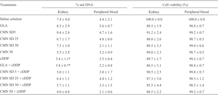
Cell viability observed in the kidney and peripheral blood was greater than 70% in all of the analysed groups, in accordance with recommendations for performing a comet assay analysis (Azqueta and Collins, 2013), as shown in Table 2. In the comet assay, the extent of DNA damage was assessed based on the tail intensity parameter (% tail DNA). No genotoxic effects of CMN SD, unmodified CMN or GLA were observed in kidney or peripheral blood cells (Table 2). The results regarding % tail DNA observed in the animals treated with cDDP revealed a significant decrease in DNA migration compared to the saline solution group in renal tissue, but not in peripheral blood (Table 2). The re-sults for the cDDP group indicated the formation of crosslinks with DNA. Treatment with CMN SD or unmodi-fied CMN in association with cDDP did not induce signifi-cant alterations compared to the cDDP-only group in the comet assay.
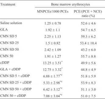
solu-tion group (Table 3), indicating that none of the treatments altered the rate of cell division in bone marrow. Table 3 shows the effect of CMN SD or unmodified CMN, either combined with cDDP or not, on the formation of micro-nuclei. CMN SD, unmodified CMN and the GLA mixture did not induce micronucleus formation. In contrast, cDDP treatment significantly increased the frequency of MNPCE compared to the saline solution group. CMN SD (at 5, 25 and 50 mg/kg b.w.) and unmodified CMN (50 mg/kg b.w.)
significantly reduced the formation of cDDP-induced micronuclei (p < 0.05). This effect occurred to a similar ex-tent under treatment with CMN SD and CMN at a dose of 50 mg/mL.
CMN SD attenuates cDDP-induced oxidative stress
Oxidative stress was evaluated by measuring the con-centrations of TBARS and GSH in renal tissue 24 hours af-ter cDDP administration. When adminisaf-tered alone, CMN Table 1- Evaluation of the variation of mass gain in rats after subacute treatment with CMN SD, unmodified CMN, cDDP and their associations.
Treatments Body weight (g)amean±standard deviation Body weight (g)bmean±standard deviation Kidney/body weight (%)
Saline solution 29.7±6.4 11.3±2.1 0.98±0.03
GLA 31.3±5.6 9.3±4.7 1.01±0.04
CMN SD 5 31.2±6.6 11.0±2.8 0.93±0.05
CMN SD 25 28.0±4.6 9.2±7.3 0.93±0.09
CMN SD 50 25.5±8.2 8.5±1.0 1.00±0.11
CMN 50 31.7±6.6 8.2±2.4 0.96±0.05
cDDP 34.0±4.3 0.6±3.9* 1.03±0.04
GLA + cDDP 27.5±6.1 0.8±2.2* 1.02±0.11
CMN SD 5 + cDDP 29.7±3.8 1.8±1.5* 0.97±0.05
CMN SD 25 + cDDP 26.7±5.8 2.3±4.8* 0.97±0.06
CMN SD 50 + cDDP 23.0±5.3 0.2±4.5* 1.00±0.09
CMN 50 + cDDP 31.8±4.7 0.1±2.5* 0.90±0.06
cDDP: cisplatin (6 mg/kg b.w.); CMN: curcumin (5, 25 and 50 mg/kg b.w.); GLA: gelucire®50-13/aerosil®; SD: solid dispersion. a: Interval 1- variation in body mass, in grams (g), between days 1 and 4 of the experimental period. b: Interval 2 -variation in body mass, in grams (g), between day 4 and 5 of the experimental period. The results represent the mean±standard deviation for each group (six animals/group). *Significantly different from saline solution group, assessed by ANOVA and Tukey’spost hoctest (p < 0.05).
Table 2- Tail Intensity (% tail DNA) and cell viability (expressed as % in relation to saline solution group) in cells of kidney and peripheral blood after subacute treatment with CMN SD, unmodified CMN, cDDP and their associations, analyzed in the comet assay.
Treatments % tail DNA Cell viability (%)
Kidney Peripheral blood Kidney Peripheral blood
Saline solution 7.4±4.0 4.4±2.1 100.0±0.0 100.0±0.0
GLA 8.3±2.9 2.6±0.7 88.5±1.9 98.8±0.7
CMN SD5 9.4±2.8 4.7±1.6 91.2±2.4 99.2±0.7
CMN SD 25 6.7±1.7 4.8±0.6 88.0±2.6 98.7±0.5
CMN SD 50 7.5±3.0 2.1±1.1 88.5±3.3 99.0±0.6
CMN 50 5.5±2.8 3.2±0.9 89.0±2.3 98.7±0.5
cDDP 3.4±1.1* 2.5±0.4 89.7±1.7 99.2±0.7
GLA + cDDP 3.8±0.7* 2.2±0.8 86.5±3.1 98.8±0.7
CMN SD 5 + cDDP 5.0±1.1 3.0±1.7 90.5±2.5 99.8±0.7
CMN SD 25 + cDDP 6.4±1.1 4.4±1.2 87.3±3.6 98.5±1.2
CMN SD 50 + cDDP 5.7±1.1 3.3±1.5 85.5±4.4 98.5±1.4
CMN 50 + cDDP 4.0±0.8 2.1±0.6 88.5±2.2 99.2±0.7
SD and unmodified CMN did not alter the GSH and TBARS concentrations detected in renal tissue (Table 4). cDDP significantly increased TBARS levels compared to the saline solution group but did not alter GSH levels. CMN SD or unmodified CMN, administered together with cDDP, was able to maintain the TBARS levels observed in the saline solution group (p > 0.05) (Table 4). There was no significant difference between the groups treated with GLA together with cDDPvs.cDDP alone.
Tp53mRNA levels are affected by CMN SD
Figure 1 shows the effects of CMN SD (50 mg/kg b.w.) and unmodified CMN (50 mg/kg b.w.), either alone or in association with cDDP (6 mg/kg b.w.), on theTp53
mRNA levels in kidney tissue. The results showed that CMN SD, unmodified CMN and cDDP did not alterTp53
gene expression compared to the saline solution group. However, when either CMN SD or unmodified CMN was administered together with cDDP, Tp53 expression was up-regulated compared to saline solution group. There was no difference in the levels ofTp53mRNA in kidney cells when comparing the CMN SD and unmodified CMN groups.
Discussion
With the objective of evaluating whether CMN SD could induce chromosomal damage or interfere with the recognized antioxidant and antigenotoxic properties of un-Table 3 - Frequency of micronucleated polychromatic erythrocytes
(MNPCE) and the percentage (%) of PCE/(PCE + NCE) in 500 erythro-cytes in the bone morrow of Wistar rats treated with CMN SD, unmodified CMN, cDDP or their associations, analyzed in the micronucleus test.
Treatment Bone marrow erythrocytes
MNPCEs/1000 PCEs PCE/(PCE + NCE) ratio (%)
Saline solution 1.25±0.78 52.6±4.6
GLA 1.92±1.1 54.7±6.5
CMN SD 5 2.25±1.13 59.3±6.2
CMN SD 25 1.5±0.82 53.4±10.4
CMN SD 50 2.42±1.09 43.2±6.0
CMN 50 1.91±1.27 55.7±8.8
cDDP 13.25±3.51* 49.9±5.6
GLA + cDDP 12.75±3.32* 44.8±4.9
CMN SD 5 + cDDP 6.88±1.77*# 51.8±5.9
CMN SD 25 + cDDP 5.33±2.98*# 53.9±8.3
CMN SD 50 + cDDP 6.42±3.12*# 51.1±3.0
CMN 50 + cDDP 7.08±3.04*# 51.0±7.5
Saline solution or cDDP was administered intraperitoneally 30 min after the last gavage of CMN SD or unmodified CMN. cDDP: cisplatin (6 mg/kg b.w.); CMN: curcumin (5, 25 and 50 mg/kg b.w.); GLA: gelucire®50-13/aerosil®; SD: solid dispersion. The results represent the mean±standard deviation for each group (six animals/group).*Signifi-cantly different from saline solution group. #Signifianimals/group).*Signifi-cantly different from cDDP group, assessed by ANOVA and Tukey’spost hoctest (p < 0.05).
Table 4- Evaluation of reduced glutathione (GSH) and thiobarbituric-acid-reactive substances (TBARS) in the kidney of Wistar rats treated with CMN SD, unmodified CMN, cDDP or their associations.
Treatments GSH (nmol/mg
protein)
TBARS (nmol/mg protein)
Saline solution 18.9±0.4 0.249±0.011
GLA 17.7±2.6 0.226±0.023
CMN SD5 14.2±1.3 0.237±0.018
CMN SD 25 15.3±1.1 0.232±0.026
CMN SD 50 17.2±1.7 0.247±0.018
CMN 50 14.4±1.2 0.236±0.018
cDDP 15.2±3.5 0.302±0.026*
GLA+ cDDP 18.5±0.6 0.286±0.014*
CMN SD 5 + cDDP 16.6±1.3 0.215±0.009#
CMN SD 25 + cDDP 14.0±1.7 0.200±0.011#
CMN SD 50 + cDDP 19.1±2.5 0.218±0.018#
CMN 50 + cDDP 17.0±1.4 0.222±0.064#
Saline solution or cDDP was administered intraperitoneally 30 min after the last gavage of CMN SD or unmodified CMN. cDDP: cisplatin (6 mg/kg b.w.); CMN: curcumin (5, 25 and 50 mg/kg b.w.); GLA: gelucire®50-13/aerosil®; SD: solid dispersion. The results represent the mean±standard deviation for each group (six animals/group).*Signifi-cantly different from saline solution and GLA groups. #Signifianimals/group).*Signifi-cantly dif-ferent from cDDP group, assessed by ANOVA and Tukey’spost hoctest (p < 0.05).
modified CMN, we performed anin vivocomparative anal-ysis between CMN SD and unmodified CMN, by measur-ing DNA damage, evaluatmeasur-ing oxidative stress and analysing Tp53 mRNA levels. Our results showed that CMN SD decreased chromosomal damage induced by cDDP, up-regulated Tp53expression when administered together with cDDP, and attenuated cDDP-induced oxida-tive stress. There were no significant differences observed between the effects of CMN SD and unmodified CMN for any of the parameters evaluated in this study.
cDDP was used as the damage-inducing agent in this study due its genotoxic and nephrotoxic effects, and the kidney was evaluated as a target organ. The genotoxic mechanisms of cDDP involve chromosomal damage, as demonstrated by the induction of micronuclei (Guptaet al., 2011); the formation of cDDP-DNA crosslinks, as shown by the decrease in the percentage of DNA in the tail (Stang and Witte, 2009), and the regulation ofTp53mRNA and p53 protein levels (Yuanet al., 2011). Thein vivo mecha-nisms of cDDP nephrotoxicity are mainly related to the in-duction of oxidative stress: cDDP increases free radical production (Ognjanovicet al., 2012) and decreases antioxi-dant enzyme activity (Badaryet al., 2005).
According to Wolfseggeret al.(2009), the changes in total mass of an animal and the relationship between organ weights and total mass of an animal can be used as an indi-cation of the toxicity of a compound under evaluation. Ani-mals treated with CMN SD and unmodified CMN showed no difference in total mass compared to the saline solution group. However, there was a significant reduction in body mass of the rats treated with cDDP (6 mg/kg b.w.) com-pared to the saline solution group. Other studies in rodents have also shown a decrease in body mass following cDDP administration at the same dose as applied in this study (Zhanget al., 1999). This is most likely due to cytotoxic ef-fects of cDDP.
Assessment of chromosomal damage was performed via the micronucleus test in erythrocytes from bone mar-row, and DNA damage was evaluated using the comet as-say in kidney and peripheral blood samples. These two tests are frequently employed to evaluate the genotoxic and mutagenic effects of physical or chemical agents, where by the comet assay can detect initial lesions in DNA, and the micronucleus assay can detect chromosomal breaks and losses (Bowenet al., 2011; Collins, 2015). DNA lesions de-tected by the comet assay can be single- and double-strand breaks, alkaline-labile sites and DNA and DNA-protein crosslinks. Single- or double-strand breaks and al-kaline-labile sites are further identified in the comet assay as an increase in DNA migration, while DNA-DNA and DNA-protein crosslinks are detected as a decrease in DNA migration (Nesslanyet al., 2007).
CMN SD did not induce DNA or chromosomal dam-age in any of the analysed tissues, suggesting that CMN SD did not induce genotoxicity in these. Regarding the
anti-genotoxicity action of CMN SD, we saw that it reduced micronucleus formation in the bone marrow cells of rats ex-posed to cDDP, but did not reduce the formation of cDDP-DNA crosslinks observed in kidney. The antigenotoxicity of CMN SD was similar to that of unmodified CMN in this study, a finding that is comparable to that reported in other studies involving CMN (Mendonçaet al., 2009; Celiket al., 2013). These results furthermore suggest that the pro-tective mechanism of CMN SD is not related to a reduction in the formation of cDDP-DNA crosslinks, since CMN SD did not interfere with the mechanism of cDDP genoto-xicity. It seems, however, related with the reduction of cDDP-induced breaks and loss of chromosomes.
Various studies have demonstrated the relevance of oxidative stress in cDDP-induced cellular damage. Oxida-tive stress can cause DNA damage, resulting in strand breaks, alterations in gene expression, and mutations (Cookeet al., 2003). Some antioxidant agents may exert their protective effects by increasing the capacity of cellu-lar antioxidant defense systems, or via the sequestration of reactive species (Costaet al., 2012), and the protective ef-fects of CMN, as well as its antigenotoxicity activity are of-ten related to its antioxidant properties.
In this study, the evaluated oxidative stress parame-ters were the GSH concentration and TBARS formation in renal tissue. It is generally accepted that the mechanism by which cDDP induces oxidative stress in renal tissue in-volves the induction of lipid peroxidation (Ognjanovicet al., 2012), and the antioxidant properties of CMN are re-lated to its ability to modulate the concentrations of GSH and TBARS (Biswaset al., 2005; Kauret al., 2006). Some findings suggest that CMN could be useful in reducing the nephrotoxicity of cDDP (Swamyet al., 2012), and our re-sults showed that CMN SD, when administered together with cDDP, was able to maintain the TBARS levels ob-served in the saline solution group. These results suggest that CMN SD, processed via spray dry technology, can pro-tect against cDDP-induced lipid peroxidation in the kidney and maintain TBARS at basal levels.
In addition, we assessed the expression ofTp53 in kidney cells because the involvement of p53 protein has been implicated in cDDP toxicity in normal cells, as ob-served in nephrotoxicity (Jiang and Dong, 2008). Like un-modified CMN, CMN SD increased the mRNA levels of
Tp53when administered together with cDDP in renal tis-sue, compared to the saline group; and they did not alter the
been of great relevance for the development of new drugs (Hornberget al., 2014). Our findings demonstrate that the technique of producing a solid dispersion containing CMN did not affect the antigenotoxic effects of this compound, and CMN SD showed effects similar to those of unmodi-fied CMN for all of the evaluated parameters. In conclu-sion, CMN SD maintained the protective and antioxidant effects of unmodified CMN with the advantage of being chemically water soluble. Thus, the optimization of the physical and chemical properties of CMN SD may increase its potential for therapeutic use.
Acknowledgments
This research was supported by CAPES (Coorde-nação de Aperfeiçoamento de Pessoal de Ensino Superior), CNPq (Conselho Nacional de Desenvolvimento Científico e Tecnológico) and the São Paulo Research Foundation (FAPESP), grants #2008/53947-7 and #2008/10482-4. LMM was sponsored by a fellowship from the São Paulo Research Foundation FAPESP (grant #2008/10482-4).
References
Aggarwal BB and Harikumar KB (2009) Potential therapeutic ef-fects of curcumin, the anti-inflammatory agent, against neurodegenerative, cardiovascular, pulmonary, metabolic, autoimmune and neoplastic diseases. Int J Biochem Cell Biol 41:40-59.
Aggarwal BB, Yuan W, Li S and Gupta SC (2013) Curcumin-free turmeric exhibits anti-inflammatory and anticancer activi-ties: Identification of novel components of turmeric. Mol Nutr Food Res 57:1529-1542.
Antunes LM, Araujo MC, Dias FL and Takahashi CS (1999) Modulatory effects of curcumin on the chromosomal dam-age induced by doxorubicin in Chinese hamster ovary cells. Teratog Carcinog Mutagen 19:1-8.
Antunes LM, Darin JD and Bianchi MdL (2001) Effects of the an-tioxidants curcumin or selenium on cisplatin-induced nephrotoxicity and lipid peroxidation in rats. Pharmacol Res 43:145-150.
Azqueta A and Collins AR (2013) The essential comet assay: A comprehensive guide to measuring DNA damage and repair. Arch Toxicol 87:949-968.
Badary OA, Abdel-Macsoud S, Ahmed WA and Owieda GH (2005) Naringenin attenuates cisplatin nephrotoxicity in rats. Life Sci 76:2125-2135.
Barzegar A and Moosavi-Movahedi AA (2011) Intracellular ROS protection efficiency and free radical-scavenging activity of curcumin. PLoS One 6:e26012.
Biswas SK, McClure D, Jimenez LA, Megson IL and Rahman I (2005) Curcumin induces glutathione biosynthesis and in-hibits NF-kappaB activation and interleukin-8 release in al-veolar epithelial cells: Mechanism of free radical scaveng-ing activity. Antioxid Redox Signal 7:32-41.
Bowen DE, Witwell JH, Lillford L, Henderson D, Kidd D, Mc Garry S, Pearce G, Beevers C and Kirkland DJ (2011) Eval-uation of a multi-endpoint assay in rats, combining the bone-marrow micronucleus test, the Comet assay and the
flow-cytometric peripheral blood micronucleus test. Mutat Res 722:7-19.
Buege JA and Aust SD (1978) Microsomal lipid peroxidation. Methods Enzymol 52:302-310.
Celik A, Eke D, Ekinci SY and Yildirim S (2013) The protective role of curcumin on perfluorooctane sulfonate-induced genotoxicity: Single cell gel electrophoresis and micronu-cleus test. Food Chem Toxicol 53:249-255.
Collins AR (2015) The comet assay: A heavenly method! Muta-genesis 30:1-4.
Cooke MS, Evans MD, Dizdaroglu M and Lunec J (2003) Oxida-tive DNA damage: Mechanisms, mutation, and disease. FASEB J 17:1195-1214.
Costa LA, Badawi A and El-Sohemy A (2012) Nutrigenetics and modulation of oxidative stress. Ann Nutr Metab 60:27-36. Duvoix A, Blasius R, Delhalle S, Schenekenburger M, Morceau
F, Henry E, Dicato M and Diederich M (2005) Chemo-preventive and therapeutic effects of curcumin. Cancer Lett 223:181-190.
Esatbeyoglu T, Ulbrich K, Rehberg C, Rohn S and Rimbach G (2015) Thermal stability, antioxidant, and anti-inflamma-tory activity of curcumin and its degradation product 4-vinyl guaiacol. Food Funct 6:887-893.
Ganta S, Devalapally H and Amiji M (2010) Curcumin enhances oral bioavailability and anti-tumor therapeutic efficacy of paclitaxel upon administration in nanoemulsion formula-tion. J Pharm Sci 99:4630-4641.
Goel A, Kunnumakkara AB and Aggarwal BB (2008) Curcumin as “Curecumin”: From kitchen to clinic. Biochem Phar-macol 75:787-809.
Gupta V, Agrawal RC and Trivedi P (2011) Reduction in cisplatin genotoxicity (micronucleus formation) in non target cells of mice by protransfersome gel formulation used for manage-ment of cutaneous squamous cell carcinoma. Acta Pharm 61:63-71.
Hartree EF (1972) Determination of protein: A modification of the Lowry method that gives a linear photometric response. Anal Biochem 48:422-427.
Hornberg JJ, Laursen M, Brenden N, Persson M, Thougaard AV, Toff DB and Mow T (2014) Exploratory toxicology as an in-tegrated part of drug discovery. Part I: Why and how. Drug Discov Today 19:1131-1136.
Jiang M and Dong Z (2008) Regulation and pathological role of p53 in cisplatin nephrotoxicity. J Pharmacol Exp Ther 327:300-307.
Kaur G, Tirkey N, Bharrhan S, Chanana V, Rishi P and Chopra K (2006) Inhibition of oxidative stress and cytokine activity by curcumin in amelioration of endotoxin-induced experimen-tal hepatoxicity in rodents. Clin Exp Immunol 145:313-321. Livak KJ and Schmittgen TD (2001) Analysis of relative gene
ex-pression data using real-time quantitative PCR and the 2(-Delta Delta C(T)) method. Methods 25:402-408. Mendonça LM, Dos Santos GC, Antonucci GA, Dos Santos AC,
Bianchi ML and Antunes LM (2009) Evaluation of the cytotoxicity and genotoxicity of curcumin in PC12 cells. Mutat Res 675:29-34.
Nair VD, Yuen T, Olanow CW and Sealfon SC (2004) Early sin-gle cell bifurcation of pro- and antiapoptotic states during oxidative stress. J Biol Chem 279:27494-27501.
Nesslany F, Zennouche N, Simar-Meintieres S, Talahari I, Nkili-Mboui EN and Marzin D (2007) In vivoComet assay on iso-lated kidney cells to distinguish genotoxic carcinogens from epigenetic carcinogens or cytotoxic compounds. Mutat Res 630:28-41.
Ognjanovic BI, Djordjevic NZ, Matic MM, Obradovic JM, Mladenovic JM, Stajin AS and Saicic ZS (2012) Lipid peroxidative damage on Cisplatin exposure and alterations in antioxidant defense system in rat kidneys: A possible pro-tective effect of selenium. Int J Mol Sci 13:1790-1803. Olfert ED and McWilliam AA (1993) Guide to the care and use of
experimental animals. 2nd edition. Canadian Council on Animal Care, Ottawa, 211 p.
Premkumar K, Kavitha S, Santhiya ST, Ramesh AR and Suwan-teerangkul J (2004) Interactive effects of saffron with garlic and curcumin against cyclophosphamide induced genoto-xicity in mice. Asia Pac J Clin Nutr 13:292-294.
Schmid W (1975) The micronucleus test. Mutat Res 31:9-15. Sedlak J and Lindsay RH (1968) Estimation of total,
protein-bound, and nonprotein sulfhydryl groups in tissue with Ellman’s reagent. Anal Biochem 25:192-205.
Seo SW, Han HK, Chun MK and Choi HK (2012) Preparation and pharmacokinetic evaluation of curcumin solid dispersion us-ing Solutol(R) HS15 as a carrier. Int J Pharm 424:18-25. Serpeloni JM, Batista BL, Angeli JP, Barcelos GR, Bianchi Mde
L, Barbosa FJ and Antunes LM (2013) Antigenotoxic prop-erties of chlorophyll b against cisplatin-induced DNA dam-age and its relationship with distribution of platinum and magnesium in vivo. J Toxicol Environ Health 76:345-353. Singh NP, McCoy MT, Tice RR and Schneider EL (1988) A
sim-ple technique for quantitation of low levels of DNA damage in individual cells. Exp Cell Res 175:184-191.
Singh U, Barik A, Singh BG and Priyadarsini KI (2011) Reactions of reactive oxygen species (ROS) with curcumin analogues: Structure-activity relationship. Free Radic Res 45:317-325. Somparn P, Phisalaphong C, Nakornchai S, Unchern S and Morales NP (2007) Comparative antioxidant activities of curcumin and its demethoxy and hydrogenated derivatives. Biol Pharm Bull 30:74-78.
Sreejayan and Rao MN (1994) Curcuminoids as potent inhibitors of lipid peroxidation. J Pharm Pharmacol 46:1013-1016. Stang A and Witte I (2009) Performance of the comet assay in a
high-throughput version. Mutat Res 675:5-10.
Swamy AV, Gulliaya S, Thippeswamy A, Koti BC and Manjula DV (2012) Cardioprotective effect of curcumin against doxorubicin-induced myocardial toxicity in albino rats. In-dian J Pharmacol 44:73-77.
Tice RR, Agurell E, Anderson D, Burlinson B, Hartmann A, Kobayashi H, Miyamae Y, Rojas E, Ryu JC and Sasaki YF (2000) Single cell gel/comet assay: Guidelines for in vitro and in vivo genetic toxicology testing. Environ Mol Mutagen 35:206-221.
Vasconcelos T, Sarmento B and Costa P (2007) Solid dispersions as strategy to improve oral bioavailability of poor water sol-uble drugs. Drug Discov Today 12:1068-1075.
Wahlstrom B and Blennow G (1978) A study on the fate of curcumin in the rat. Acta Pharmacol Toxicol 43:86-92. Wolfsegger MJ, Jaki T, Dietrich B, Kunzler JA and Barker K
(2009) A note on statistical analysis of organ weights in non-clinical toxicological studies. Toxicol Appl Pharmacol 240:117-122.
Yallapu MM, Jaggi M and Chauhan SC (2012) Curcumin nano-formulations: A future nanomedicine for cancer. Drug Discov Today 17:71-80.
Yu WG, Xu G, Ren GJ, Xu X, Yuan HQ, Qi XL and Tian KL (2011) Preventive action of curcumin in experimental acute pancreatitis in mouse. Indian J Med Res 134:717-724. Yuan JM, Li XD, Liu ZY, Hou GQ, Kang JH, Huang DY and Du
SX (2011) Cisplatin induces apoptosis via upregulating Wrap53 in U-2OS osteosarcoma cells. Asian Pac J Cancer Prev 12:3465-3469.
Zhang JG, Viale M, Esposito M and Lindup WE (1999) Tiopronin protects against the nephrotoxicity of cisplatin in the rat. Hum Exp Toxicol 18:713-717.
Zhou H, Beevers CS and Huang S (2011) The targets of curcumin. Curr Drug Targets 12:332-347.
Associate Editor: Daisy Maria Fávero Salvadori