Vol.59: e16160151, January-December 2016 http://dx.doi.org/10.1590/1678-4324-2016160151
ISSN 1678-4324 Online Edition
BRAZILIAN ARCHIVES OF BIOLOGY AND TECHNOLOGY
A N I N T E R N A T I O N A L J O U R N A L
Antioxidant Status, Lipid Peroxidation and
Testis-histoarchitecture Induced by Lead Nitrate and Mercury
Chloride in Male Rats
Hatice Bas
1*;
Suna Kalender
2.
1Bozok University Faculty of Arts and Science – Department of Biology, Turkey;2Gazi University, Gazi Education Faculty, Science Education, Ankara, Turkey;
ABSTRACT
This study was done to evaluate the effects of lead nitrate and mercury chloride in testis tissues of Wistar rats. Lead nitrate and mercury chloride are widely used heavy metals in industry. Oral lead and mercury administrations to adult male rats at doses 45 mg/kg bw and 0.02 mg/kg bw, respectively for 4 weeks caused a significant increasing in MDA levels and antioxidant enzyme activities (SOD, CAT, GPx and GST). The MDA levels and acivities of antioxidant enzymes was lower in rats that were administrated by lead nitrate than mercury chloride treated group. Light microscopic analyses revealed that lead nitrate and mercury chloride induced numerous histopathological changes in testis tissues of rats. Histopathological observations of the testis tissues showed that mercury chloride caused more harmful effects than lead nitrate, too. The results indicate that lead nitrate and mercury chloride have reproductive toxicity, in male rats at the tested doses. The effect which we observed applying the lead nitrate and mercury chloride together, was more greater than when we used them alone.
Key Words: Antioxidant enzymes, lead, mercury, oxidative stress, testis
*
Authors for correspondence: htc.haticebas@gmail.com
INTRODUCTION
Humans and animals were exposed to
environmental pollutants at different stages of life
[1]. Environmental pollutants such as heavy metals
are the main factors responsible for oxidative stress.Intoxication by heavy metals constitutes
serious threats to human health [2]. Järup [3]
indicated that heavy metals such as lead, cadmium and mercury may evoke behavioral disturbances, learning and concentration difficulties and diminished intellectual capacity.
The highest lead intake in adults is through lungs
and gastrointestinal tract [4]. Lead accumulates
primarily in the bones, liver and kidney of animals. Depending on the level of exposure, lead can harm the nervous and immune systems, cause reproductive impairment and alter calcium
homeostasis [5]. Mercury intoxication can result
from inhalation, ingestion, or absorption through the skin. Previous studies demonstrated that mercury caused renal toxicity, neurotoxicity,
reproductive toxicity and hematotoxic effects [6].
Mercury, may cause accidental and occupational exposures and serious damage in various organs in
human and experimental animals [7].
Oxidative stress is induced by reactive oxygen species (ROS). Extreme levels of ROS can negatively impact on sperm quality, motility and increased sperm DNA damage. Therefore, equilibrium is required between the generation of ROS and antioxidant scavenging activity in the
male reproductive organs [8]. Cells have developed
defence mechanisms, including enzymatic
systems, against oxidative damage. Enzymatic defence systems involve enzymes such as superoxide dismutase (SOD), catalase (CAT), glutathione peroxidase (GPx) and
glutathione-S-transferase (GST) [9]. SOD catalyzes the
dismutation of superoxide radicals to hydrogen
peroxide and molecular oxygen [10].CAT catalyzes
the reduction of H2O2 to water and oxygen and
protects the cell against H2O2 induced oxidative
damage [11]. The GPx and GST catalyse the
reduction of H2O2 to H2O and the conjugation of
electrophilic substrates to the thiol group of GSH
so it produces less toxic forms, respectively [12,13].
Male germ cells are more susceptible to oxidative stress than other cells, because they have higher polyunsaturated fatty acids in their membranes than somatic cells. Oxidative stress plays an important role in the etiology of defective sperm
formation, function, sperm count profile and male
infertility [14]. So, in this study, we investigated
the possible adverse effects of lead nitrate and mercury chloride on the testis tissue of rats in terms of oxidative stress. For this purpose, rats were given lead nitrate and mercury chloride by oral gavage for 4 weeks, then malondialdehyde (MDA) levels, SOD, CAT, GPx and GST activities also histopathological changes of testis tissues were assessed.
MATERIALS AND METHODS
CHEMICALS
The heavy metals lead nitrate and mercury chloride, and all the other chemicals were purchased from Sigma Aldrich. Lead nitrate and mercury chloride were dissolved in distilled water [15,16].
ANIMALS
Sexually mature male Wistar rats were obtained from the Gazi University Laboratory Animals Growing and Experimental Research Center. The rats were acclimated to the laboratory environment
(18-22 oC and a 12 h light/ dark cycle) for 10 days
before use. All animals were housed in plastic cages and given standard laboratory chow and water ad libitum.
EXPERIMENTAL PROCEDURE
The animal treatment protocol was approved by the Gazi University Animal Experiments Local
Ethics Committee (Protocol no: G.U.ET – 13.011).
All animal experiments were performed
accordancing to the international guidelines for care and use of laboratory animals.
Rats were randomly divided into four treatment groups, each with six rats. The first group served as a control and was administered 1ml/kg b.w (body weight) distilled water. The second group was dosed with lead nitrate dissolved in distilled water by gavage at the dose level of 45 mg/kg b.w
(1/50 LD50) [
16,17
]. The third group was treated with mercury chloride at the dose level of 0.02
mg/kg b.w (1/50 LD50) [
15
At the end of the 4th week, the rats were weighed and sacrificed and the testis tissues were excised.
HISTOLOGICAL EXAMINATION
The organs were fixed in Bouin’s solution. Then
samples were processed using a graded ethanol series and embedded in paraffin. Paraffin sections
4-6 μ thick were stained with haematoxylin and
eosin for histopathological examination and photographed using a light microscope (Olympus BX51, Tokyo, Japan) with an attached camera (Olympus E-330, Olympus Optical Co. Ltd., Japan). Ten slides were prepared from each testis.All sections were evaluated for the degree of separating of cells from basal region, edema in
interstitial tissue, degenerative changes in
seminiferous tubules and decreasing number of spermatogenic cells. The severity of changes was assessed for each slide by scoring using a scaleof no (-), mild (+), moderate (++) and severe (+++) damage.
MEASUREMENT OF MDA LEVELS AND ANTIOXIDANT ENZYME ACTIVITIES
Testis MDA levels were assayed using the thiobarbituric acid test as described by Ohkawa et
al. [18] at 532 nm. The level of MDA is defined as
nmol/mgprotein.
The SOD activity was estimated in tissue homogenate by the method of Marklund and
Marklund [19] at 440 nm. Testis GST activity was
determined according to the procedure of Habig et
al. [20] at 340 nm. CAT and GPx activities were
assayed by the method of Aebi [21], Paglia and
Valentine [22] respectively at 240 nm. The
activities of GST and GPx were defined as nmol/mgprotein, CAT activity was defined as μmol/mgprotein, SOD activity was defined as U/mgprotein.
STATISTICAL ANALYSIS
The data are expressed as the mean ± SD and were analyzed by a one-way analysis of variance (ANOVA) test followed by Tukey. The level of significance was set at P < 0.05. The program SPSS for Windows 20.0 was used to analyze the data. Dependence of various data sets was characterized by calculating their Pearson’s correlation. The closer the coefficient is to either -1 or -1 the stronger the correlation between the variables.
RESULTS
Malondialdehyde (MDA) Levels
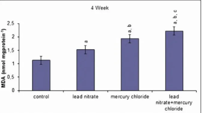
When the lead nitrate and mercury chloride treated groups were compared with the control group at
the end of 4th week, there were significantly
increasing in the MDA levels in the testis tissues. The MDA levels were statistically significantly higher in the mercury chloride treated group compared to lead nitrate treated group. Treated with combination of lead nitrate and mercury chloride caused more harmfull effects than use of them alone (P < 0.05, Fig. 1).
Figure 1: Effects of subacute treatment of lead nitrate and mercury chloride on MDA levels in the testis tissues of rats. Each bar represents mean±SD of six animals in each group. Significance at P<0.05. a
Comparison of control and other groups. bComparison of lead nitrate group and other groups. cComparison of mercury chloride group and other groups
Superoxide Dismutase (SOD) Activity
Figure 2: Effects of subacute treatment of lead nitrate and mercury chloride on SOD activities in the testis tissues of rats. Each bar represents mean±SD of six animals in each group. Significance at P<0.05. a
Comparison of control and other groups. bComparison of lead nitrate group and other groups. cComparison of mercury chloride group and other groups
Catalase (CAT) Activity
A significant increasing in CAT activity was observed in the lead nitrate and mercury chloride treated groups compared with the control group. Also significant increasing were observed in the mercury chloride treated group compared with the lead nitrate treated group in testis tissues. Lead nitrate+mercury chloride group has the highest CAT activity between all groups (P < 0.05, Fig. 3).
Figure 3: Effects of subacute treatment of lead nitrate and mercury chloride on CAT activities in the testis tissues of rats. Each bar represents mean±SD of six animals in each group. Significance at P<0.05. a
Comparison of control and other groups. bComparison of lead nitrate group and other groups. cComparison of mercury chloride group and other groups
Glutathione Peroxidase (GPx) Activity
The lead nitrate and mercury chloride groups showed significantly increasing in the GPx activity compared with the control group. We observed more increasing in mercury chloride treated group than lead nitrate treated group in testis tissues. The highest GPx activity was observed in lead nitrate + mercury chloride group among all the groups (P < 0.05, Fig. 4).
Figure 4: Effects of subacute treatment of lead nitrate and mercury chloride on GPx activities in the testis tissues of rats. Each bar represents mean±SD of six animals in each group. Significance at P<0.05. a
Comparison of control and other groups. bComparison of lead nitrate group and other groups. cComparison of mercury chloride group and other groups
Glutathione-S-transferase (GST) Activity
GST activity was statistically significantly increased in the lead nitrate and mercury chloride traeted groups compared to the control group. Mercury chloride increased the GST activity more than lead nitrate. A significant increasing in GST activity were observed in the lead nitrate+mercury chloride group compared with the all groups. (P < 0.05, Fig. 5).
Figure 5: Effects of subacute treatment of lead nitrate and mercury chloride on GST activities in the testis tissues of rats. Each bar represents mean±SD of six animals in each group. Significance at P<0.05. a
Comparison of control and other groups. bComparison of lead nitrate group and other groups. cComparison of mercury chloride group and other groups
Correlation Coefficients
Correlation coefficients illustrate quantitative measure of some type of correlation and dependence, meaning statistical relationships between two or more variables or observed data
values. Correlation coefficients (RP) between
MDA concentration and other parameters were shown in Table 1. The closer the coefficient is to either -1 or 1 the stronger the correlation between the variables. Strong positive correlation was observed between MDA values and activities of antioxidant enzymes.
Table 1:Correlation coefficients (RP) between MDA concentration and other parameters. RP> 0.9 values are in bold, 0.9 >RP> 0.8 values are in italics.
Malondialdehyde
SOD 0.982a
GPx 0.914a
GST 0.876a
a Significant correlation at p < 0.05.
Histological Examination
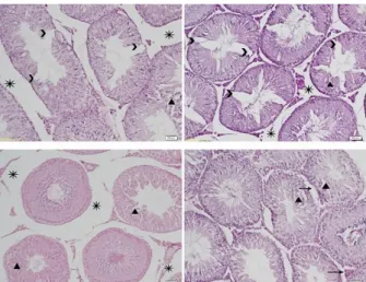
The histopathological assessments performed on testicular sections from the control and treatment groups are presented in Figures 6, 7, 8 and 9. The testicular architecture of the control animals was
normal, as characterized by complete
spermatogenesis (Fig. 6). Distinct
histopathological abnormalities were observed in the testis tissues of lead nitrate and mercury chloride treated rats (Figs. 7 and 8). An increment in the separating of cells from basal region, edema in interstitial tissue, degenerative changes in seminiferous tubules and decreasing number of spermatogenic cells were observed in the testis tissues in lead nitrate + mercury chloride treated group (Fig. 9). Table 2 shows the percentages of treated rats that showed abnormalities observed in the testis tissues.
Figure 6:Testicular sections of control rats showing seminiferous tubules (S) and interstitial tissue (I), x200, H&E
Figure 7:Testicular sections of lead nitrate treated rats showing separating of cells from basal region (→), edema in interstitial tissue (*), degenerative changes in
seminiferous tubules (▲) and decreasing number of
spermatogenic cells (>), x200, H&E
Figure 8: Testicular sections of mercury chloride treated rats showing separating of cells from basal
region (→), edema in interstitial tissue (*), degenerative changes in seminiferous tubules (▲) and decreasing
number of spermatogenic cells (>), x200, H&E
Figure 9: Testicular sections of lead nitrate+mercury chloride treated rats showing separating of cells from
basal region (→), edema in interstitial tissue (*), degenerative changes in seminiferous tubules (▲) and
Table 2: Grading of the histopathological changes in the testis sections
Groups
Pathology
Control Lead nitrate Mercury chloride Lead nitrate +
Mercury chloride
separating of cells from basal region
- + ++ ++
edema in interstitial
tissue
- + ++ +++
degenerative changes in seminiferous tubules
- ++ ++ +++
decreasing number of spermatogenic cells
- + + +++
Scoring was done as follows: (-) none, (+) mild, (++) moderate, (+++) severe
DISCUSSION
There are many studies which have indicated that
lead and mercury exposure could cause
biochemical and physiological dysfunctions in
experimental animals and humans [23,24]. Recently,
oxidative stress has become the focus of interest as
a potential cause of male infertility [8].Reactive
oxygen species (ROS) are the subject of intense research because of their effects on cellular
pathogenesis [25]. ROS are supposed to play one of
the key roles in the development of testis toxicity which is a sporadic and challenging issue for
pharmaceutical drug development [26,27]. So, we
studied the harmfull effects of lead nitrate and mercury chloride on testis tissues in terms of oxidative stress.
The measure of MDA could be useful diagnostic tool for estimation of oxidative stress. MDA is one of the products of peroxidized polyunsaturated fatty acids. So, increased MDA level is an
important indicator of lipid peroxidation
[9,28,29].Previous studies show that heavy metals increased MDA levels in several rat tissues [24,30,31]. In our study, MDA levels also increased in the lead nitrate and mercury chloride treated groups. So, it might be due to lipid peroxidation effects of lead and mercury. Our result reveals that lead nitrate+mercury chloride treated rats had the highest concentration of MDA in the testicular tissues which indicates the generation of LPO and subsequently loss of membrane structure and function. Our results are thus in agreement with
the findings of Xia et al. [28]. Apaydın et al. [32]
who reported an increase in testicular MDA levels in lead-treated rats relative to the control group.
Xenobiotics exposure induced the lipid
peroxidation and changed the activities of the antioxidant enzymes; SOD, CAT, GPx and GST. These scavenger enzymes are considered as the
part of first line defence against ROS [33]. As
shown in numerous studies, increased
concentrations of MDA correlate with changing of
enzyme activities [11]. Activities of SOD, GPx,
CAT and GST are proven indicators of oxidative
stress [34,35]. Also, these antioxidant enzymes are
potential targets for heavy metal toxicity [36,37]. So
we determined these parameters for understanding the toxicity of lead nitrate and mercury chloride. In our study the activities of CAT, SOD, GPx, and GST significantly enhanced in testis in lead nitrate and mercury chloride treated rats compared with normal rats. This finding is supported with the data
of Apaydın et al. [32
]. Mercury chloride showed more toxicity than lead nitrate except GST activity. In combination with lead nitrate and mercury chloride caused more damages than use of them alone. Oxidative damage of proteins by lead nitrate and mercury chloride exposure may lead to the structural alteration and functional inactivation of many enzymes and cell signaling
receptors. Antioxidant enzyme activity
changesreported in the present study may be due to the productionof ROS. Because, Table 1 shows that MDA values and activities of antioxidant enzymes had strong positive correlation (For SOD
RP: 0.982, for CAT RP: 0.993, for GPx RP: 0.914
and for GST RP: 0.876). Also, previous
mercurymay affect the cellular antioxidant defense and alter activities of antioxidant enzymes by inhibitingfunctional sulfhydryl groups, because they have high affinityfor sulfhydryl groups in
these enzymes resulting in toxic effects [5,36].
In previous studies, it was reported that heavy metals increased level of lipid peroxidation and also caused many histopathological alterations in
several tissues like liver, kidney and brain [31,36].
Heavy metals can pass through the blood testis barrier and induces testicular damage including degeneration of the spermatogenic and Leydig
cells [30]. Heavy metals may also affect male
reproductive function [14,38]. It has been
demonstrated that mercury caused necrosis and disintegration of spermatocytes from basal
membrane in testis tissues [39] and lead caused
necrosis in seminifer tubules, degenerative
changes and edema in interstitial tissue [32]. Lead
nitrate and mercury chloride induced sever
testicular toxicity as shown in the
histopathological results, which associated with marked changes of biochemical results. In the present study, our investigation demonstrated that exposure to lead nitrate and mercury chloride induced histopathological changes of testis in
concentrations of 1/50 LD50. The
histomorphological alterations such as separating of cells from basal region, edema in interstitial tissue, degenerative changes in seminiferous tubules and decreasing number of spermatogenic cells were shown of the testis of lead nitrate and mercury chloride treated groups. The results showed the histological changes were more abundant when compared mercury chloride treated
rats with lead nitrate exposed rats. Demir et al [9]
indicated in their study that, an imbalance between ROS production and cellular antioxidant defences has been reported to occur in several pathological conditions. These pathological alterations which we obtained in this study in lead nitrate and mercury chloride treated groups may be due to increased ROS production which caused oxidative stress. In addition, light microscopic findings support the results of the MDA and antioxidant enzyme activity assays.
CONCLUSION
The present study demonstrated that oxidative stress generated by lead nitrate and mercury chloride resulted in incresing of morphological alterations with the increasing in MDA levels and
SOD, CAT, GPx and GST activities. Thus, present study indicates that a low doses of lead nitrate and mercury chloride cause testicular toxicity in male rats. It may be related to oxidative effects of mercuric chloride and lead nitrate on testis cell membrane and also testis tissues. More incresing in SOD, CAT, GST, GPx activities, MDA levels and histopathological changes were determined in mercury chloride group than lead nitrate group. Also we can say according to the data of this paper, treated with combination of lead nitrate and mercury chloride caused more harmfull effects than use of them alone.
Conflict of Interest
The authors declare that they have no conflict of interest.
REFERENCES
1- Kalender S, Apaydın FG, Demir F, Bas H.Lead
nitrate induced oxidative stress in brain tissues of rats: protective effect of sodium selenite. GU J Sci.2014; 27(3): 883-889.
2- Karaboduk H, Uzunhisarcıklı M, Kalender Y.
Protective role of sodium selenite and vitamin E on mercury chloride-induced cardiotoxicity in male rats. Braz Arch Biol Technol. 2015; 58(2): 229-238. 3- Jarup L. Hazards of heavy metal contamination. Br
Med Bull. 2003; 68: 167–182.
4- Janicka M, Binkowski LJ, Blaszczyk M, Paluch J, Wojtas W, Massanyi P, Stawarz R. Cadmium, lead and mercury concentrations and their influence on morphological parameters in blood donors from different age groups from southern Poland. J Trace Elem Med Biol.2015; 29: 342–346.
5- Rainio MJ, Eeva T, Lilley T, Stauffer J, Ruuskanen S. Effects of early-life lead exposure on oxidative status and phagocytosis activity in great tits (Parus major). Comp Biochem Physiol C. 2015; 167: 24– 34.
6- Apaydın FG, Bas H, Kalender S, Kalender
Y.Subacute effects of low dose lead nitrate and mercury chloride exposure on kidney of rats.
Environ Toxicol Pharmacol. 2016; 41: 219-224.
7- Çelikoğlu E, Aslantürk A, Kalender Y.Vitamin E
and sodium selenite against mercuric chloride-induced lung toxicity in the rats. Braz Arch Biol Technol. 2015; 58(4): 587-594.
8- Mehrotra A, Katiyar DK, Agarwal A, Das V, Pant KK. Role of total antioxidant capacity and lipid peroxidation in fertile and infertile men. Biomed Res. 2013; 24: 347-352.
and quercetin. Pestic Biochem Physiol. 2011; 99: 77-81.
10- Kalender S, Apaydın FG, Baş H, Kalender Y.
Protective effects of sodium selenite on lead nitrate-induced hepatotoxicity in diabetic and non-diabetic rat rats. Environ Toxicol Pharmacol. 2015; 40: 568-574.
11- Renugadevi J, Prabu SM. Cadmium-induced hepatotoxicity in rats and the protective effect of naringenin. Exp Toxicol Pathol. 2010; 62: 171-181. 12- Messarah M, Klibet F, Boumendjel A, Abdennour C, Bouzerna N, Boulakoud MS, El Feki A. Hepatoproctective role and antioxidant capacity of selenium on arsenic-induced liver injury in rats. Exp Toxicol Pathol. 2012; 64: 167–174.
13- Baş H, Kalender S, Pandır D. In vitro effects of
quercetin on oxidative stress mediated in human erythrocytes by benzoic acid and citric acid. Folia Biol-Krakow. 2014; 62: 59–66.
14- Acharya UR, Mishra M, Patro J, Panda MK. Effect of vitamins C and E on spermatogenesis in mice exposed to cadmium. Reprod Toxicol. 2008; 25: 84– 88.
15- Yole M, Wickstrom M, Blakley B. Cell death and cytotoxic effects in YAC-1 lymphoma cells following exposure to various forms of mercury.Toxicology. 2007; 231: 40-57.
16- Sharma V, Sharma A, Kansal L. The effect of oral administration of Allium sativum extracts on lead nitrate induced toxicity in male mice.Food Chem Toxicol. 2010; 48: 928-936.
17- Plastunov B, Zub S. Lipid peroxidation processes and antioxidant defense under lead intoxication and iodine-deficient in experiment. Annales Universitatis Mariae Curie Sklodowska Lublin-pol.
2008; 21: 215–217.
18- Ohkawa H, Ohishi N, Yagi K. Assay for lipid peroxides in animal tissues by thiobarbituric acid reaction. Anal Biochem. 1979; 95: 351-358. 19- Marklund S, Marklund G. Involvement of the
superoxide anion radical in the autoxidation of pyrogallol and a convenient assay for superoxide dismutase. Eur J Biochem. 1974; 47: 469-474. 20- Habig WH, Pabst MJ, Jakoby WB.
Glutathione-S-transferases: the first enzymatic step in mercapturic acid formation. J Biol Chem. 1974; 249: 7130-7139. 21- Aebi H. Catalase in vitro. Methods Enzymol. 1984;
105: 121-126.
22- Paglia DE, Valentine WN. Studies on the quantative and qualitative characterization of glutathione peroxidase.J Lab Clin Med. 1967; 70: 158-165.
23- Etchevers A, Tertre AL, Lucas J, Bretin P, Oulhote Y, Bot BL, Glorennec P. Environmental determinants of different blood lead levels in children: A quantile analysis from a nationwide survey. Environ Int. 2015; 74: 152–159.
24- Baş H, Kalender Y. Nephrotoxic effects of lead
nitrate exposure in diabetic and nondiabetic rats: involvement of oxidative stress and protective role of sodium selenite. Environ Toxicol.in press; doi: 10.1002/tox.22130
25- Kalender Y, Kaya S, Durak D, Uzun FG, Demir F.Protective effects of catechin and quercetin on antioxidantstatus, lipid peroxidation and testis-histoarchitectureinduced by chlorpyrifos in male rats.Environ Toxicol Pharmacol. 2012; 33: 141– 148.
26- Sasaki JC, Chapin RE, Hall DG, Breslin W, Moffit J, Saldutti L, Enright B, Seger M, Jarvi K, Hixon M, Mitchard T, Kim JH.Incidence and nature of testicular toxicity findings in pharmaceutical development. Birth Defects Res B Dev Reprod Toxicol.2011; 92: 511–525.
27- Dirican EK, Kalender Y. Dichlorvos-induced testicular toxicity in male rats and the protective role of vitamin C and E. Exp Tox Pathol. 2012; 64: 820-830.
28- Xia D, Yu X, Liao S, Shao Q, Mou H, Ma W. Protective effect of Smilax glabra extract against lead-induced oxidative stress in rats. J Ethnopharmacol. 2010; 130: 414-420.
29- Uzun FG, Kalender Y. Chloropyrifos-induced hepatotoxicity and hematological changes in rats: The role of quercetin and catechin. Food Chem Toxicol. 2013; 55: 549-556.
30- Sainath SB, Meena R, Supriya CH, Reddy KP, Reddy PS. Protective role of Centella asiatica on lead-induced oxidative stress and suppressed reproductive health in male rats. Environ Toxicol Pharmacol. 2011; 32: 146-154.
31- Apaydın FG, Kalender S, Demir F, Bas H. Effects
of sodium selenite supplementation on leadnitrate-induced oxidative stress in lung tissue ofdiabetic and non-diabetic rats. GU J Sci.2014;27(2): 847-853.
32- Apaydin FG, Kalender S, Bas H, Demir F, Kalender Y.Lead nitrate induced testicular toxicity in diabetic and non-diabetic rats: protective role of sodium selenite. Braz Arch Biol Technol.2015; 58(1): 68-74. 33- Gharagozloo P, Aitken RJ. The role of sperm oxidative stress in male infertility and the significance of oral antioxidant therapy. Hum Reprod. 2011; 26: 1628–1640.
34- Pathak N, Khandelwal S. Oxidative stress and apoptotic changes in murine splenocytes exposed to cadmium. Toxicol. 2006a; 220: 26–36.
37- Patra RC, Rautray AK, Swarup D. Oxidative stress in lead and cadmium toxicity and its amelioration.
Vet MedInternat.2011; 2011: 1-9.
38- Al-Attar AM. Antioxidant effect of vitamin E treatment on some heavy metals-induced renal and testicular injuries in male mice. Saudi J Biol Sci.
2011; 18: 63–72.
39- Orisakwe OE, Afonne OJ, Nwobodo E, Asomungha L, Dioka CE. Low-dose mercury induces testicular damage protected by zinc in mice. Eur J Obstet Gynecol Reprod Biol. 2001; 95: 92–96.