Aluana Maria da Costa Dal Vechio(a)
Fernanda Salgueiredo Giudice(a) Felipe Fornias Sperandio(a) Andrea Mantesso(a)
Décio dos Santos Pinto Junior(a)
(a) Department of Oral Diagnosis and Oral
Pathology, School of Dentistry, University of São Paulo, São Paulo - SP - Brazil.
Corresponding author:
Aluana Maria da Costa Dal Vechio University of São Paulo
Department of Oral Pathology Av. Prof. Lineu Prestes, 2227 CEP: 05508-900
São Paulo - SP - Brazil E-mail: [email protected]
Received for publication on Nov 17, 2010 Accepted for publication on May 03, 2011
Vimentin expression and the influence
of Matrigel in cell lines of head and
neck squamous cell carcinoma
Abstract: Vimentin is a cytoeskeletal intermediate ilament protein com-monly observed in mesenchymal cells; however, it can also be found in malignant epithelial cells. It is demonstrated in several carcinomas, such as those of the cervix, breast and bladder, in which it is widely used as a marker of the epithelial to mesenchymal transition that takes place dur-ing embryogenesis and metastasis. Vimentin is associated with tumors that show a high degree of invasiveness, being detected in invasion front cells. Its expression seems to be inluenced by the tumor microenviron-ment. The aim of this study was to evaluate vimentin expression in head and neck squamous cell carcinoma (HNSCC) cell lines, and to investigate the contribution of the microenvironment to its expression. HNSCC cell lines (HN6, HN30 and HN31) and an immortalized nontumorigenic cell line (HaCaT) were submitted to a three-dimensional assay with gel. Cytoplasmatic staining of the HN6 cell line cultured without Matri-gel and of the HN30 and HN31 cell lines cultured with MatriMatri-gel was demonstrated through immunohistochemistry. Western Blotting revealed a signiicant decrease in vimentin expression for the HN6 cell line and a signiicant increase for the HN30 and HN31 cell lines cultured with Matrigel. The results suggest that vimentin can be expressed in HNSCC cells and its presence is inluenced by the microenvironment of a tumor.
Descriptors: Carcinoma, Squamous Cell; Vimentin; Extracellular Matrix.
Introduction
Head and neck squamous cell carcinoma (HNSCC) is the sixth most common cancer worldwide. It represents 90% of all head and neck ma-lignancies and causes more deaths than any other oral disease.1,2
It is known that cancer progression is marked by genetic changes that control the interaction between the cells and the extracellular matrix. Thus, some signaling pathways can be stimulated, or even suppressed, thereby inducing cell proliferation3,4 and replacing the epithelial
pheno-type of intercellular tight junctions and polarity by a more mesenchymal phenotype with reduced cell-cell adhesions, altered shape, expression of mesenchymal cellular markers and enhanced cell motility.5-7
These morphological changes deine the epithelial-mesenchymal tran-sition (EMT), which is recognized as a hallmark of tumor progression, that characterizes highly invasive and metastatic carcinomas.7,8
Conflict of interest statement:
Proteins related to invasion, cell proliferation and metastasis are being evaluated exhaustively. Vi-mentin is a type III intermediate ilament protein, present in most mesenchymal cells, that is expressed when tumor cells undergo EMT.9-11 This protein
contributes to the aggressive pattern of cancer cells. It is widely studied in carcinomas of the breast, cer-vix and bladder,12,13 and has been suggested to be a
predictor of the recurrence and invasive potential of prostate cancer cells.14
The extracellular matrix controls cell behavior and inluences cell development, migration, prolif-eration, morphology and function, thereby creating an anchor for several other structures.15 It is known
that neoplastic cells have abnormal interactions with their environments.16
Matrigel is a reconstituted basement membrane that biologically mimics this environment and stimulates cell differentiation.17 Although the
two-dimensional model is the usual method used for in
vitro cell culturing, and provides a convenient and
fast means for research, it represents a limited tool for analyzing speciic tissue functions and signaling pathways.18
Therefore, the purpose of the present investiga-tion was to evaluate vimentin expression in HNSCC cell lines and to investigate the inluence of Matrigel upon its expression.
Methodology
This study was approved by the Ethics Commit-tee of the School of Dentistry of the University of São Paulo (protocol 134/05). All experiments were performed in triplicate.
Cell lines and culture conditions
The HNSCC cell lines [HN6 (base of tongue), HN30 (pharynx), HN31 (lymph node metastatic cells)]19 and HaCaT, an immortalized
nontumori-genic human skin keratinocyte cell line, were cul-tured in DMEM (Sigma-Aldrich, St. Louis, USA) supplemented with 10% fetal bovine serum (Gibco, Grand Island, USA), and 1% antibiotic-antimycotic solution (Sigma-Aldrich, St. Louis, USA). The cells were maintained in a 5% CO2-humidiied incuba-tor.
Immunohistochemistry and hematoxylin-eosin staining
For two-dimensional assay, the cell lines were cultured in 75 cm² culture lasks until reaching 80% conluence. Then, they were removed (trypsiniza-tion - Trypsin Tryple Express - Gibco, Grand Island, USA), ixed in 4% paraformaldehyde (Synth, Di-adema, Brazil) and dehydrated in a graded ethanol series (Synth, Diadema, Brazil). HistoGelΤΜ (Rich-ard-Allan Scientiic, Kalamazoo, USA) was added and the samples were incubated in a graded ethanol series, ethanol/xylene (1:1), xylene (Synth, Diadema, Brazil) and parafin, to obtain cell blocks.
In terms of a three-dimensional cell culture mod-el, the cell lines were seeded in 75 cm² culture lasks, cultivated until reaching 80% conluence, removed (trypsinization - Trypsin Tryple Express - Gibco, Grand Island, USA) and resuspended in 1:3 Matri-gel (BD MatriMatri-gel Matrix - cat. Number 356231 - Two Oak Park, Bedford, USA) for 72 hours. Then, the samples were ixed following the same protocol as in the two-dimensional assay.
Three-µm thick tissue sections were deparaf-inized and re-hydrated in a graded ethanol series (Synth, Diadema, Brazil). One slide was stained with Hematoxylin and Eosin (Synth, Diadema, Brazil). Vimentin expression was detected by the avidin-bio-tin peroxidase method. Sections were deparafinized and, for antigen retrieval, were treated with citric acid (10 mM, pH 6.0 - Sigma-Aldrich, St. Louis, USA) by heating at 95 ºC for 30 min. Endogenous peroxidase was then blocked with hydrogen perox-ide and sections were incubated with anti-vimentin (cloneV9, concentration of 1:200, Dako, Carpinte-ria, USA). Diaminobenzidine (Dako, CarpinteCarpinte-ria, USA) was used as the chromogen, followed by coun-terstaining with Mayer’s hematoxylin (Synth, Di-adema, Brazil). Negative controls were obtained by omitting primary antibodies.
Western blotting
(pH 7.4), 1 mmol/L EDTA, 150 mmol/L NaCl, 1% Triton X-100, 1% DOC, 0.1% SDS (Sigma-Aldrich, St. Louis, USA) with freshly added protease inhibitor cocktail (Sigma-Aldrich, St. Louis, USA)] at 4 °C for 20 min and scraped so the lysate could be collected in a microfuge tube. The sample was cleared by cen-trifugation at 13,000 rpm for 25 min at 4 ºC, and the supernatant was collected and stored at −80 ºC for posterior quantiication. Protein concentrations of all samples were determined using the Pierce BCA method (Pierce Biotechnology, Rockford, USA), fol-lowing the manufacturer’s protocol.
In the case of the three-dimensional cell culture model, the cell lines were cultivated in 75 cm² cul-ture lasks until reaching 80% conluence, removed (trypsinization - Trypsin Tryple Express - Gibco, Grand Island, USA) and resuspended in 1:3 Matri-gel for 72 hours. Cold 1X PBS (Sigma-Aldrich, St. Louis, USA) was added, the sample was cleared by centrifugation at 13,000 rpm for 25 min at 4 ºC and the supernatant was discarded. This step was repeated thrice. The cell pellet was incubated in an ice-cold lysis buffer for 20 min at 4 °C and clari-ied by centrifugation at 13,000 rpm for 25 min at 4 ºC. Supernatants (total cell lysate) were collected and stored at −80 ºC for posterior quantiication, as previously stated.
For Western blotting analysis, 20 µg of protein were loaded onto 10% polyacrylamide gels and transferred to a polyvinylidene luoride membrane (PVDF membrane) (Biorad Laboratories, Hercules, USA). Non-speciic binding sites on the membrane were blocked by incubation in a blocking buffer (5% non-fat dry milk, 1% Tween-20 in 20 mmol/L Tris buffered saline (pH 7.6 - Sigma-Aldrich, St. Louis, USA) for 2 hours. Blots were probed overnight with primary antibody vimentin (clone V9, concentration of 1:200, Sigma-Aldrich, St. Louis, USA), or for 2 hours with primary antibody beta-actin (the ACTB gene is one of six different actin isoforms which have been identiied in humans) (Sigma-Aldrich, St. Lou-is, USA), and for 90 minutes with peroxidase-con-jugated secondary antibody (goat anti-mouse IgG-HRP, Santa Cruz Biotechnology, Santa Cruz, USA). A bound antibody was detected by a colorimetric method using an Opti 4CN kit (Biorad
Laborato-ries, Hercules, USA). Relative band intensities were determined using densitometry software (NIH, Im-age J 1.42, Bethesda, USA). Statistical analyses, to compare each group, were estimated using Student’s
t-test. Data were considered signiicant if the P value was < 0.05.
Results
Histological findings
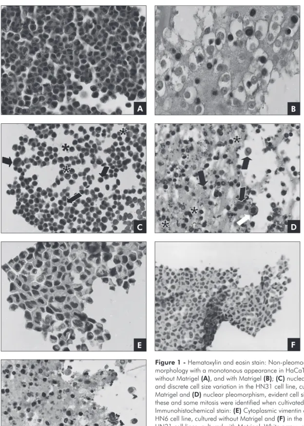
The HaCaT cell line, cultured either with or without Matrigel, demonstrated a non-pleomor-phic, monotonous appearance (Figures 1A and 1B). On the other hand, the HNSCC cell lines cultured without Matrigel exhibited nuclear pleomorphism, discrete cell size variation and absence of mitosis (Figure 1C). The same morphological characteris-tics were found when these cell lines were cultivated with Matrigel, although they were more evident and some mitotic cells could be identiied (Figure 1D).
Vimentin immunohistochemical expression Vimentin expression was not observed in the Ha-CaT, HN30 and HN31 cell lines cultured without Matrigel. In contrast, scattered cells from the HN6 cell line showed cytoplasmic positivity (Figure 1E). When cultured with Matrigel, the HaCaT and HN6 cell lines demonstrated no vimentin expression while the HN30 and HN31 cell lines showed immu-nopositivity (Figures 1F and 1G).
Vimentin intracellular levels
Western blotting conirmed the results shown by immunohistochemistry. A signiicant decrease in vimentin expression levels for the HN6 cell line (p = 0.022), and a signiicant increase for the HN30 (p = 0.027) and HN31 (p = 0.030) cell lines cultured with Matrigel, were noted. HaCaT showed no ex-pression when cultured with Matrigel. Additionally, only the HN6 cell line showed evident vimentin ex-pression levels when cultured without Matrigel (Fig-ure 2).
Discussion
Figure 1 - Hematoxylin and eosin stain: Non-pleomorphic cell morphology with a monotonous appearance in HaCaT cell line cultured without Matrigel (A), and with Matrigel (B); (C) nuclear pleomorphism and discrete cell size variation in the HN31 cell line, cultured without Matrigel and (D) nuclear pleomorphism, evident cell size variation: these and some mitosis were identified when cultivated with Matrigel. Immunohistochemical stain: (E) Cytoplasmic vimentin expression in the HN6 cell line, cultured without Matrigel and (F) in the HN30 and (G) HN31 cell lines, cultured with Matrigel. White arrow: mitosis figure; Black arrow: variation in cell size; Asterisk: nuclear pleomorphism. Original magnification ×400.
A
E
B
F
G
microenvironments rather than on two-dimensional surfaces.20 To mimic the extracellular matrix, a
three-dimensional cell culture with Matrigel was used in this study. Matrigel contains components of the human extracellular matrix including laminin, type IV collagen, nidogen, entactin and heparan sul-fate.17
The present study showed no spindle cells com-patible with mesenchymal phenotype in the HN-SCC cell lines cultivated with Matrigel. In vivo, this phenotype change is usually described in carcinoma cells at the tumor invasion front, and is associated with more aggressive behavior.8 However,
charac-teristics also related to a more aggressive cellular pattern, like evident nuclear pleomorphism, cell size variation and mitosis, were observed in the HNSCC cell lines cultivated with Matrigel. This suggests the ability of this substrate ability to simulate the in vivo microenvironment.21
Matrigel probably stimulates growth and retains the cells in an undifferentiated state, which repre-sents an invasive phenotype.22 This hypothesis
ex-plains the evident vimentin expression in the HN30 and HN31 cell lines after being cultured with Matrigel.
The HN6 cell line has a constitutively activated epidermal growth factor receptor.19 Interestingly,
among all the studied cell lines cultured without Matrigel, only the HN6 cell line expressed the vi-mentin protein, suggesting its more aggressive phe-notype.
It is known that the vimentin protein contributes to the aggressive pattern of cancer cells because it regulates the interaction between cytoskeletal pro-teins with cell adhesion molecules. It thereby partic-ipates in cell adhesion, migration, invasion and cell signal transduction in tumor cells, tumor-associated endothelial cells and macrophages. Its highly dy-namic balances between polymerization and depo-lymerization, and its complex phosphorylation may serve as the regulation mechanisms for tumor me-tastasis and cell-cell interactions. Therefore, vimen-tin is recognized as a hallmark of tumor progression that characterizes highly invasive and metastatic carcinomas.14
On the other hand, no immunohistochemical expression and signiicant decrease in vimentin in-tracellular levels were noted for the HN6 cell line cultured with Matrigel. Although this vimentin ex-pression could have occurred subsequently, the time spent in the cell culture was probably not enough to promote it for this cell line. This time dependence can be an important factor to be considered when working with three-dimensional culture models. In this study, it was observed that Matrigel degraded after 3 days. Thus, if the cells remain in contact with the substrate for a longer time, the experiment would become 2D (conventional method of cultiva-tion) and important data from the cell-environment interaction could be lost.
Another important inding demonstrated through immunohistochemistry was that, for the cell lines that did express vimentin (HN6 cultured without Matrigel; and HN30 and HN31 cultured with Matrigel), only part of these cells showed vi-mentin staining. This could be explained by the dif-ferent degrees of cellular difdif-ferentiation. In agree-ment, Gilles et al.13 demonstrated that only part
Figure 2 - (A) Bands demonstrated in Western blotting analyses of the HNSCC cell lines, for the antibody investigat-ed (Vimentin). Beta-actin was usinvestigat-ed to control total volume of each sample. (B) Representative graph of mean values plus standard deviation. Statistically significant differences between each cell line are represented by distinct letters: (a) HN6 - p = 0.022; (b) HN30 - p = 0.027; (c) HN31 - p = 0.030.
40000
35000
30000
25000
20000
15000
5000
0 10000
HN6 HN30 HN31
p=0.022
p=0.027
p=0.030 2D
3D A
of the epithelial mammary cells express vimentin, while the most differentiated cells do not. Further-more, the lack of vimentin expression in the HaCaT cell line conirms its immortalized but nontumori-genic nature.23
Conclusions
Data from this study indicate differences
be-tween two and three-dimensional cell culture mod-els. Therefore, the distinct expression of vimentin found for each of the cell lines studied, as well as its strict relation to the extracellular matrix, indicates that the behavior of the HNSCC cell lines can be inluenced by both the extracellular microenviron-ment and the culture conditions.
References
1. Schaaij-Visser TB, Brakenhoff RH, Leemans CR, Heck AJ, Slijper M. Protein biomarker discovery for head and neck cancer. J Proteomics. 2010 Set 10;73(10):1790-803. 2. Scully C, Bagan J. Recent advances in Oral Oncol. Oral Oncol.
2007 Feb;43(2):107-15.
3. Crowe D, Hacia J, Hsieh C, Sinha U, Rice H. Molecular pathology of head and neck cancer. Histol Histopathol. 2002;17(3):909-14.
4. Hanahan D, Weinberg RA. The hallmarks of cancer. Cell. 2000 Jan 7;100(1):57-70.
5. Ruiz P, Gunthert U. The cellular basis of metastasis. World J Urol. 1996;14(3):141-50.
6. Boyer B, Valles AM, Edme N. Induction and regulation of epithelial-mesenchymal transitions. Biochem Pharmacol. 2000 Oct 15;60(8):1091-99.
7. Hay ED. The mesenchymal cell, its role in the embryo, and the remarkable signaling mechanisms that create it. Dev Dyn. 2005 Jul;233(3):706-20.
8. Birchmeier C, Birchmeier W, Brand-Saberi B. Epithelial-mesenchymal transition in cancer progression. Acta Anat. 1996;156(3):217-26.
9. Araujo VC, Pinto Junior DS, Sousa SO, Nunes FD, Araujo NS. Vimentin in oral squamous cell carcinoma. Eur Arch Otorhinolaryngol. 1993;250(2):105-9.
10. Ivaskaa J, Pallarib HM, Nevoa J, Eriksson JE. Novel functions of vimentin in cell adhesion, migration, and signaling. Exp Cell Res. 2007 Jun 10;313(10):2050-62.
11. Mendez MG, Kojima SI, Goldman RD. Vimentin induces changes in cell shape, motility, and adhesion during the epithe-lial to mesenchymal transition. FASEB J. 2010 Jun;24(6):1838-51.
12. Taki M, Kamata N, Yokoyama K, Fujimoto R, Tsutsumi S, Nagayama M. Down-regulation of Wnt-4 and up-reg-ulation of Wnt-5a expression by epithelial-mesenchymal transition in human squamous carcinoma cells. Cancer Sci. 2003 Jul;94(7):593-7.
13. Gilles C, Polette M, Zahm JM, Tournier JM, Volders L, Foi-dart JM, et al. Vimentin contributes to human mammary epithelial cell migration. J Cell Sci. 1999 Dec;112(Pt 24):4615-25.
14. Liu LK, Jiang XY, Zhou XX, Wang DM, Song XL, Jiang HB. Upregulation of vimentin and aberrant expression of E-cadherin/beta-catenin complex in oral squamous cell car-cinomas: correlation with the clinicopathological features and patient outcome. Mod Pathol. 2010 Feb;23(2):213-24. 15. LeBleu V, MacDonald B, Kalluri R. Structure and Function of
Basement Membranes. Exp Biol Med. 2007 Oct;232(9):1121-9 16. Boudreau N, Bissel M. Extracellular matrix signaling: intera-tion of form and funcintera-tion in normal and malignant cells. Curr Opin Cell Biol. 1998 Oct;10(5):640-6.
17. Kleinman HK, McGarvey ML, Hassell JR, Star VL, Cannon FB, Laurie GW, et al. Basement membrane complexes with biological activities. Biochemistry. 1986 Jan 28;25(2):312-8. 18. Schmeichel K, Bissell M. Modeling tissue-specific signaling
and organ function in three dimensions. J Cell Sci. 2003 Jun 15;116(Pt 12):2377-88.
19. Cardinali M, Pietraszkiewicz H, Ensley J, Robbins K. Tyrosine phosphorylation as a marker for aberrantly regulated growth-promoting pathways in cell lines derived from head and neck malignancies. Int J Cancer. 1995 Mar 29;61(1):98-103. 20. Griffith L, Swartz M. Capturing complex 3D tissue physiology
in vitro. Nat Rev Mol Cell Biol. 2006 Mar;7(3):211-24. 21. Kujan O, Oliver RJ, Khattab A, Roberts SA, Thakker N, Sloan
P. Evaluation of a new binary system of grading oral epithelial dysplasia for prediction of malignant transformation. Oral Oncol. 2006 Nov;42(10):987-93.
22. Hughes CS, Postovit LM, Lajoie GA. Matrigel: a complex protein mixture required for optimal growth of cell culture. Proteomics. 2010 May;10(9):1886-90.