Faculdade de Ciências da Universidade de Lisboa Departamento de Biologia Animal
THE ROLE OF PERIPHERAL BLOOD
MONONUCLEAR CELLS IN ATHEROGENESIS
Cláudia Simões Martins Bispo
Mestrado em Biologia Humana e Ambiente 2011
-Faculdade de Ciências da Universidade de Lisboa Departamento de Biologia Animal
THE ROLE OF PERIPHERAL BLOOD
MONONUCLEAR CELLS IN ATHEROGENESIS
Dissertação orientada por:
Professora Doutora Ana Maria Viegas Crespo Professora Doutora Luciana Maria Gonçalves da Costa
Cláudia Simões Martins Bispo
Mestrado em Biologia Humana e Ambiente 2011
2
AGRADECIMENTOS
Em primeiro lugar gostaria de agradecer à Doutora Luciana Costa por me ter dado a oportunidade de trabalhar neste projecto e dado todo o apoio e orientação durante este ano. Pelo conhecimento transmitido, sugestões e também pelos merecidos “puxões de orelhas” quando necessários nas meticulosas revisões desta tese.
Agradeço à Doutora Ana Crespo por todo o apoio, preocupação e disponibilidade que demonstrou durante todo este percurso.
Um agradecimento especial à Liliana por todo o conhecimento transmitido, orientação e incentivo na realização deste trabalho. Obrigada por toda a paciência e compreensão, sobretudo nesta fase final.
Um agradecimento especial à Arminda por ter sido desde o primeiro momento um grande apoio nesta nova fase da minha vida, obrigada por todos os bons conselhos.
A todo o pessoal de laboratório do LEMES um enorme obrigado por todas as dicas e ajuda na integração nesta vida de bancada. Assim como a todos que se voluntariaram para que fosse possível realizar as minhas experiências. Muito obrigada!
À Ana Patrícia, a minha companheira de almoços, por ter estado sempre presente para todos os desabafos ou pelas palavras de encorajamento.
Ao pessoal da FCUL, colegas de licenciatura e mestrandos de BHA, obrigada pela vossa amizade e companheirismo, estes foram certamente os melhores anos da minha vida.
Aos meus pais por incutirem em mim o gosto de aprender e realização pessoal, entre outros valores que regem a minha vida. À minha família, pela sua confiança, carinho e compreensão durante este tempo de ausência.
Ao meu irmão Eduardo, que sem o perceber não me deixa desanimar e encoraja sempre a seguir em frente ultrapassando todos os obstáculos. Obrigada pela amizade e por todos os bons momentos. Sem ti certamente tudo teria sido mais difícil.
2
ABSTRACT
Atherosclerosis (ATH) is recognized as a chronic inflammatory condition and it is the leading cause of cardiovascular disease. Atherogenesis is characterized by the infiltration of low density lipoprotein (LDL) through the endothelial layer, and migration of activated peripheral blood lymphocytes (PBL) and monocytes (PBMN) that contribute to a pro-inflammatory state in specific locations. However, the functional interaction between immunity and LDL metabolism is still not fully understood. One hypothesis for the etiopathogeny of ATH may be associated with an ongoing inflammatory process caused by a pro-oxidant/anti-oxidant imbalance induced by metals such as iron (Fe) or copper (Cu). Interestingly, ceruloplasmin (Cp) is a multicopper oxidase with a relevant role in Fe metabolism and oxidation of LDL, but also an acute-phase protein involved in the inflammatory process.
Herein, we intended to study by flow cytometry the effect of putative pro-atherogenic immune stimuli on the expression of Cp at the surface of human peripheral blood mononuclear cells (PBMC), and search for a putative association with LDL oxidation. Additionally, a population of Familial Hypercholesterolemia (FH) patients was used as a clinical model of ATH to clarify the physiologic interplay between inflammation, Fe/Cu and lipid metabolism at systemic level in this disease.
The obtained results showed that higher cell surface expression of Cp was consistently observed in PBMN compared to PBL, in activated vs non-activated cells and in non-T cells vs T cells. Also, PBMC surface expression of Cp was differently modulated by several tested treatments. In particular, various modulators caused opposite effects on Cp expression of PBMN compared to PBL, suggesting a specific cell-type regulation for this protein. Specifically, it was observed that different sources of Fe might activate specific regulation mechanisms of Cp. Until now, due to technical limitations it was not possible to demonstrate an association between PBMC surface Cp expression and oxidation of LDL, but this issue should be addressed in future experiments. Of notice, IL-1β which was found to significantly increase PBMN surface Cp expression in vitro, was increased in serum from FH patients and positively correlated with total cholesterol and ApoB. However, the fact that no correlation was found between IL-1β and serum Cp (sCp)/oxidated LDL suggest that at systemic level, IL-1β may not be a contributing factor for oxidation of LDL by sCp. However, the role of PBMC surface Cp expression on LDL oxidation, namely in specific inflammatory states, remains to be established.
In summary, the results of this study demonstrate that specific pro-atherogenic conditions are involved in the modulation of Cp expression at surface of PBMC and thus, suggest a possible a role of Cp in ATH.
2
SUMÁRIO
A aterosclerose (AT) é a principal causa de doenças cardiovasculares (DCV). Muito embora esta doença tenha sido numa primeira fase considerada como uma simples deposição de lípidos, avanços substanciais nas áreas da ciência experimental permitiram revelar o importante papel da inflamação nesta patologia. Actualmente, a AT já não é reconhecida como uma simples consequência de envelhecimento considerando os factores de risco tradicionalmente aceites no seu envolvimento, mas antes como uma doença inflamatória crónica que pode ser convertida num evento clínico agudo por ruptura de placas ateroscleróticas e consequente trombose.
A formação da placa aterosclerótica (aterogénese) caracteriza-se por uma infiltração e activação precoce de monócitos e linfócitos (células mononucleares) que contribuem para um estado pró-inflamatório localizado em regiões de endotélio disfuncional. Adicionalmente, a passagem de lipoproteínas de baixa densidade (LDL) através da camada endotelial e a sua oxidação constituem eventos precoces da aterogénese. Contudo, os mecanismos fisiológicos envolvidos na interacção funcional entre as células imunitárias e a oxidação das LDL não estão completamente esclarecidos. Uma hipótese para a etiopatogenia da AT poderá estar associada à existência de um processo inflamatório contínuo relacionado com stress oxidativo induzido por metais, tais como ferro (Fe) ou Cobre (Cu). A ceruloplasmina (Cp) é uma proteína de fase aguda da família das multicobre oxidases com funções importantes no metabolismo do Fe, principalmente devido à sua actividade ferroxidásica. Para além disso, esta proteína apresenta potencial pró- e anti-oxidante, actividades relevantes no contexto do stress oxidativo característico deste processo.
O objectivo geral deste estudo foi investigar e caracterizar in vitro alguns dos mecanismos subjacentes à relação funcional entre inflamação, metabolismo do Fe/Cu e a homeostase lipídica, de forma a adquirir novos conhecimentos sobre a fisiopatologia da AT. Em particular, estudaram- os efeitos de status de Fe e Cu alterado, citocinas pró-inflamatórias e outros imunomoduladores na expressão de Cp à superfície de células mononucleares de sangue periférico (PBMC). Este estudo foi realizado através da utilização de citometria de fluxo, que possibilitou caracterizar a expressão da Cp membranar em subpopulações leucocitárias específicas de monócitos (PBMN) e linfócitos (PBL) de sangue periférico. Posteriormente, avaliou-se a possível associação entre a modulação da expressão de Cp membranar nas condições testadas e a oxidação de LDL. Adicionalmente, realizou-se a caracterização laboratorial de uma população de doentes diagnosticados com Hipercolesterolemia Familiar (HF), como um modelo clínico de AT. Neste contexto, pretendeu-se complementar os estudos in vitro procurando a existência de associações entre os parâmetros do metabolismo do Fe/Cu, lípidos e inflamação medidos a nível sistémico.
Os resultados obtidos através de citometria de fluxo mostraram consistentemente que a expressão de Cp à superfície de PBMN é maior comparativamente com PBL e que em ambas as populações as células activadas apresentam maiores níveis de expressão desta proteína que as suas
2
homólogas não activadas. Além disso, mostrou-se que dentro das subpopulações de PBL, são as células não-T que possuem maior expressão de Cp à sua superfície em comparação com as células T. Por outro lado, os resultados obtidos através das experiências de modulação mostraram que a Cp membranar das duas populações celulares em estudo é diferentemente regulada na presença dos diversos estímulos. Em condições de status de Fe alterado foi observado que a Cp à superfície de PBMC é diferentemente regulada nos dois tipos celulares testados, e parecem existir mecanismos de regulação específicos consoante o dador de Fe utilizado. Adicionalmente, a alteração do status de Cu não produziu modificações significativas na expressão de Cp à superfície de PBMC. Estas observações não corroboraram resultados obtidos anteriormente por outros autores e devem-se provavelmente ao curto espaço de tempo de incubação que não possibilitou visualizar o efeito de variação da concentração deste metal na estabilidade da Cp.
Em relação aos tratamentos com as citocinas e outros imunomodulatores observou-se de uma forma geral que a interleucina (IL)-1β, lipopolissacarídeo, e forbol 12-miristato 13-acetato induziram o aumento da expressão de Cp à superfície dos PBMN enquanto que a diminuíram nos PBL, sugerindo que a Cp membranar destas populações celulares poderá ser regulada especificamente devido às funções particulares e diferenças de cada tipo celular durante a resposta inflamatória. Os tratamentos com interferão-γ e IL-2 diminuíram a expressão de Cp à superfície de PBMN e PBL em incubações de longa duração, enquanto os tratamentos efectuados com os mediadores IL-6, IL-8, factor estimulador de colónia de macrófagos e o factor de necrose tumoral-α não resultaram variações na expressão membranar de Cp em ambos os tipos celulares. Provavelmente, a expressão de Cp à superfície de PBMC poderá ser nestes casos modulada em curtos espaços de tempo, pelo que as condições experimentais utilizadas não permitiram confirmar esta hipótese. A influência de espécies reactivas de azoto na expressão de Cp à superfície de PBMC foi igualmente testada realizando o tratamento com S-nitroso-N-acetilpenicilamina(dador de óxido nítrico), verificando-se que na presença deste tipo de condições de stress oxidativo são especificamente os PBMN que aumentam a expressão de Cp à sua superfície. Estes resultados sugerem que em estado pró-inflamatórios e de stress oxidativo a Cp expressa à superfície dos PBMN parece estar mais associado ao papel de proteína de fase aguda descrito para a forma circulante da Cp enquanto nos PBL esta proteína parece estar mais associada à regulação de status de Fe intracelular de modo a permitir a proliferação deste tipo de células após estimulação. Posteriormente, avaliou-se uma possível associação entre a indução de Cp à superfície de PBMC e oxidação das LDL no meio. Contudo, limitações técnicas impediram a confirmação desta hipótese neste trabalho. No entanto, a detecção de níveis de LDL oxidada (oxLDL) no sobrenadante de culturas em que Cp membranar de PBMN fora induzida por IL-1β, sugere a existência de potencial pró-oxidante na forma membranar de Cp. Esta observação carece contudo de confirmação após optimização de procedimento experimental utilizado.
2
Os resultados decorrentes da caracterização clínica do grupo de indivíduos diagnosticados com HF revelaram que estes doentes apresentam diferenças nos parâmetros relacionados com o metabolismo dos lípidos, consistentes com o seu diagnóstico molecular que mostra a existência de mutações genéticas em componentes do metabolismo lipídico. Nestes doentes foram ainda encontrados valores mais elevados de ferritina, oxLDL e IL-1β comparativamente com os indivíduos saudáveis. Além disso, nos doentes HF observou-se a existência de correlações positivas entre Cp circulante (sCp) e oxLDL, concordante com potencial pró-oxidante desta proteína, e entre sCp e proteína C reactiva - duas proteínas de fase aguda presentes em processos inflamatórios envolvidos na progressão desta doença. Foi também encontrada a correlação positiva de IL-1β com níveis de colesterol total e ApoB, parâmetros associados com risco aumentado de DCV. Contudo, o facto de não ter sido observada nenhuma correlação entre os níveis de IL-1β e sCp / oxLDL nestes indivíduos sugere que a nível sistémico a IL-1β parece não contribuir para o aumento de acção pró-oxidante da sCp. No entanto não é de excluir a possibilidade de um possível papel fisiológico de Cp expressa à superfície das PBMC a nível local nas placas ateroscleróticas.
Em conclusão, os resultados deste estudo demonstram que modulatores específicos de Fe/Cu e mediadores inflamatórios putativamente associados a estados pró-aterogénicos podem estar, pelo menos em parte, envolvidos na modulação da forma membranar da Cp em PBMC, sugerindo um possível papel da Cp à superfície dessas células durante a aterogénese.
Palavras Chave: Aterosclerose - Ceruloplasmina - Células Mononucleares - Metabolismo do Ferro
2
INDEX
Agradecimentos ... i
Abstract ... ii
Sumário ... iii
Index of Figures and Tables ... viii
Abbreviations ... x
Introduction ... 1
1. Atherosclerosis and Cardiovascular Disease ... 1
1.1 Inflammation and Atherosclerosis ... 2
1.2 Atherogenesis ... 4
1.3 Atherosclerotic Plaque Microenvironment ... 5
2. Oxidative Stress in Atherosclerosis ... 6
2.1 A link between the immune system, iron metabolism and oxidative stress: Ceruloplasmin... 7
Objectives ... 9
Matherials and Methods ... 10
1. Population of study ... 10
2. Isolation of peripheral blood mononuclear cells ... 10
3. In vitro modulation of cell surface ceruloplasmin expression ... 10
3.1. Iron and copper status ... 11
3.2. Cytokines and other immunomodulators ... 11
3.3. Reactive nitrogen species ... 12
4. Immunophenotyping and Flow Cytometry analysis ... 12
5. Quantification of oxidized LDL in cell culture supernatants ... 13
6. Laboratorial characterization of Familial Hypercholesterolemic patients: a model to study atherosclerosis ... 13
7. Statistical Analysis ... 13
Results ... 15
1. Ceruloplasmin expression at surface of peripheral blood mononuclear cells ... 15
2. Regulation of surface ceruloplasmin expression by iron and copper status ... 15
2.1. Modulation of iron status ... 16
2.2. Modulation of copper status ... 17
3. Regulation of surface ceruloplasmin expression by cytokines and other immunomodulators ... 18
2 3.2. IL-1β ... 20 3.3. IL-2 ... 21 3.4. IL-6 ... 22 3.5. IL-8 ... 23 3.6. M-CSF ... 23 3.7. TNF-α ... 24 3.8. LPS ... 25 3.9. PMA ... 26
4. Regulation of surface ceruloplasmin expression by reactive nitrogen species ... 26
5. Study of putative association of GPI-Cp expression and LDL oxidation ... 28
6. Biochemical and immunologic characterization of FH patients as a model to study atherosclerosis ... 28
Discussion ... 30
Future Perspectives... 40
References ... 41
2
INDEX OF FIGURES AND TABLES
Figures
Fig. 1 - Role of inflammation in all stages of atherosclerosis ... 1
Fig. 2 - Infiltration of LDL into the endothelium ... 4
Fig. 3 - Flow Cytometry analysis of surface Cp expression in non-treated PBMC ... 15
Fig. 4 - Effect on PBMC surface Cp expression after overnight incubation with Holo-Tf. ... 16
Fig. 5 - Effect on PBMC surface Cp expression after overnight incubation with Fe-NTA. ... 16
Fig. 6 - Effect on PBMC surface Cp expression after overnight incubation with DFO. ... 17
Fig. 7 - Effect on PBMC surface Cp expression after overnight incubation with CuHis ... 17
Fig. 8 - Effect on PBMC surface Cp expression after overnight incubation with BCS ... 18
Fig. 9 - Effect on PBMC surface Cp expression after 1h incubation with IFN-γ ... 18
Fig. 10 - Effect of IFN-γ on activated and non-activated PBMC surface Cp expression after 1h incubation ... 19
Fig. 11 - Effect on PBMC surface Cp expression after overnight incubation with IFN-γ ... 19
Fig. 12 - Effect on PBMC cell activation after overnight incubation with IFN-γ ... 19
Fig. 13 - Effect on PBMN surface Cp expression after overnight incubation with IL-1β ... 20
Fig. 14 - Effect on PBL surface Cp expression after overnight incubation with IL-1β. ... 20
Fig. 15 - Effect on PBMN surface Cp expression and cellular activation after overnight incubation with IL-2 ... 21
Fig. 16 - Effect on PBL surface Cp expression and cellular activation after overnight incubation with IL-2 ... 22
Fig. 17 - Effect on PBMC surface Cp expression after overnight incubation with IL-6 ... 22
Fig. 18 - Effect on PBMC surface Cp expression after overnight incubation with IL-8 ... 23
Fig. 19 - Effect on PBMC surface Cp expression after overnight incubation with M-CSF ... 24
Fig. 20 - Effect on PBMC surface Cp expression after overnight incubation with TNF-α ... 24
Fig. 21 - Effect on PBMC surface Cp expression and cellular activation after overnight incubation with LPS. ... 25
Fig. 22 - Effect on PBMC surface Cp expression after 2 h incubation with PMA.. ... 26
Fig. 23 - Effect on PBMN surface Cp expression after overnight incubation with SNAP ... 27
Fig. 24 - Effect on PBMN surface Cp expression after overnight incubation with Il-1β and SNAP ... 27
2
Tables
Table I - Risk factors for Development of ATH ... 2 Table II - Summarized list of inflammatory mediators involved in atherogenesis ... 6 Table III - Iron and copper treatments used to modify Fe/Cu status in PBMC culture ... 11 Table IV - Experimental conditions used to test the effect of cytokines and other
immunomodulators in PBMC culture ... 12 Table V - Biochemical and immunologic characterization of controls and FH patients ... 29 Table VI - Significant correlations of serum Cp or IL-1β with biochemical parameters
measured in FH patients ... 29
2
ABBREVIATIONS
Ab Antibody ApoA1 Apolipoprotein A1 ApoB Apolipoprotein B ATH Atherosclerosis BCS Bathocuproine disulphonate CHD Coronary heart disease Cp CeruloplasminCRP C-reactive protein Cu Copper
CuHis Copper histidine CVD Cardiovascular disease DFO Desferrioxamine EC Endothelial cells ELISA Enzyme-linked-immuno-sorbent-assay Fe Iron Fe-NTA Iron-nitrilotriacetate FH Familial Hypercholesterolemia GPI Glycosylphosphatidylinositol h Hours
HDL High density lipoprotein HIF Hypoxia-inducible factor
ICAM-1 Intercellular Adhesion Molecule 1 IFN-γ Interferon gama
IL Interleukin
INSA Instituto Nacional de Saúde Doutor Ricardo Jorge
LDL Low density lipoprotein Lp(a) Lipoprotein (a)
mAb Monoclonal antibody
MCP-1 Monocyte chemoattractant protein-1 M-CSF Macrophage colony-stimulating factor MFI Mean fluorescence intensity
MI Myocardial infarction min Minutes
mL mililiter ng nanogram
NK Natural killer cells NO Nitric oxide
oxLDL Oxidized low density lipoprotein PBL Peripheral blood lymphocytes
PBMC Peripheral blood mononuclear cells PBMN Peripheral blood monocytes PMA Phorbol-12-myristate-13-acetate RNS Reactive nitrogen species
ROS Reactive oxygen species sCp secreted Ceruloplasmin SMC Smooth muscle cells
SNAP S-nitroso-N-acetylpenicillamine Tf Transferrin
Th T helper cells
TNF-α Tumor necrosis factor alpha
INTRODUCTION
1. Atherosclerosis and Cardiovascular Disease
Cardiovascular disease (CVD) is the main cause of death in Western societies, being responsible for approximately 50% of all deaths [1]. Also, the economic and social difficulties caused by CVD are immense, constituting the greatest single cause of adult disability and a serious economic burden [2].
CVD is a collective term that comprises the clinical manifestations of coronary heart disease (CHD) and cerebrovascular disease, both of which have as a primary cause Atherosclerosis (ATH). [3]
The development of CVD involves genetic factors as well other acquired and modifiable risk factors. Smoking, hypercholesterolemia, diabetes mellitus and hypertension, among others are some of the main players in the development of this disease; however the major underlying cause leading to CVD is indeed ATH. [2, 4]
ATH is considered to be a complex multifactorial disease [5] that begins in the first decade of life, generally affecting the large arteries [2] as fatty streak lesions start to form in the artery wall. These early lesions are characterized by inflammation, lipid accumulation, cell death and fibrosis. Over time, they will mature and develop new characteristics as continued recruitment of mononuclear cells (Figure 1A), proliferation and migration of smooth muscle cells (SMC) occurs [2, 6]. This type of lesions, are then called atheromas or atherosclerotic plaques (Figure 1B) [7].
Clinical complications of ATH can arise from plaques causing flow-limiting stenoses, as significant narrowing of the arterial lumen occurs [8]. The most severe clinical events happen however after the rupture of a plaque (Figure 1C). This usually ensues when the combined efforts of the physical forces of blood flow and intensified inflammatory activation (that may lead to local proteolysis) disrupt the arterial wall [3, 6, 7]. These actions will eventually expose prothrombotic material from the interior of the plaque to the blood, triggering a thrombus that can cause sudden thrombotic occlusion of the artery at the site of disruption and the subsequent blockage of blood flow. In the heart, this leads to myocardial infarction (MI) and heart failure, whereas in the arteries that perfuse the brain, it can cause ischemic stroke and transient ischemic attacks [6].
Fig. 1 - Role of inflammation in all stages of atherosclerosis. A) Leukocyte recruitment to the nascent atherosclerotic lesion. B) T
2
Table I - Risk factors for Development of ATH
(From Greaves et al. [2])
1.1 Inflammation and Atherosclerosis
During the last decades, experimental, clinical, and epidemiological studies have shed light on several key aspects related to this disease. Although ATH was formerly considered a bland lipid storage disease, the substantial advances in basic and experimental science have illuminated the pivotal role of inflammation in this disease [9], particularly the role of monocytes/macrophages and lymphocytes, and the defined series of changes that occur in the vessel during atherogenesis. It is now clear that ATH is not simply an inevitable degenerative consequence of ageing, but rather a chronic inflammatory condition that can be converted into an acute clinical event by plaque rupture and thrombosis [1].
Multiple independent pathways of evidence now identify inflammation as a key regulatory process that links multiple risk factors for ATH and its complications with altered arterial biology [10]. In this context, epidemiologic studies have identified many genetic and environmental risk factors (Table I) that seem to cause an impact on the inflammatory and immunological response of the organism [11]. Thus, cardiovascular risk factors are essentially injuries or insults to which the body adapts by continuously triggering a response throughout years of exposure [12].
This kind of compensatory response occurs preferentially at sites of hemodynamic strain [13], segments with low average shear but high oscillatory shear stress [14]. The normal homeostatic properties of the endothelium are then altered, and it becomes dysfunctional. Thus, the different forms of injury increase the adhesiveness of the endothelium with respect to leukocytes or platelets, as well as its permeability. They also induce the endothelium to have pro-coagulant instead of anticoagulant properties and to form vasoactive molecules, cytokines, and growth factors. If the offending agent or agents are not successfully dealt with by the immune system, and the inflammation as a chance to progress and continue indefinitely, the inflammatory response converts from a protective to an injurious response [15].
Such constant or repetitive injury can also stimulate the affected tissue to repair or wall off the damage by means of a fibroproliferative response, which, when excessive, diminishes the functional capacity of the tissue or organ and becomes part of the disease process. Because of this process, initially, the only cells thought to proliferate during expansion of atherosclerotic lesions were SMC [15]. However, as it is, most of the cells found in atheromas are blood-borne immune cells, and the remainder are then vascular endothelial and SMC [7]. Therefore, replication of monocyte-derived macrophages and T cells is probably equal importance during
2
the expansion of atheromas [16]. The ability of macrophages to produce cytokines (such as tumor necrosis factor α (TNF-α), interleukin (IL)-1 and transforming growth factor β), proteolytic enzymes (particularly metalloproteinases), and growth factors (such as platelet-derived growth factor and insulin-like growth factor I) may be critical in the role of these cells during the damage and repair that ensues as the lesions progress [15].
Many of the immune cells in the atheroma exhibit signs of activation and produce pro-inflammatory cytokines [7]. Activated macrophages express class II histocompatibility antigens that allow them to present antigens to T lymphocytes [17]. Thus, it is not surprising that cell-mediated immune responses may be involved in atherogenesis, since both CD4 and CD8 T cells are present in the lesions at all stages of the process [18].This smoldering inflammatory state may lead to detectable blood levels of pro-inflammatory cytokines and other acute phase reactants. C-reactive protein (CRP) and IL-6 are elevated in patients with unstable angina and MI; high levels of these factors also predict a poor outcome [19, 20]. Furthermore, other inflammatory markers appear elevated in these patients, including IL-7, IL-8, soluble CD40 ligand, and the CRP-related protein pentraxin-3 [21-23].
The fact that several different inflammatory markers with different biological activities contribute to significantly increase the risk for CVD makes it unlikely that any of these particular markers actually causes the disease. Instead, the elevated levels may reflect an exacerbated inflammatory state in the atheroma, which promotes or causes plaque rupture, thrombosis, and ischemia [7]. In regard to possible causes of endothelial dysfunction, high levels of serum cholesterol are probably unique in being sufficient to drive the development of ATH in humans and experimental animals, even in the absence of other recognizable risk factors [11].
Free radical formation caused by cigarette smoking, hypertension, and diabetes mellitus causes focal endothelial activation in large- and medium-sized arteries [24]. Of these risk factors, smoking was shown to be one of the main factors in enhancing the risk of development of ATH, as it increases the lifetime risk in two-fold [25]. Likewise, the lack of exercise and the associated adiposity, in addition to a high intake of saturated fats and a low intake of certain vitamins, are commonly associated with an increased CHD risk in the general population [26]. Also, genetic alterations, as elevated plasma homocysteine concentrations that increases collagen production [27] and decreases the availability of nitric oxide [28], infectious microorganisms such as herpes viruses or Chlamydia pneumoniae and/or combinations of these, and other factors [29, 30], can contribute to atherogenesis [15].
The mechanism of action of these factors is, at least in part, thought to be associated with altered plasma levels of lipids and atherogenic lipoproteins. In fact, high levels of low-density lipoprotein (LDL) cholesterol and low levels of high-density lipoprotein (HDL) cholesterol have consistently been shown to be connected with CHD risk. [26]
2
1.2 Atherogenesis
The early phase of atherosclerotic disease is best explained as an inflammatory response elicited by retention of lipoproteins in the arterial intima [7]
at sites of dysfunctional endothelium, where disturbed flow dynamics cause modifications at the cellular and molecular level [14]. This occurs when plasma levels of the cholesterol-rich very low-density lipoprotein and LDL rise, and the lipoproteins infiltrate the artery wall to an extent that exceeds the capacity for elimination, and so they are retained in the extracellular matrix (Figure 2) [24].
The oxidation of LDL is believed to occur locally within the arterial wall after retention [31]. Once LDL enters the vascular wall, the lipoproteins lose the protection from plasma anti-oxidants [32], thus their modification is promoted by oxidation, glycation (in diabetes), aggregation, association with proteoglycans, or incorporation into immune complexes [33, 34]. Presumably reactive oxygen species (ROS) produced by endothelial cells (EC), macrophages, proteins and several enzymes which occur in human atherosclerotic lesions have an important role at this stage [32].
EC and vascular SMC have been shown to oxidize LDL in culture [35-38], as have a number of enzymes [39-43] and proteins, such as ceruloplasmin (Cp) [44]. In some experimental situations, it was observed that administration of anti-oxidants can delay the progression of atherosclerotic lesions that develop in the face of hyperlipidemia, however human clinical trials have repeatedly failed to corroborate the concept that anti-oxidant vitamin therapy can improve clinical outcomes [9].
The oxidative stress generated within the intima [11] is a major cause of injury to the endothelium and underlying smooth muscle [45] and eventually evokes an inflammatory response in the arterial wall [11]. This response triggers the endothelial expression of adhesion molecules which cause blood cells rolling on the vascular surface to adhere at the site of activation [46] (such as vascular cell adhesion molecule-1 (VCAM-1) [47] and P-selectin [48]) and chemoattractant factors (such as monocyte chemoattractant protein 1 (MCP-1) and IL-8) that direct the migration and diapedesis of circulating monocytes and other leukocytes into the vascular intima of the arterial wall [49, 50] to neutralize or remove the offending agents [15]. Ultimately, this culminates in the formation of a fibrous plaque in that site [51], as illustrated in Fig. 1A and 1B. If the immune response is not successful, it can continue indefinitely. In doing so, the inflammatory response stimulates migration and proliferation of SMC that become intermixed with the area of inflammation to form an intermediate lesion [15].
The degree to which LDL is modified can vary greatly [45, 52] but once modified it can be internalized by macrophages by means of the scavenger receptors on the surfaces of these cells
Fig. 2 - Infiltration of LDL into the endothelium. When it is retained in the
intima, it can undergo oxidative modification(From Hanson et al. [7])
2
[33, 34, 45, 53]. The internalization leads to the formation of lipid peroxides and facilitates the accumulation of cholesterol esters, resulting in their cellular activation and modification into foam cells [15]. Because these scavenger receptors are not down-regulated by increases of intracellular cholesterol levels, as it happens with the native LDL receptor, foam cells continually engulf modified LDL [54] until their eventual death, contributing to the amplification of the inflammatory process and the formation of the necrotic core found in advanced lesions [11].
Removal and sequestration of modified LDL are important parts of the initial, protective role of macrophages in the inflammatory response [52, 53] and minimize the effects of modified LDL on EC and SMC [15]. In fact, oxidized LDL (oxLDL) has numerous atherogenic properties on its own [55] and the bioactive phospholipids released by its modification can activate EC [56]. Furthermore, in addition to its ability to injure these cells [45], modified LDL is chemotactic on itself for other monocytes and can up-regulate the expression of genes for macrophage colony-stimulating factor (M-CSF) [57] and MCP-1 [58] derived from EC [15].
Thus, a vicious circle of inflammation, modification of lipoproteins, and further inflammation can be maintained in the artery by the presence of these lipids [15]. Specifically, mediators of inflammation such as TNF-α, IL-1 and M-CSF increase binding of LDL to endothelium and smooth muscle, and increase the transcription of the LDL-receptor gene [59].
1.3 Atherosclerotic Plaque Microenvironment
Within the inflamed arterial intima an altered cellular microenvironment is established, allowing the progression of ATH lesions [60, 61]. Interactions between macrophages foam cells, T helper (Th) 1 and Th2 cells eventually establishes the chronic inflammatory process [11].
A variety of different chemokines and cytokines mediates chemotactic recruitment of adherent monocytes and lymphocytes to the forming lesion [7], and can exert both pro- and antiatherogenic effects on each of the cellular elements of the vessel wall [11].
The ubiquitous monocyte, the precursor of macrophages in all tissues, is present in every phase of atherogenesis. Also, it’s when T cells are activated by binding of antigens processed and presented by macrophages that the cell activation results in the secretion of cytokines, including interferon γ (IFN-γ) and TNF-α and β, that amplifies the inflammatory response. [6]
The unending entry, the survival, and replication of mononuclear cells in atherosclerotic plaques are dependent, in part, on factors such as M-CSF and granulocyte–macrophage colony-stimulating factor for monocytes and IL-2 for lymphocytes. Continued exposure to M-CSF allows macrophages to survive in vitro and possibly to multiply within the atherosclerotic plaques in vivo. In contrast, inflammatory cytokines such as IFN-γ activate macrophages and under certain circumstances induce them to undergo programmed cell death (apoptosis) [15].
But considering these effects in a global manner it was observed that the presence of M-CSF [62], TNF-α and IFN-γ [1, 32] in plaques changes expression of macrophages scavenger receptors, that will allow these cells to continually engulf modified lipoproteins leading to their accumulation in the cytoplasm until their death [6] and in this way become involved in the necrotic cores characteristic of advanced, complicated lesions [15].
When considering the role of inflammation in the development and progression of ATH it is also important to distinguish between local inflammation, shown in the activation of cells within the plaque microenvironment itself, and systemic inflammation, as evidenced by acute-phase proteins production and circulating pro-inflammatory mediators. Locally produced inflammatory mediators include the products
of activated macrophages and T cells, such as TNF-α, IL-1β, IL-8, IL-18 and MCP-1, all of which are produced in response to modified forms of LDL and/or bacterial products. Systemic mediators and markers of inflammation include IL-6, IL-1β and CRP [2]. Table II lists some of the cytokines and inflammatory mediators that influence the development of atherosclerotic lesions in animal models of ATH [63].
2. Oxidative Stress in Atherosclerosis
In general, initiation and progression of ATH is best explained as an inflammatory response elicited by retention of lipoproteins in the arterial intima [7]. However, the mechanisms by which LDL is oxidized in the arterial wall are still not fully understood, but they might involve the cumulative effect of ROS or reactive nitrogen species (RNS) generated by inflammatory cells and enzymatic oxidation [64] during the response against the previous referred risk factors. For example, when in a hypertensive state the individual experiences a rise in the formation of hydrogen peroxide and free radicals such as superoxide anion and hydroxyl radicals in the plasma [45, 65]. These substances reduce the formation of nitric oxide (NO) by the endothelium [66], increase leukocyte adhesion and peripheral resistance [15].
Others authors proposed that the oxidative modification of LDL might be due to the existence of catalytically active transition metal ions [64] in the plaque microenvironment. The extensive LDL oxidation characteristic of atherosclerotic lesions may require a source of iron (Fe) or copper (Cu) as catalyst for the oxidation [67]. In fact, the interior of advanced human atherosclerotic plaques is a highly pro-oxidant environment containing redox-active Fe and Cu
Table II - Summarized list of inflammatory mediators involved in atherogenesis (From Greaves & Channon [2])
2
ions, that in vitro induces lipid peroxidation [68, 69], but as Cu is more potent than Fe in catalyzing cellular LDL oxidation [70] it is possible it could account for the initiation of oxidation of LDL present in the arterial wall [71].
2.1 A link between the immune system, iron metabolism and oxidative stress: Ceruloplasmin
Cp is an abundant, blue plasma protein (metalloenzyme) which accounts for 95% of total circulating Cu in healthy adults [72]. Cp is a multifunctional protein with several functions that may play contradictory roles [73]. In fact, the protein is a multicopper oxidase and it possesses both anti-oxidant and pro-oxidant activities. It is also an acute-phase protein involved in the inflammatory process [67].
As a result of alternative splicing [74] there are two isoforms of Cp, one secreted mainly by the liver (sCp) and other anchored to the membrane by a glycosylphosphatidylinositol (GPI) group (GPI-Cp) in various cell types [75, 76], including monocytes [77]. Previously, it was demonstrated by our research group that GPI-Cp is also expressed in peripheral blood lymphocytes (PBL), reinforcing the intimate connection between Cp and the immune system [78] and its putative involvement during atherogenesis.
Cp is able to oxidise different substrates such as biogenic amines, phenols and Fe. On the other hand, Cp’s anti-oxidant activity was also reported and it seems that this activity has a crucial importance in the inflammatory process and acute phase response [67]. This feature is evidenced not only by the capacity of Cp to act as a scavenger of superoxide anion radicals and other ROS [79], but also through the inhibition of Fenton reactions, by conversion of Fe2+ in to Fe3+ (a non-toxic form) due to the ferroxidase activity of Cp [67]. This contribution by Cp might help activated macrophages to drive Fe out of infected cells [80] and is thought to be an important mechanism in host defense by driving Fe homeostasis in a unfavourable way to the infectious organism [81]. Furthermore, Cp ferroxidase activity is thought to enhanced Fe efflux, by increasing the binding of Fe to transferrin (Tf), thus providing a negative Fe gradient from the perspective of the cell [82]. Observations in Cp-/- animals and in human patients with hereditary Cp deficiency, also supports this theory as it leads to an accumulation of Fe in the spleen, liver, and brain but with an overall systemic iron deficiency [83, 84]. Recently, it was reported that multicopper oxidases such as Cp are required for the Fe exporter ferroportin stabilization at cell surface, thus promoting cellular Fe efflux [85].
However, it is also known that Cp has the ability of oxidizing lipids through pro-oxidant activity. Ehrenwald et al. showed that Cp enhanced, rather than suppressed, the oxidation of LDL and that this activity was dependent on the integrity of its structure and its bound Cu [86]. Oxidative modification of Cp may result in conformational changes that dissociates the Cu
2
bound to Cp, thereby impairing its oxidase activity. In fact, free Cu may in turn promote oxidative reactions [87] with potential to cause various pathological effects, such as the decrease of NO bioavailability [86], which in turn could be an important factor in CHD since NO is responsible for inhibiting proliferation of vascular SMC, adhesion molecules expression and lipid oxidation[88].
Previous studies showed that levels serum Cp levels increase during pregnancy and in several pathological conditions including CVD [67]. Adelstain et al. were the first to correlate CVD and Cp by demonstrating that an increase in sCp was detectable after MI [89]. Afterwards, Kok et al. observed that individuals with the highest quantity of serum Cu were those who had four times higher risk of death from CHD [90]. It was also demonstrated that elevated serum LDL was associated with accelerated atherogenesis [91] and that individuals with reduced serum HDL had higher incidence of CVD. In this case, the individuals with the highest risk were those with low HDL and high Cp levels [92]. Consequently, it is believed that the contribution of Cp to the risk of CHD is not independent but rather depends on lipoprotein profile and perhaps other factors [93].
Regulation of Cp expression has been demonstrated in different cell populations [81, 94, 95]. Several cytokines and other factors induce sCp synthesis in hepatic cells, such as IL-1 and IL-6 [96], TNF-α and lipopolysaccharide (LPS). In cells of myeloid lineage, sCp synthesis was successfully induced in vitro by TNF-α in alveolar macrophages [97] and in monocytic cell lines by IFN-γ [81]. More recently, di Patti et al. reported the upregulation of Cp expression (both in the secreted and GPI-linked forms) by treatment with the pro-inflammatory cytokine IL-1β in a rat glial cellular model [94]. However, despite all these studies the physiologic functions of both Cp isoforms have not been elucidated with certainty.
Recently, it was shown that Cp and oxLDL colocalize in ATH lesions. Also, it was suggested that monocyte-derived macrophages might be the main Cp sources for LDL oxidation in the arterial wall, and that localize into the subendothelial space following endothelium activation [71].
In this study, we aim to improve our knowledge of the functional association between inflammation, lipid and Fe/Cu metabolism. Particularly, we expect to gain better insight into the role of Cp in the pathophysiology of ATH.
땠й
OBJECTIVES
The general goal of this thesis is to investigate in vitro and in vivo the underlying mechanisms relevant to the functional association of inflammation, lipid and Fe/Cu metabolism, in order to achieve a new understanding into the pathophysiology of ATH.
In order to achieve this main objective we specifically propose to:
1. Study in peripheral blood mononuclear cells (PBMC) from healthy individuals the regulation of cell surface Cp by altered Fe or Cu status, pro-inflammatory cytokines and other immunomodulators, and RNS;
2. Search for a putative association between PBMC surface Cp expression and oxidation of LDL;
3. Perform laboratorial characterization of Familial Hypercholesterolemia (FH) patients, as a clinical model of ATH;
4. Search for biochemical and immunological associations between the parameters measured in 3, in order to further understand the interaction of Fe/Cu, lipid metabolism and inflammation at systemic level.
In summary, with this study we expect to gain a better insight into the main mechanisms involved in the immune regulation of Fe/Cu metabolism related to the development of ATH, both at local and at systemic level.
땠й
MATERIALS AND METHODS
1. Population of study
Forty healthy individuals (27 females, 13 males; mean age 37.55 ± 12.36 years) and 40 FH patients (24 females, 16 males; mean age 42.85 ± 16.15 years) were included in the study.
The recruitment of healthy donors was rigorous and followed the subsequent criteria: absence of cardiovascular and lipid disorder; absence of medication and drug use; absence of drinking and smoking habits.
FH disease is one of the most common genetic disorders [98], and manifests itself by high levels of total and LDL cholesterol, with normal levels of HDL cholesterol and triglycerides. These individuals tend to develop premature ATH and have increased risk of CHD [99]. Serum samples from diagnosed FH patients were selected from the Portuguese Familial Hypercholesterolemia Study (EPHF) developed at Grupo de Investigação Cardiovascular, Unidade de I&D Departamento de Promoção da Saúde e Doenças Crónicas, Instituto Nacional de Saúde Doutor Ricardo Jorge (INSA). These individuals also had associated a family history of hypercholesterolemia and CVD [54].
Blood and serum collection took place at INSA after all participants signed an informed consent form, approved by the ethical commission of the participating institution.
2. Isolation of peripheral blood mononuclear cells
Blood from healthy volunteers was collected in citrate CPT™ vacutainer tubes (BD, Germany) and PBMC were isolated according to the manufacturer’s protocol. Briefly, tubes were centrifuged in the first 2 hours (h) after collection at 2800 rpm for 30 minutes (min) at room temperature to separate the PBMC from erythrocytes and granulocytes by the Ficoll inserted below a gel layer.
The resulting mixture of PBMC and plasma was recovered and then cells were pelleted (1500 rpm, 10 min). Residual erythrocytes were excluded using a lysis solution (10 min, 37 °C), and finally PBMC were washed with PBS. Assessment of cell viability was performed by Trypan blue exclusion method, using a Neubauer chamber before pelleted cells were cultured. Aliquots of plasma samples were stored at -80ºC for future laboratorial characterization.
3. In vitro modulation of cell surface ceruloplasmin expression
After isolation of PBMC from healthy volunteers, the cells were cultured in the presence/absence of a specific treatment depending on which condition to be tested, and
땠й
incubated in triplicates (n=3) at 37 ºC in a humidified incubator with 5% CO2 atmosphere for
each described time.
After the incubation period, PBMC were washed and pelleted (1500 rpm, 10 min) from the supernatants and ressuspended in Staining Buffer, the appropriate solution for immunophenotyping and Flow Cytometry analysis. The supernatants from the cultures were collected, alliquoted and stored at −80°C for future Western Blot analysis if needed.
3.1. Iron and copper status
Several compounds were used in order to increase or remove Fe or Cu ions from medium of PBMC culture. Increased Cu status was induced using Cu-Histidine (CuHis), which is a stable complex, while Cu depletion was achieved with Bathocuproine disulphonate (BCS), a specific Cu chelator. On the other hand, alterations of Fe status were induced on cell culture medium using Holotransferrin (Holo-Tf), a high affinity Fe saturated transferrin (responsible by the preferential and physiological way of Fe delivery in a wide range of cell types [100]). Also, a non-transferrin bound Fe compound was tested, 'free' Fe provided in the form of Fe-nitrilotriacetate (Fe-NTA, a stable complex). Fe depletion was achieved with Fe(III) chelator desferrioxamine (DFO).
The tested conditions were as follows:
Table III – Iron and copper treatments used to modify Fe/Cu status in
PBMC culture
Concentration tested Incubation time
Holo-Tf 2,5; 5 mg/mL 20 h
Fe-NTA 10, 50, 100 µM 18 h
DFO 10, 50, 100 µM 18 h, 20 h
CuHis 10, 50, 100 µM 18 h
BCS 100, 200 µM 18 h
3.2. Cytokines and other immunomodulators
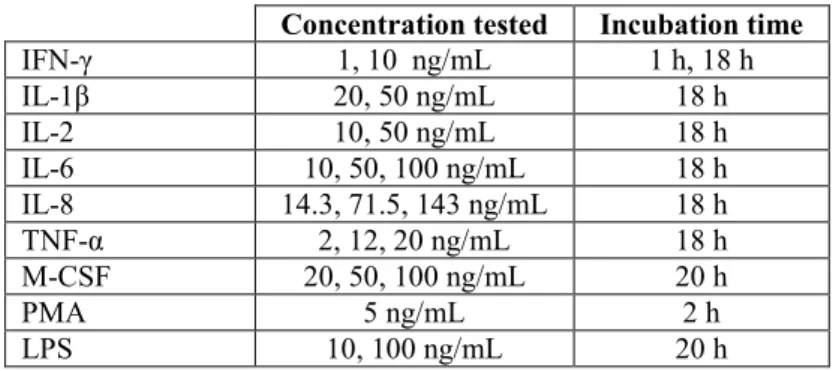
In order to study the effect of several putative pro-atherogenic stimuli on Cp expression at surface of PBMC, IFN-γ, IL-1β, IL-2, IL-6, IL-8, TNF-α (BD Pharmigen, USA) and M-CSF (R&D Systems) were used. Also, the inflammatory stimulators phorbol-12-myristate-13-acetate (PMA; Sigma-Aldrich, USA) and LPS from Escherichia coli (Sigma-Aldrich, USA) were tested. Below are described the conditions used for each treatment:
2
Table IV – Experimental conditions used to test the effect of cytokines and
other immunomodulators in PBMC culture
Concentration tested Incubation time
IFN-γ 1, 10 ng/mL 1 h, 18 h IL-1β 20, 50 ng/mL 18 h IL-2 10, 50 ng/mL 18 h IL-6 10, 50, 100 ng/mL 18 h IL-8 14.3, 71.5, 143 ng/mL 18 h TNF-α 2, 12, 20 ng/mL 18 h M-CSF 20, 50, 100 ng/mL 20 h PMA 5 ng/mL 2 h LPS 10, 100 ng/mL 20 h
3.3. Reactive nitrogen species
In the context of pro-atherogenic conditions present in the microenvironment of atherosclerotic plaques, ROS and RNS are very important to establish the pro-oxidant/anti-oxidant setting encountered at these sites.
To test the influence of specific RNS conditions on cell surface Cp expression of PBMC, cells from healthy donors were cultured overnight in the presence/absence of the NO donor S-nitroso-N-acetylpenicillamine (SNAP; Invitrogen, USA) at 100 and 500 µM concentrations.
4. Immunophenotyping and Flow Cytometry analysis
After the specific incubations, treated and non-treated PBMC were plated in 96-well round-bottomed microtiter plates (Nunclon, Denmark) at 5×10⁵ cells/well and incubated for 15 min with AB-Human Serum, blocking the Fc-receptor binding sites for reduction of non-specific reactions. Afterwards, cells were stained for Cp using the rabbit anti-Human Cp (KOMA Biotech, Korea) as primary antibody (Ab) for 45 min, washed once with Staining Buffer and spun 250 X g at 4ºC for 5 min. Subsequently the cells were incubated for 45 min with the secondary Ab, a goat F(ab′)2 anti-rabbit FITC-conjugated (Rockland, USA) along with monoclonal Abs (mAb) CD45-PerCP, CD14-APC, CD3-PE and CD69-PerCP.
After staining, the cells were washed once with Staining Buffer, spun 250 X g, 4ºC for 5 min and ressuspended in the same solution, transferred to cytometer tubes and analyzed in a FACSCalibur Flow Cytometer (BD Biosciences, USA).
To determine Cp mean fluorescence intensity (MFI) in specific cell subsets, fluorochrome-conjugated mAbs against CD45, CD14, CD3 and CD69 were used. MAb CD45-PerCP allows for lymphocyte gating and combined with mAb CD3-PE allows the positive or negative selection of T cells. Also, mAb CD45-PerCP with CD14-APC were used for monocyte gating. In each case, mAb CD69-PerCP was used to gate activated and non-activated cells. All mAb were from BD Pharmigen, USA. Non-treated stained cells were used as negative control, to
筐й
determine autofluorescence in each assay. CellQuest (BD Biosciences, USA) was used for data acquisition, and measurement of Cp expression was performed using Weasel v.2.7.4 software.
5. Quantification of oxidized LDL in cell culture supernatants
PBMC were isolated from healthy donors and PBMN e PBL were incubated overnight with RPMI medium in the presence/absence of 20 ng/mL of IL-1β and human LDL (Sigma-Aldrich, USA) reconstituted in nitrogen-saturated solution, to certify the LDL was in its native state and minimize its spontaneous oxidative modification not related to the effect of Cp.
The concentration of oxLDL in the culture supernatants was measured by a sandwich Enzyme-Linked-Immuno-Sorbent-Assay (ELISA) (oxLDL ELISA kit, K7810, ImmunDiagnostik AG, Germany) according to the kit protocol and measured spectrophotometrically with ELISA plate reader Milenia™ Kinetic Analyzer (Diagnostic Products Corporation, USA).
6. Laboratorial characterization of Familial Hypercholesterolemic patients: a model to study atherosclerosis
The groups of healthy individuals and FH patients was biochemically characterized through the determination of concentration levels of total cholesterol, HDL and LDL cholesterol, triglycerides, apolipoprotein A1 (ApoA1), apolipoprotein B (ApoB), Lipoprotein (a) (Lp(a)), Fe, Tf, ferritin, CRP and sCp, which were measured in serum samples (stored at -20ºC). Determination of lipid profile was performed by an enzymatic colorimetric method following the manufacturer’s instructions in an automated chemistry analyzer (Cobas Integra 400, Roche). Determination of CRP, Fe, Ferritin, Tf, sCp levels were executed in the same equipment with respective method. All these analysis were performed at Unidade Laboratorial Integrada (INSA).
Additionally, the levels of oxLDL and IL-1β were quantified in serum samples using the following immunoenzymatic assays (ELISAs): oxLDL ELISA Kit (ref. K7810, ImmunDiagnostik AG, Germany) and BD OptEIA™ ELISA IL-1β Kit (BD, USA) respectively, following the manufacturers’ instructions.
7. Statistical Analysis
All data analysis was performed using Microsoft™ Office Excel and SPSS v.17.
Results from each modulation experiment are presented as means ± Standard deviation in the respective figure. After the normality assessment was performed, to compared the means of two
筐й
related samples Student’s t-test was used. In alternative it was employed the non-parametric test Wilcoxon.
When comparing the two independent samples from FH patients and control group, after normality assessment was performed the parametric Student’s t test was employed. In alternative, the non-parametric test Mann-Whitney was used. Spearman correlation coefficient was used to analyze the relationship between the laboratorial characterization parameters of these two groups.
In all comparisons, the significance level to reject the null hypothesis was 5%, that is, p- values < 0.05 were considered statistical significant.
2
RESULTS
1. Ceruloplasmin expression at surface of peripheral blood mononuclear cells
Cp is usually described as a multifunctional protein with both anti-oxidant and pro-oxidant activities, being of crucial importance during the inflammatory process and acute phase response [67]. The intimate connection between Cp and the immune system [78] and possible role of Cp during atherogenesis is justified due to its existence in various cell types [76], including PBMN [74] and PBL [78]. Thus, in order to understand the possible physiological role of Cp expressed at the surface of specific leukocyte populations, this protein expression was analyzed at surface of PBMN and PBL isolated from healthy blood donors, using flow cytometry analysis.
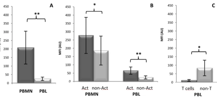
Results obtained after overnight incubation of PBMC with culture medium alone (RPMI-1640 medium) showed consistently higher cell surface expression of Cp in PBMN compared to PBL (Figure 3A) and in activated vs non-activated cells (Figure 3B). Also, analysis of Cp expression at surface of PBL subpopulations, showed that non-T cells have higher surface Cp expression compared to T cells (Figure 3C).
Fig. 3 –Flow Cytometry analysis of surface Cp expression in non-treated PBMC. A) Surface Cp expression
in PBMN and PBL (n=16); B) Surface Cp expression in PBMN and PBL depending on their activation state (n=12); C) Surface Cp expression in PBL subpopulations (n=16), (*p < 0.05, **p < 0.01).
2. Regulation of surface ceruloplasmin expression by iron and copper status
Human atherosclerotic plaques contain elevated levels of both Cu and Fe [68]. In order to test if Fe and/or Cu status influence the expression of surface Cp in PBMC, cells were treated in presence/absence of these metals under different experimental conditions.
0 50 100 150 200 250 300 350 400 450 1 M F I (A U ) PBMN PBL ** A 0 50 100 150 200 250 300 350 400 450 1 M F I (A U )
Act non-Act Act. non-Act
PBMN PBL ** * B 0 50 100 150 200 250 300 350 400 450 1 M F I (A U ) T cells non-T PBL * C
л
2.1. Modulation of iron status
2.1.1. Holotransferrin
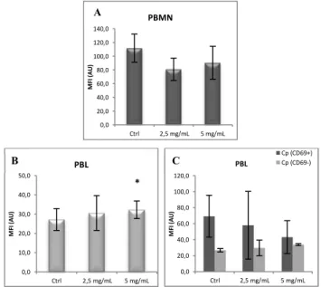
PBMC incubation with holo-Tf caused a 10% decrease in the percentage of Cp+ PBMN (data not shown), while a concomitantly non-statistically significant decrease in Cp surface expression was observed on this cells (Figure 4A).
On the other hand, PBL treated with holo-Tf showed upregulation of surface Cp expression in a dose dependent manner, being statistically significant for 5 mg/mL holo-Tf (Figure 4B) due to the upregulation of Cp expression in activated cells (Figure 4C). Also, a slight increase in the number of activated cells and Cp+ cells was observed in this cellular population (data not shown).
Fig. 4 -Effect on PBMC surface Cp expression after overnight incubation with Holo-Tf.
Flow cytometry results of Cp expression at surface of A) PBMN, B) PBL and C) activated and non-activated PBL incubated overnight with Holo-Tf (Ctrl: non-treated cells, n=3, *p<0.05).
2.1.2. Iron-NTA
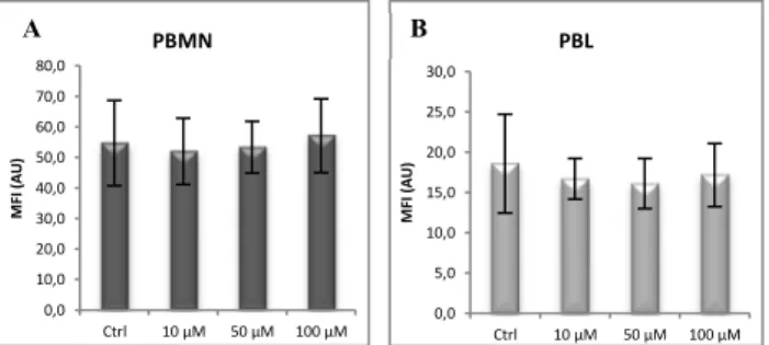
As observed in Figure 5, Fe-NTA treatment did not show any significant effect on Cp surface expression in PBMN and PBL (Figure 5A and 5B, respectively). In fact, only a trend to decrease in Cp expression at surface of PBMN could be observed, particularly at 100 µM Fe-NTA. 0,0 20,0 40,0 60,0 80,0 100,0 120,0 140,0 Ctrl 2,5 mg/mL 5 mg/mL M F I (A U ) PBMN 0,0 10,0 20,0 30,0 40,0 50,0 Ctrl 2,5 mg/mL 5 mg/mL M F I (A U ) PBL * 0,0 20,0 40,0 60,0 80,0 100,0 120,0 Ctrl 2,5 mg/mL 5 mg/mL M F I (A U ) PBL Cp (CD69+) Cp (CD69-) 0,0 50,0 100,0 150,0 200,0 Ctrl 50 µM 100 µM 200 µM M F I (A U ) PBMN 0,0 5,0 10,0 15,0 20,0 25,0 30,0 Ctrl 50 µM 100 µM 200 µM M F I (A U ) PBL A B
Fig. 5 - Effect on PBMC surface Cp expression after overnight incubation with Fe-NTA.
Results of flow cytometry analysis of surface Cp expression in A) PBMN and B) PBL incubated overnight with Fe-NTA (Ctrl: non-treated cells, n=3).
A
̠п
2.1.3. Desferrioxamine
Incubation of PBMC with the Fe chelator DFO showed a trend to an increased surface Cp expression in both cell populations analyzed. In PBMN the upregulation of surface Cp expression was statistically significant at 100 µM DFO (Figure 6A).
In PBL, no statistically significant differences were observed between cells treated with DFO and non-treated. However, a trend to an increase of surface Cp expression in a dose dependent manner was observed (Figure 6B), with a concomitantly increase in the percentage of activated and Cp+ cells (data not shown).
Fig. 6- Effect on PBMC surface Cp expression after overnight incubation with DFO. Flow
cytometry results of Cp expression at surface of A) PBMN and B) PBL in cell cultures incubated overnight with DFO (Ctrl: non-treated cells, n=3, *p<0.05).
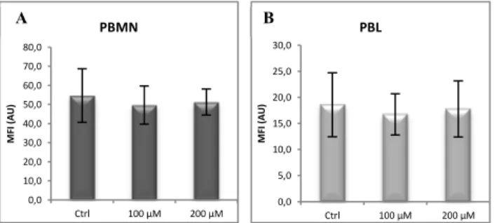
2.2. Modulation of copper status
Modulation of Cu status in vitro did not have any significant effect in surface Cp expression both in PBMN (Figure 7A) and PBL (Figure 7B). This finding was also observed in both PBMN (Figure 8A) and PBL (Figure 8B) when BCS Cu chelator treatment was tested.
Fig. 7- Effect on PBMC surface Cp expression after overnight incubation with CuHis.
Results of flow cytometry analysis of surface Cp expression in A) PBMN and B) PBL incubated overnight with CuHis (Ctrl: non-treated cells, n=3).
0,0 200,0 400,0 600,0 800,0 1000,0 1200,0 Ctrl 50 μM 100 μM M F I (A U ) PBMN * 0,0 10,0 20,0 30,0 40,0 50,0 Ctrl 50 μM 100 μM M F I (A U ) PBL 0,0 10,0 20,0 30,0 40,0 50,0 60,0 70,0 80,0 Ctrl 10 µM 50 µM 100 µM M F I (A U ) PBMN 0,0 5,0 10,0 15,0 20,0 25,0 30,0 Ctrl 10 µM 50 µM 100 µM M F I (A U ) PBL A B A B
р
Fig. 8- Effect on PBMC surface Cp expression after overnight incubation with BCS.
Results of flow cytometry analysis of surface Cp expression in A) PBMN and B) PBL incubated overnight with BCS (Ctrl: non-treated cells, n=3).
3. Regulation of surface ceruloplasmin expression by cytokines and other immunomodulators
In order to test the hypothesis of Cp immunomodulation by pro-atherogenic stimuli, measurement of Cp surface expression of PBMC cultured in the presence of several cytokines and immunomodulators putatively associated to atherogenesis, was performed.
3.1. IFN-γ
IFN-γ is produced locally on atherosclerotic plaques [101] and promotes macrophage and EC activation. It also inhibits cell proliferation, collagen production, and cholesterol efflux [102]. Importantly, IFN-γ has been often considered a major pro-atherogenic cytokine [103].
In this study, PBMC incubated for 1 h in culture medium in the presence of IFN-γ showed a slight increase of surface Cp in PBMN (Figure 9A) and PBL (Figure 9B). However, these differences were not statistically significant.
Fig. 9 - Effect on PBMC surface Cp expression after 1h incubation with IFN-γ. Flow
cytometry results of Cp expression at surface of A) PBMN and B) PBL incubated for 1 h with INF-γ (Ctrl: non-treated cells, n=3).
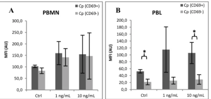
Observing Cp expression at surface of PBMN and PBL depending on their state of activation showed that while in PBMN the upregulation of surface Cp expression was due to both activated and non-activated cells (Figure 10A), in PBL the upregulation of Cp is mostly associated with activated cells (Figure 10B).
0,0 10,0 20,0 30,0 40,0 50,0 60,0 70,0 80,0 Ctrl 100 µM 200 µM M F I (A U ) PBMN 0,0 5,0 10,0 15,0 20,0 25,0 30,0 Ctrl 100 µM 200 µM M F I (A U ) PBL 0,0 50,0 100,0 150,0 200,0 250,0 Ctrl 1 ng/mL 10 ng/mL M F I (A U ) PBMN 0,0 10,0 20,0 30,0 40,0 50,0 Ctrl 1 ng/mL 10 ng/mL M F I (A U ) PBL A B A B
р
Fig. 10- Effect of IFN-γ on activated and non-activated PBMC surface Cp expression after 1h incubation. Flow cytometry results of Cp expression at surface of activated and non-activated A)
PBMN and B) PBL after 1 h incubation with INF-γ (Ctrl: non-treated cells, n=3).
On the other hand, in PBMC incubated in the same conditions but for a longer period of time (18 h), a decrease of Cp expression at cell surface was observed. Figure 11 shows the decreased expression in total PBMN (Figure 11A) and PBL (Figure 11B), being the decreased Cp expression in PBL statistically significant for 10 ng/mL (Figure 11B).
Fig. 11 - Effect on PBMC surface Cp expression after overnight incubation with IFN-γ. Flow
cytometry results of Cp expression at surface of A) PBMN and B) PBL incubated overnight with INF-γ (Ctrl: non-treated cells, n=3, *p<0.05).
Furthermore, in the presence of IFN-γ PBMN cell activation increased in a dose dependent manner (Figure 12A), while the percentage of activated PBL was not largely affected by this cytokine (Figure 12B).
Fig. 12 - Effect on PBMC cell activation after overnight incubation with IFN-γ. Flow
cytometry analysis of CD69+ expression at surface of A) PBMN and B) PBL in cell cultures incubated overnight with INF-γ (Ctrl: non-treated cells, n=3).
0,0 50,0 100,0 150,0 200,0 250,0 300,0 Ctrl 1 ng/mL 10 ng/mL M F I (A U ) PBMN Cp (CD69+) Cp (CD69-) 0,0 20,0 40,0 60,0 80,0 100,0 120,0 140,0 160,0 180,0 200,0 Ctrl 1 ng/mL 10 ng/mL M F I (A U ) PBL Cp (CD69+) Cp (CD69-) * * 0,0 50,0 100,0 150,0 200,0 250,0 Ctrl 1 ng/mL 10 ng/mL M F I (A U ) PBMN 0,0 5,0 10,0 15,0 20,0 25,0 30,0 Ctrl 1 ng/mL 10 ng/mL M F I (A U ) PBL * 0% 10% 20% 30% 40% 50% 60% 70% 80% 90% 100% Ctrl 1 ng/mL 10 ng/mL PBMN # cells CD69-# cells CD69+ 0% 10% 20% 30% 40% 50% 60% 70% 80% 90% 100% Ctrl 1 ng/mL 10 ng/mL PBL # cells CD69-# cells CD69+ A B A B A B
̠п
3.2. IL-1β
IL-1β is known as a lymphocyte activating and B-cell differentiation factor [104], but it was recently associated with CVD [105] and with ATH progression [106]. In this context, the effect of IL-1β on PBMC surface Cp expression was tested.
When PBMN were incubated with IL-1β at 20 and 50 ng/mL concentrations, an increase of surface Cp expression was observed in these cells (Figure 13A). Also, in this cell population, the number of activated cells increased with the treatment (data not shown), being surface Cp upregulation mostly associated with activated PBMN (Figure 13B).
Fig. 13 - Effect on PBMN surface Cp expression after overnight incubation with IL-1β.
Flow cytometry results of Cp expression at surface of A) PBMN and B) activated and non-activated PBMN in cell cultures incubated overnight with IL-1β (Ctrl: non-treated cells, n=3, *p<0.05)
In total PBL, no significant effect of IL-1β on surface Cp expression was observed when comparing treated and non-treated cells (Figure 14A). However, surface Cp expression showed a tendency to decrease in a dose-dependent manner when analyzing activated vs non-activated PBL (Figure 14B). The percentage of Cp+ PBL was not affected by this interleukin and only a minimal increase in cell activation could be observed in PBL (data not shown). Regarding PBL subpopulations, a significant decrease of surface Cp expression was observed in non-T cells when in the presence of 50 ng/mL IL-1β (Figure 14C). Concomitantly, a small but steady increase in the percentage of activated non-T cells was observed along a small but constant decline in Cp+ cells was observed (data not shown).
Fig. 14 - Effect on PBL surface Cp expression after overnight incubation with IL-1β. Flow cytometry results of Cp
expression at surface of A) PBL, B) PBL depending on their activation state and C) PBL subpopulations incubated overnight with IL-1β (Ctrl: non-treated cells, n=3, *p<0.05, **p<0.01).
0,0 100,0 200,0 300,0 400,0 500,0 Ctrl 20 ng/mL 50 ng/mL M F I (A U ) PBMN * 0,0 50,0 100,0 150,0 200,0 250,0 300,0 350,0 400,0 Ctrl 20 ng/mL 50 ng/mL M F I (A U ) PBMN Cp (CD69+) Cp (CD69-) * * * 0,0 10,0 20,0 30,0 40,0 50,0 60,0 70,0 Ctrl 20 ng/mL 50 ng/mL M F I (A U ) PBL 0,0 10,0 20,0 30,0 40,0 50,0 60,0 70,0 80,0 Ctrl 20 ng/mL 50 ng/mL M F I (A U ) PBL Cp (CD69+) Cp (CD69-) ** * * 0,0 20,0 40,0 60,0 80,0 100,0 120,0 140,0 160,0 Ctrl 20 ng/mL 50 ng/mL M F I (A U ) PBL T cells non-T cells Autofluo * A B A B C
![Table I - Risk factors for Development of ATH (From Greaves et al. [2])](https://thumb-eu.123doks.com/thumbv2/123dok_br/15616006.1054288/14.892.482.768.403.660/table-i-risk-factors-development-ath-greaves-et.webp)

![Table II - Summarized list of inflammatory mediators involved in atherogenesis (From Greaves & Channon [2])](https://thumb-eu.123doks.com/thumbv2/123dok_br/15616006.1054288/18.892.456.769.413.570/table-summarized-inflammatory-mediators-involved-atherogenesis-greaves-channon.webp)