online | memorias.ioc.fiocruz.br
Differential expression of the costimulatory molecules CD86, CD28,
CD152 and PD-1 correlates with the host-parasite outcome in leprosy
Maria de Lourdes Palermo1, Maria Ângela Bianconcini Trindade2, Alberto José da Silva Duarte1, Camila Rodrigues Cacere1, Gil Benard3,4/+
1Laboratório de Investigação Médica 56 3Laboratório de Investigação Médica 53, Divisão de Clínica Dermatológica,
Faculdade de Medicina 4Laboratório de Micologia Médica, Instituto de Medicina Tropical, Universidade de São Paulo, São Paulo, SP, Brasil 2Instituto de Saúde, Secretaria de Estado da Saúde de São Paulo, São Paulo, SP, Brasil
Leprosy is a spectral disease exhibiting two polar sides, namely, lepromatous leprosy (LL) characterised by impaired T-cell responses and tuberculoid leprosy in which T-cell responses are strong. Proper T-cell activation requires signalling through costimulatory molecules expressed by antigen presenting cells and their ligands on T-cells. We studied the influence of costimulatory molecules on the immune responses of subjects along the leprosy spectrum. The expression of the costimulatory molecules was evaluated in in vitro-stimulated peripheral blood mononuclear cells of lepromatous and tuberculoid patients and healthy exposed individuals (contacts). We show that LL patients have defective monocyte CD86 expression, which likely contributes to the impairment of the antigen presentation process and to patients anergy. Accordingly, CD86 but not CD80 blockade inhibited the lymphopro-liferative response to Mycobacterium leprae. Consistent with the LL anergy, there was reduced expression of the positive signalling costimulatory molecules CD28 and CD86 on the T-cells in these patients. In contrast, tubercu-loid leprosy patients displayed increased expression of the negative signalling molecules CD152 and programmed death-1 (PD-1), which represents a probable means of modulating an exacerbated immune response and avoiding immunopathology. Notably, the contacts exhibited proper CD86 and CD28 expression but not exacerbated CD152 or PD-1 expression, suggesting that they tend to develop a balanced immunity without requiring immunosuppressive costimulatory signalling.
Key words: Mycobacterium leprae - costimulatory molecules - CD80 - CD86 - CD152 - PD-1
Although the prevalence of leprosy has been decreas-ing all over the world, hyper-endemic areas persist where the transmission of leprosy is still out of control (WHO 2010). Leprosy, caused by the bacillus Mycobacterium leprae, is a chronic, incapacitating disease that affects the peripheral nerves and skin. Leprosy manifests as a spectral disease. The lepromatous form of the disease, comprising both borderline lepromatous leprosy (BL) and lepromatous leprosy (LL), is a consequence of the impaired immune cellular response of the patient and is characterised by an antigen-specific anergy, a Th-2 pat-tern of immune response and a lack of control of bacilli multiplication. The tuberculoid form of the disease, com-prising both borderline tuberculoid (BT) and tuberculoid tuberculoid (TT) leprosy, corresponds to the more benign form of the disease and is characterised by a strong gran-ulomatous response and the control of bacilli multiplica-tion (Yamamura et al. 1991, Sinsimer et al. 2010).
Thus, a protective immune response in leprosy is considered to rely on the cellular arm of the immune system, specifically on the generation of helper T-cells
Financial support: FAPESP, CNPq, CAPES, Fundação Paulista contra a Hanseníase
+ Corresponding authors: [email protected] Received 3 March 2012
Accepted 27 July 2012
CD86 and CD28 are constitutively expressed by APC and T-cells, respectively, CD80 and CD152 are expressed only following 24-48 h of activation of the APC and T-cell (Lenschow et al. 1996, Bour-Jordan et al. 2011). A model of T-cell costimulation has thus been proposed in which the distinct structures and binding properties of CD86 and CD80 significantly enhance the activating and inhibitory functions of CD28 and CD152, respec-tively (Collins et al. 2002). This seems to be relevant to the regulation of the immune responses in several clini-cal conditions. For example, the increased expression of CD86 by monocytes was shown to play a key role in the exacerbated inflammatory response of multiple sclerosis (Filion et al. 2003); conversely, the defective expression of CD86 on monocytes was shown to be crucial in urae-mia-associated immunodeficiency (Girndt et al. 2001).
However, other costimulatory molecules have been described that influence the subsequent adaptive im-mune responses, such as programmed death-1 (PD-1) and inducible costimulatory protein (ICOS) (Green-wald et al. 2005). These molecules have been shown to play major roles in triggering autoimmunity or pe-ripheral tolerance and directing the protective immune response to pathogens or facilitating the persistence of the parasite leading to chronic immune activation (Zhu et al. 2011, Bour-Jordan et al. 2011).
This study aimed to verify the influence of costim-ulatory molecules on the immune response of subjects along the leprosy spectrum. The expression levels of the costimulatory molecules CD80, CD86, CD28, CD152, PD-1 and ICOS were evaluated in in vitro-stimulated peripheral blood mononuclear cells (PBMCs) from both lepromatous and tuberculoid patients and healthy exposed individuals.
PATienTS, MATeRiALS AnD MeThoDS
Patients and healthy contacts - A total of 28 con-secutive, newly diagnosed leprosy patients admitted to our service were analysed. All patients were studied be-fore the start of the specific therapy; they were all human immunodeficiency virus-negative and had no other in-fectious comorbidities. Some of the patients had partici-pated in a previous study (Palermo et al. 2012). Fourteen had tuberculoid leprosy (age range 43 ± 5 years, 4 female and 10 male subjects), 11 of whom presented with the BT and three of whom presented TT. The other 14 patients had LL (age range 38 ± 4 years, 6 female and 8 male subjects); of these patients, 10 had BL and four presented polar LL. A control group composed of 10 healthy M. leprae-exposed individuals (contacts, age range 32 ± 4 years) was selected from among persons living in close contact with the LL patients. Exposure was defined by a positive response in the lymphoproliferation as-say with M. leprae antigen, as described below (Table). The study was approved by the Ethical Committee of the Clinics Hospital, University of São Paulo Medical School (protocol 0955/08). Informed consent was ob-tained from all participants.
PBMC isolation and cultures - PBMCs were cultured as previously described (Cacere et al. 2008). Briefly, PB-MCs were isolated from heparinised peripheral blood
by a density gradient and then resuspended in RPMI medium supplemented with gentamicin (40 µg/mL) and 10% human AB serum (Sigma-Aldrich, St. Louis, MO, USA). PBMCs (2.5 x 10⁵/well) were cultivated at 37ºC
and 5% CO2 in the presence of medium only, a cell wall preparation from M. leprae (MLCwA)(10 µg/mL) or, as a positive control, phytohaemagglutinin (PHA) (5 µg/ mL) (Sigma, Saint Louis, MO, USA). For the lymphop-roliferation assays, cells were incubated for three days (PHA) or six days (MLCwA) and then pulsed for an additional 18 h with 1.0 µCi/well [3H]-thymidine (Ra-diochemical Centre, Amersham, UK) before harvest-ing. Cell-bound radioactivity was measured using a
β-counter (Perkin-Elmer, Boston, MA, USA). In some
experiments, the following blocking monoclonal
anti-bodies were added to the cultures, at 10 μg/mL, from
the start of the culture (day 0), as described elsewhere (Cacere et al. 2008): anti-CD152 (IgG2a, clone BI3) (BD Pharmingen, San Diego, CA, USA), anti-CD80 (IgG1,6 clone 2D10.4) (eBioscience, San Diego, CA, USA) and anti-CD86 (IgG2b, clone IT2.2) (eBioscience).
The expression of costimulatory molecules by mono-cytes and T-cells - For T-cell staining, PBMCs, which were isolated as described above from both the con-tacts and the leprosy patients, were cultured in 24-well plates for four days and the cells were then carefully collected. Day 4 was considered to be optimal for the expression of CD152 and the B7 family of molecules on T-cells according to preliminary time-course ex-periments and previous reports (Cacere et al. 2008). The cells were washed twice with cold phosphate buffered saline (PBS) and then re-suspended at 1 x 10⁶ cells/mL in
PBS/bovine serum albumin (BSA) 1% buffer. Aliquots containing 2.5 x 10⁵ cells were distributed to cytometer
tubes and the Fc receptors were blocked by incubation for 20 min with human immunoglobulin (50 mg/mL) (Baxter Biosciences, Vienna, Austria). After washing with PBS-BSA 1%, 10 µL of the following antibodies was added: anti-CD3 (tri-colour, Caltag) and anti-CD28 (PE, Caltag), anti-CD152 (Cy-chrome, Pharmingen), anti-PD-1 (FITC, eBioscience), anti-ICOS (PE, eBio-science), anti-CD80 (FITC, Caltag) or anti-CD86 (PE, Caltag). To analyse the expression of CD86 and CD80 on peripheral blood monocytes, CD14+ cells were isolated
from the entire PBMC population by positive selection with anti-CD14-conjugated beads (EasySep, StemCell Technologies). The purity of the population was always
≥ 98%. The purified CD14+ population was allowed to
rest in 24-well plates for 48 h at 37ºC and 5% CO2, a step that was necessary for the cells to return to a rest-ing state after the column passage. After the 24 h restrest-ing period, MLCwA or Candida Metabolic Antigen (Sanofi-Pasteur, Paris, France) (5 µg/mL), a control non-related antigen, was added and the incubation was continued for 4 h. Monocytes were carefully collected from the wells through several washes with cold RPMI using a 1 mL pipette and the cells were washed again with PBS/BSA 1%, resuspended in PBS/BSA 1% buffer at 106 cells/mL
both T-cell and monocyte staining, the tubes containing the labelled cells were gently vortexed and incubated in the dark for 30 min at room temperature, resuspended in 500 µL of PBS/BSA 1% buffer, washed in PBS to re-move unbound antibody and finally suspended in PBS-azide. In each experiment, one tube remained without mAb addition as an autofluorescence control. In addi-tion, each protocol was set up with colour compensation controls. Cells were immediately acquired and analysed on a Coulter flow cytometer using the software System II (Coulter Epics XL-MCL, Hialeah, FL, USA). The results were expressed as percentages of costimulatory mole-cule-expressing cells and mean fluorescence intensities (MFIs) per at least 10,000 CD3+ or CD14+ gated cells.
Statistical analysis - The comparison of data from among the three groups was performed using one-way analysis of variance and the Newman-Keuls multiple comparison test. The differences were considered sig-nificant when p < 0.05. GraphPad Prism 5.0 software was used (Graph Pad Software Inc San Diego, CA).
ReSuLTS
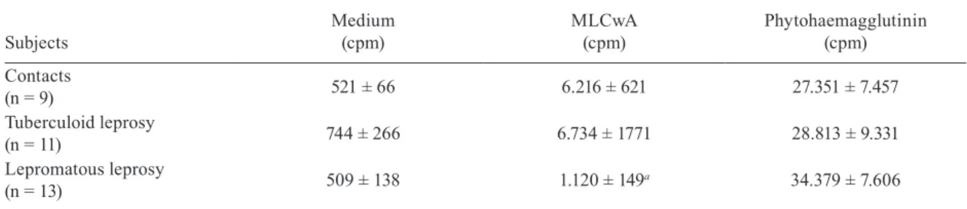
Proliferative responses - The Table shows that the PBMCs of patients with the lepromatous form of leprosy were anergic to MLCwA, while positive responses were observed for the tuberculoid patients and contacts. The responses to PHA were not significantly different among the three groups.
CD86 and CD80 expression by CD14+ cells -
Signifi-cantly fewer monocytes (CD14+ cells) from lepromatous
patients (~50%) expressed the costimulatory molecule CD86 compared with tuberculoid patients and contacts (~80%) after 4 h of culture in the presence of medium only or MLCwA (Fig. 1A). There were no significant dif-ferences in CD86 expression when the monocytes were challenged with a control antigen from Candida spp. The analysis of the CD86 MFI data revealed a similar trend; monocytes from lepromatous patients expressed the mol-ecule at lower densities in the presence of medium only or MLCwA (Fig. 1B). In contrast, CD80 was poorly ex-pressed by monocytes (< 10%) that were either unchal-lenged or chalunchal-lenged with the antigens, with no significant differences among the three groups (data not shown).
The expression of costimulatory molecules by CD3+
cells - CD28 was similarly expressed by high percent-ages of CD3+ cells from all three groups of patients (Fig.
2C); however, the MFI differed among the groups (Fig. 2D). CD3+ cells from unstimulated and
MLCwA-stimu-lated cultures of PBMCs from both lepromatous patients
TABLE
Lymphocyte proliferative responses
Subjects
Medium (cpm)
MLCwA (cpm)
Phytohaemagglutinin (cpm)
Contacts
(n = 9) 521 ± 66 6.216 ± 621 27.351 ± 7.457
Tuberculoid leprosy
(n = 11) 744 ± 266 6.734 ± 1771 28.813 ± 9.331
Lepromatous leprosy
(n = 13) 509 ± 138 1.120 ± 149
a 34.379 ± 7.606
a: p < 0.01 vs. contacts and tuberculoid leprosy (one-way analysis of variance with Newmans-Keuls post-test); cpm: counts per minute; MLCwA: Mycobacterium leprae antigen.
Fig. 1: expression of CD86 on monocytes (CD14+ cells) from
and contacts expressed CD28 at lower intensities than those from tuberculoid patients. The CD28 MFI was comparable in CD3+ cells stimulated with PHA, which
was used as a positive control.
As observed above for CD14+ cells, CD80 expression
in unstimulated and MLCwA-stimulated CD3+ cells was
poor (< 10%) in all three groups (data not shown). CD86 was expressed by higher numbers of CD3+ cells, but with
no significant differences among the three groups (Fig. 2A). However, the MFI of CD86 in lepromatous patients’ CD3+ cells was significantly reduced compared with the
MFIs from tuberculoid patients and contacts in all three experimental conditions (Fig. 2B).
The expression of the negative signalling molecules CD152 and PD-1 also differed among groups (Fig. 3). Higher proportions of unstimulated and PHA and ML-CwA-stimulated CD3+ cells from tuberculoid patients
expressed CD152 with a higher MFI than the same cells from lepromatous patients and contacts (Fig. 3A, B). With regard to PD-1, significantly higher proportions of CD3+ cells from both lepromatous and tuberculoid
pa-tients expressed this molecule compared with contacts either when cultured in medium only or in the presence of MLCwA (Fig. 3C). With PHA, the patients’ CD3+ cells
also expressed higher levels than the cells from contacts, although the differences did not reach statistical signifi-cance. PD-1 MFI did not differ among groups (Fig. 3D). ICOS expression was low in CD3+ cells and was not
different among groups in any of the experimental con-ditions studied (data not shown).
Blocking experiments - We tested the effect of anti-CD152, anti-CD86 and anti-CD80 blocking antibodies in PBMC cultures from six donors, three tuberculoid pa-tients and three contacts who had positive proliferative responses to MLCwA. As shown in Fig. 4, CD86 block-ade reduced the proliferative response to MLCwA to less than 50% of the control cultures while CD80 blockade had no effect; CD152 blockade slightly but significantly increased the response.
DiSCuSSion
Our data demonstrate that CD86, but not CD80, seems to play a critical role in the presentation of leprosy anti-gens by monocytes. The blockade of CD86 signalling, but not the blockade of CD80 signalling, with a neutralising monoclonal antibody resulted in significant inhibition of the proliferative response to M. leprae antigens. Accord-ingly, CD86, but not CD80, was differentially expressed among contacts and patients; the former was highly pressed by monocytes from both healthy individuals ex-posed to M. leprae (contacts) and patients at the tubercu-loid pole of leprosy, while the latter was poorly expressed by both patients and contacts. In contrast, monocytes from patients at the LL pole exhibited a striking deficien-cy in the expression of CD86. This deficiendeficien-cy may help to explain the well-described differences in the T-cell responses between lepromatous and tuberculoid patients (Yamamura et al. 1991). In fact, unlike the tuberculoid patients, our lepromatous patients were unable to mount strong cellular immune responses against M. leprae anti-gens, as demonstrated in vivo by the poor granulomatous
Fig. 2: expression of CD86 and CD28 on T-lymphocytes (CD3+ cells)
inflammatory response and high bacillary load reported in a previous study (Palermo et al. 2012) and as observed in vitro in this study by the reduced antigen-specific pro-liferative responses. These findings may be explained by the reduced expression of CD86 during the early stage of recognition and presentation of M. leprae-derived an-tigens in lepromatous lesions, which interferes with the formation of the immune synapse that is required to ef-ficiently activate the T-cells and generate effector T-cells (Lanzavecchia & Sallusto 2000).
Previous data on CD80 and CD86 in leprosy are scarce and controversial, suggesting either a role for CD80 in the anergy of lepromatous patients or enhanced CD80 expression as a predictor of reaction states (Agre-wala et al. 1998, Schlienger et al. 1998, Santos et al. 2007). Differences in the methods and the types of cells analysed and in the nature of the antigen used may ac-count for the different results.
Several mechanisms may be put forward to explain the deficient CD86 expression by LL patients. T-regs have been shown to down-modulate the expression of CD80 and CD86 on APCs (Cederbom et al. 2000), likely through direct cell-to-cell interactions. We have previously dem-onstrated that these patients have increased numbers of T-regs, both in situ and in vitro (Palermo et al. 2012). IL-10 has been shown to down-modulate CD86 expression on monocytes (Creery et al. 1996). In LL, the monocytes are exposed to highly IL-10-enriched microenvironments because in this form of leprosy, IL-10 expression is sig-nificantly enhanced in both in situ and in vitro-stimulated PBMCs; in contrast, in tuberculoid leprosy, the IL-10 lev-els are low (Moubasher et al. 1998, Palermo et al. 2012). Alternatively, the process of monocyte invasion by M. leprae by itself could down-modulate CD86 expression on the host cell, as has been demonstrated with Mycobac-terium tuberculosis infection (Castaño et al. 2011).
Differential costimulatory molecule expression by T-cells was also verified among our lepromatous and tuberculoid patients and contacts. Molecules that either signal for (CD86 and CD28) or reduce (CD152 and PD-1) T-cell activation were evaluated. Probably as a con-
Fig. 3: expression of CD152 and programmed death-1 (PD-1) on T-lymphocytes (CD3+ cells) from tuberculoid (n= 9-14) and
leproma-tous (n= 10-11) leprosy patients and healthy exposed contacts (n= 8) stimulated with phytohemaglutinnin (PHA) or a Mycobacterium leprae antigen (MLCwA) or unstimulated (medium). Results are pre-sented as mean ± standard error of the means of the percentage of expressing cells (A, C) and mean fluorescence intensity (MFI) of the molecule (B, D). *: p< 0.05; **: p < 0.01.
Fig. 4: effect of anti-CD80, anti-CD86 and anti-CD152 blocking monoclonal antibodies on the lymphocyte proliferative response to
Mycobacterium leprae antigen (MLCwA) of six subjects responders
sequence of defective APC function, T-cells from lep-romatous patients were driven to an anergic state and exhibited reduced expression of both CD86 and CD28, especially when compared with tuberculoid patients. The latter exhibited the highest levels of expression, especially regarding CD28 expression, which was even significantly higher than even that of the contacts. This would indicate an on-going exacerbated T-cell response. However, T-cells from these patients also exhibited en-hanced CD152 expression, possibly preventing an un-controlled immune reaction. The net result would be an effective immune response capable of limiting bacillary multiplication and the dissemination of lesions without causing immunopathology. The expression of CD152 by the T-cells from contacts was significantly lower than that from tuberculoid patients, which suggests that distinct immune regulatory mechanisms take place in tuberculoid patients’ and contacts’ M. leprae-specific responses. Consistent with the fact that CD152 expres-sion is cell-activation dependent, when resting-state molecules found in intracellular reservoirs are rapidly mobilised to the cell surface (Schneider et al. 2006), T-cells from lepromatous patients exhibited low levels of expression of this molecule. Moreover, corroborating with its inhibitory function in leprosy, the blockade of CD152 resulted in a slight but significant enhancement of the M. leprae-specific proliferative responses.
Interestingly, the pattern of expression of the inhibi-tory molecule PD-1 differed from that of CD152. In fact, it has been suggested that PD-1 and CD152 play comple-mentary and non-overlapping roles in peripheral T-cell hypo-responsiveness (Fife & Bluestone 2008). CD152 may predominantly control the T-cell response at the induction phase, while PD-1 may be responsible for the maintenance of the tolerant or anergic state. PD-1 intrin-sically inhibits the function of effector T-cells (Fife et al. 2006) and it may also act by affecting the stability of their ligation with DCs (Fife et al. 2009). A significantly higher number of T-cells expressed PD-1 in tuberculoid and lepromatous patients than in contacts, thus serving as a marker of disease. In fact, PD-1 is highly expressed in situations of high and constant antigen exposure, such as chronic viral infections or malignancy (Barber et al. 2006, Zhu et al. 2011). Chronic exposure to mycobacte-rial antigens is indeed a feature of leprosy patients due to the protracted evolution of the disease.
Finally, the T-cell expression levels of CD80 and ICOS were also examined and indicated low levels of expression (data not shown), likely suggesting that they play minor roles in the regulation of the immune re-sponse in leprosy.
Based on our findings, a model can be built that proposes that, in LL, the defective APC function due to reduced CD86 expression by these cells results in an abnormal T-cell activation characterised by reduced antigen-specific proliferative responses and the deficient expression of CD28 and CD86 on T-cells. CD152 is not required for the induction of this anergic state. However, the chronic exposure to M. leprae antigens enhances PD-1 expression, which reinforces the state of tolerance against the bacillus. Consequently, patients develop a
dis-seminated multi-bacillary disease. In tuberculoid lepro-sy, proficient APC function exists, positive proliferative responses are detected and high expression levels of posi-tive signalling molecules on T-cells (CD28 and CD86) are detected. However, these positive signalling molecules need to be counter-regulated by the enhanced expression of inhibitory molecules (CD152 and PD-1), thus allow-ing a balanced effective immune response to be reached, i.e., control of the parasite burden without causing im-mune pathology. In contacts, there is no deficiency in the antigen-presenting process and a balanced immune response “naturally” develops because enhanced expres-sion of negative signalling molecules is not required. A better understanding of the immune regulation of the host immune response in leprosy may allow for immune interventions that would impact the prognosis of this still poorly controlled infectious disease.
ReFeRenCeS
Agrewala JN, Kumar B, Vohra H 1998. Potential role of B7-1 and CD28 molecules in immunosuppression in leprosy. Clin Exp Im-munol 111: 56-63.
Barber DL, Wherry EJ, Masopust D, Zhu B, Allison JP, Sharpe AH, Freeman GJ, Ahmed R 2006. Restoring function in exhausted CD8 T cells during chronic viral infection. Nature439: 682-687.
Bhatia S, Edidin M, Almo SC, Nathenson SG 2006. B7-1 and B7-2: similar costimulatory ligands with different biochemical, oligo-meric and signaling properties. Immunol Lett104: 70-75.
Bour-Jordan H, Esensten JH, Martinez-Llordella M, Penaranda C, Stumpf M, Bluestone JA 2011. Intrinsic and extrinsic control of peripheral T-cell tolerance by costimulatory molecules of the CD28/B7 family. Immunol Rev 241: 180-205.
Cacere CR, Mendes-Giannini MJ, Fontes CJ, Kono A, Duarte AJS, Benard G 2008. Altered expression of the costimulatory mol-ecules CD80, CD86, CD152, PD-1 and ICOS on T-cells from paracoccidioidomycosis patients: lack of correlation with T-cell hyporesponsiveness. Clin Immunol129: 341-349.
Castaño D, Barrera LF, Rojas M 2011. Mycobacterium tuberculosis
alters the differentiation of monocytes into macrophages in vitro.
Cell Immunol268: 60-67.
Cederbom L, Hall H, Ivars F 2000. CD4+CD25+ regulatory T-cells
down-regulate co-stimulatory molecules on antigen-presenting cells. Eur J Immunol30: 1538-1543.
Collins AV, Brodie DW, Gilbert RJ, Iaboni A, Manso-Sancho R, Walse B, Stuart DI, van der Merwe PA, Davis SJ 2002. The interaction properties of costimulatory molecules revisited. Im-munity17: 201-210.
Creery WD, Diaz-Mitoma F, Filion L, Kumar A 1996. Differen-tial modulation of B7-1 and B7-2 isoform expression on human monocytes by cytokines which influence the development of T helper cell phenotype. Eur J Immunol 26: 1273-1277.
Fife BT, Bluestone JA 2008. Control of peripheral T-cell tolerance and autoimmunity via the CTLA-4 and PD-1 pathways. Immunol Rev224: 166-182.
Fife BT, Guleria I, Gubbels Bupp M, Eagar TN, Tang Q, Bour-Jordan H, Yagita H, Azuma M, Sayegh MH, Bluestone JA 2006. Insulin-induced remission in new-onset NOD mice is maintained by the PD-1-PD-L1 pathway. J Exp Med203: 2737-2747.
Filion LG, Matusevicius D, Graziani-Bowering GM, Kumar A, Freedman MS 2003. Monocyte-derived IL-12, CD86 (B7-2) and CD40L expression in relapsing and progressive multiple sclero-sis. Clin Immunol106: 127-138.
Girndt M, Sester M, Sester U, Kaul H, Köhler H 2001. Defective expres-sion of B7-2 (CD86) on monocytes of dialysis patients correlates to the uremia-associated immune defect. Kidney Int59: 1382-1389.
Greenwald RJ, Freeman GJ, Sharpe AH 2005. The B7 family revis-ited. Annu Rev Immunol 23: 515-548.
Lanzavecchia A, Sallusto F 2000. From synapses to immunological memory: the role of sustained T cell stimulation. Curr Opin Im-munol12: 92-98.
Lenschow DJ, Walunas TL, Bluestone JA 1996. CD28/B7 system of T cell costimulation. Annu Rev Immunol 14:233-258.
Moubasher AD, Kamel NA, Zedan H, Raheem DD 1998. Cytokines in leprosy. I. Serum cytokine profile in leprosy. Int J Dermatol 37: 733-740.
Mueller DL, Jenkins MK, Schwartz RH 1989. Clonal expansion ver-sus functional clonal inactivation: a costimulatory signalling pathway determines the outcome of T cell antigen receptor oc-cupancy. Annu Rev Immunol7: 445-480.
Murray RA, Siddiqui MR, Mendillo M, Krahenbuhl J, Kaplan G 2007. Mycobacterium leprae inhibits dendritic cell activation and maturation. J Immunol178: 338-344.
Palermo ML, Pagliari C, Trindade MAB, Yamashitafuji TM, Duarte AJS, Cacere CR, Benard G 2012. Increased expression of regula-tory T cells and down regularegula-tory molecules in lepromatous lep-rosy. Am J Trop Med Hyg 86: 878-883.
Santos DO, Castro HC, Bourguignon SC, Bastos OM, Rodrigues CR, Van Heuverswyn H, Nery JA, Miranda A 2007. Expression of B7-1 costimulatory molecules in patients with multibacillary lep-rosy and reactional states. Clin Exp Dermatol32: 75-80.
Schlienger K, Uyemura K, Jullien D, Sieling PA, Rea TH, Linsley PS, Modlin RL 1998. B7-1, but not CD28, is crucial for the mainte-nance of the CD4+ T-cell responses in human leprosy. J Immunol 161: 2407-2413.
Schneider H, Downey J, Smith A, Zinselmeyer BH, Rush C, Brewer JM, Wei B, Hogg N, Garside P, Rudd CE 2006. Reversal of the TCR stop signal by CTLA-4. Science313: 1972-1975.
Sinsimer D, Fallows D, Peixoto B, Krahenbuhl J, Kaplan G, Manca C 2010. Mycobacterium leprae actively modulates the cytokine response in naïve human monocytes. Infect Immun78: 293-300.
WHO - World Health Organization 2010. Leprosy - global situation 2010. Wkly Epidemiol Rec85: 337-348.
Yamamura M, Uyemura K, Deans RJ, Weinberg K, Rea TH, Bloom BR, Modlin RL 1991. Defining protective responses to patho-gens: cytokine profiles in leprosy lesions. Science254: 277-279.