PROCEEDINGS
OF THE
X
thINTERNATIONAL
PEAR SYMPOSIUM
ISHS Section Pome and Stone Fruits ISHS Working Group on European and Asian Pears
Acta Horticulturae 800
Microstructural Changes of 'Rocha' Pear Following Storage under
Controlled Atmosphere
S.C. Fonseca and F.X. Malcata Escola Superior de Biotecnologia Universidade Cat6lica Portuguesa 4200-072 Porto
Portugal
A.C. Galvis-Sanchez Departamento de Quimica Universidade de Aveiro
Campus Universitario de Santiago 3810-193 Aveiro
Portugal
Abstract
This research effort was aimed at evaluating the influence of storage (for 4 mo at 2°C), under various controlled atmospheres (viz. 1.9 kPa O2+4.9 kPa CO2, 1.9 kPa O2+0.5 kPa C02 and 1.9 kPa O2 +0 kPa C02), on the microstructure of 'Rocha' pear. Toward this goal, the morphology of cellular disassembly, as well as the quantity of granules and intercellular space using scanning electron microscopy (SEM), were tentatively correlated with sensory and instrumental firmness, by 1, 6 and 8 d of exposure to air, at room temperature, after storage. A specific methodology, based on panel evaluation of SEM images, was developed and statistically validated. The degree of cellular disassembly increased throughout exposure time to air at room temperature. Pears stored under 1.9 kPa O2+0.5 kPa CO2 yielded a degree of cellular disassembly similar to that of the control.
INTRODUCTION
Fruits are typically characterized by a complex cellular structure - which is known to directly influence texture, as perceived by the consumer (Aguilera, 2000; Kalab, 1995) - which may hamper marketability. Microstructure can be characterized via scanning electron microscopy (SEM). The possibility of observation of (quasi) three-dimensional images, and the ease of use make SEM an interesting analytical tool for microstructural studies. However, the quality of SEM images is strongly dependent on preliminary procedures of sample preparation, because analytical artefacts may be inadvertently introduced; fresh fruits are indeed living structures - which are extremely fragile, so cutting of thin sections may disrupt the tissues to be observed themselves. Additionally, the very high water content thereof creates difficulties to sample drying. Therefore, one must normally resort to alternative preparation procedures, viz. freezing. Another limitation of SEM is the difficulty to carry out image analysis according to objective parameters - in attempts to quantify structural features, due to the complexity of SEM images. Novel methodologies are thus urged in this field of study that will eventually be able to take full advantage of SEM.
The main objective of this work was thus to evaluate the effects of controlled atmospheres, with different composition, upon the microstructure of 'Rocha' pear throughout storage - in attempts to better understand the influence of the overhead storage atmosphere on the fruit final quality. To achieve this goal, three sub-objectives were put forward and duly pursued: i) to test a sample preparation procedure, tailored for 'Rocha' pear analysis by SEM; ii) to develop a methodology to quantify SEM images of 'Rocha' pear samples; and iii) to relate microstructural parameters to sensory and instrumental quality parameters of 'Rocha' pears.
Plant Material and Handling
'Rocha' pears were grown at Alcoba<;a, Portugal, under controlled cultural practices. Immediately following harvest, fruits were transported to Escola Superior de
Proc.
x
thIS an PearEds.: A.D. Webster and C.M. Oliveira
Biotecnologia (Porto, Portugal). Pears free of defects were carefully cleaned, and cooled overnight to 4°C, under 93±2% relative humidity. On the following day, pears were placed in plastic containers (with a capacity of 7 L, and containing ca. 2.0 kg of fruits each). The (hermetically sealed) lids of those containers were potted to two rubber tubes for selected gas flushing; one of those tubes extended from the inlet to the bottom of the container, so as to facilitate uniform flushing of the gas mixture. The containers were weighted and stored in a Fitoclima chamber (model D 1200 PH, from Aralab, Portugal!.. according to the experimental design detailed below.
Experimental Design
Pears in containers were stored under constant temperature and relative humidity. and were flushed with four distinct atmospheres: 1.9 kPa O2
+
4.9 kPa CO2 (balance witt N2), 1.9 kPa O2+
0.5 kPa C02 (balance with N2), 1.9 kPa O2 (balance with N2) and air (control). The temperature therein was kept at 2±0.5°C, and the relative humidity a: 93±2%. The atmospheres were maintained via customized gas mixtures, storedm
pressurized bottles prepared in advance (by Gasin, Portugal). For each modified atmosphere, a bottle of the appropriate gas mixture was connected by polyamide tubingtC'a flowboard inside the chamber. Each flowboard delivered a constant (and predefmed. gas flow rate - 13 ml.min-1(determined so that the respiration process would Il((
significantly affect the O2 and CO2 partial pressures inside), to each of four containers. Before the experiment started, each flowboard output was criteriously calibrated and regulated, so as to guarantee a constant, preset gas flow rate. Gas streams were previous~ humidified via bubbling through deionized water, in order to avoid weight losses of the product due to dehydration. Throughout the whole storage period, the pear weight loss was below 1%(w/w). Fruits were removed from storage by 4 mo, and allowed to ripen IT: air at room temperature (19±1°C) for 8 d. Ten pears were taken at random from each storage condition, and assessed in terms of microstructural parameters, by 1, 6 and 8 d of exposure to air at room temperature.
Image Acquisition and Analysis
Samples of pears were taken randomly at the time of harvest, and after 4 mo of storage under each storage condition, followed by exposure to air at room temperature for distinct periods (1, 6 and 8 d). The peel was removed, and the flesh was cut into small. oriented blocks (1 x 5 x 5 mm) with a razor blade, immersed in liquid nitrogen and stored at -80°C. Frozen pear samples were mounted on stubs in a cold stage at -22°C (so as
w
maintain the frozen state), and observed by low vacuum SEM using a JSM-5600LY instrument (JEOL, Japan), operated at an accelerating voltage of 15 kY. Images were taken in representative parts of the sample, and observed at magnifications of 55x and 200x. A methodology to analyze SEM images, based on techniques currently used in sensory analysis via a trained panel, was specifically designed and tested (Langton, 1995,_ In order to prepare the evaluation sheet and to select the panelists, three steps took place as described below:1. Identification of descriptors and definition of scales. A large number of people (N=20) were exposed to SEM images taken at random, and were asked to use their own terminology in attempts to individually describe the aforementioned images. The most consensual terms were thus selected, viz. cellular disassembly (CD), quantity of granules (G) and intercellular space (IS); a sheet was thus built, bearing continuous 1-9 intensity rating scales (1=absence, 3=very little, 5=moderately, 7=very much and 9=extremely intense) for each descriptor.
II. Evaluation of discriminatory and reproducibility capacities of panelists. A set of four images - two of which were the same (with one rotated 90°, and flipped horizontally relative to the other), and the other two of which were representative of the scale extremes, was presented to 14 potential panelists; they were then asked to obserw each image, and make an assessment in terms of previously selected characteristics. The objective here was to assess the capacity of the various (potential) panel members to
distinguish different images apart from similar images.
III. Selection of panel. The selection of the panel members was according to the higher scores of the capacity demonstrated thereby in step II.
After these three preliminary steps were taken, the following methodology was chosen for assessment, by the panellists, of the images taken from pear samples after 4 mo of storage, at different combinations of storage conditions and time of exposure to air: (i) select a set of four images of the same time at room temperature, one for each storage condition, under the same magnification; (ii) place each set of four images on the same sheet, and code each image with a three digit-label as identification; and (iii) ask the selected panelists to observe each image, and make an assessment in terms of the previously selected characteristics.
Statistical Analysis
The experimental results were subject to two-factor analysis of variance, followed by application of Tukey's test to detect differences (at a significance level of 5%), using the SPSS software (USA).
RESULTS AND DISCUSSION
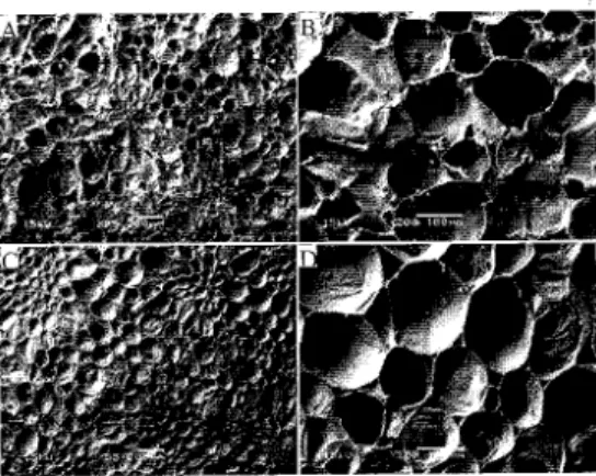
Selected scanning electron micrographs of 'Rocha' pear flesh after harvest are presented in Figure 1. Note that, prior to storage, the fruit had a turgid, ordered and well-defined cell structure. Said structure is formed by cell walls, and confers shape and form to the corresponding tissue. The bulk edible portion of most fruits corresponds to the parenchyma tissue - made of unspecialized cells, ranging in form from spherical to polyhedral. Clusters of cells, evolving from small cells in th~ center to larger elongated cells in the periphery, can be observed (Fig. lC); these clusters are called stone cells (or sclereids), and are cells that convey a gritty appearance and a grainy mouthfeel to pears (Reeve, 1970; Jewell, 1979) - the 'Rocha' variety is characterized by numerous stone cell groups (Fig. lA, C). The cells between the sclereids are roughly isodiametric, ranging from 100 to 200 11m(Fig. lD). The intercellular space filled with air is minor - which indicates a high degree of contact between cells. One possible explanation of the fact that granules are mainly detected in less mature samples (Fig. lA, B) is the presence of relatively high amounts of intracellular starch - because of the early stage of maturity of pears at harvest; starch will, in time, be converted to sugar, during maturation and ripening. This fact is in agreement with the increase in content of SS from 11.3±1.2° up to l5.0±1.0 °Brix, within 14 d of exposure to air at room temperature. Furthermore, samples of fresh pears cut into thin sections, placed on glass lamella, stained with iodine at 1%(v/v) for 5 min and observed under LM, exhibited starch granules, which appeared as dark blue spots (data not shown). This rationale agrees with Murayama et al. (2002) -who studied changes in starch content of 'La France' pears during postharvest storage, and concluded that it decreased rapidly at 20°C, and was almost negligible by 6 dafter harvest. Microstructural changes also occur as time of exposure to air at room temperature elapses - in attempts to mimic the shelf-life period; the intercellular space increased, and cells lose turgidity and thus became flatter (Fig. 1C, D).
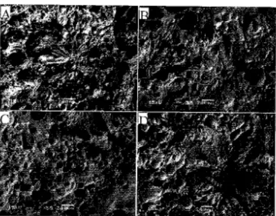
The microstructure of 'Rocha' pear flesh, after 4 mo of storage under different conditions, and exposed for 1 and 8 d to air at room temperature, is depicted in Figures 2 and 3, respectively. Inspection of these figures unfolds rather damaged structures; the cells have in fact collapsed, and present a soft and blurred cell wall. The tissue softening apparently involves cell separation and cell breakage - as also described by Brownleader et al. (1999).
Results pertaining to assessment of the discriminating ability, and the reproducibility of judgement by the panellists eventually chosen (step II), is depicted in Figure 4; the panel was indeed able to discriminate between different images, but was unable to distinguish the two similar images (Images 2 and 3, in Fig. 4). Statistical analysis of the experimental data generated by the panel, and pertaining to SEM images of 'Rocha' pear flesh, indicated that there are microstructural differences between pears
stored under distinct conditions (p=0.004), and exposed for different times to air at roor::: temperature (p=O.OOI); the interaction between these two factors was not statisticaE-.
significant (p=0.090). .
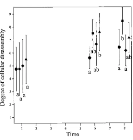
Since cellular disassembly is the parameter that demonstrated the most consisteD: behaviour, only the results concerning this parameter were considered. Cellular disassembly was higher for pears exposed for 6 and 8 d than for pears exposed for 1 d-but no differences were observed between the former times. No differences pertaining 1('
cellular disassembly were as well observed, by 1 d of exposure to air, between the fmr storage conditions (Figs. 2 and 5); however, it is clear that pears stored for 6 d under 1." kPa O2
+
4.9 kPa CO2 exhibited the higher degree of cellular disassembly, and pearsstored in air showed the lower (Fig. 5). By 8 d at room temperature, pears stored under 1.9 kPa 02
+
0.5 kPa CO2were rated as similar to those stored under air, and presenteethe lowest degree of cellular disassembly (Figs. 3 and 5).
CONCLUSIONS
SEM can be successfully applied to study microstructural changes in 'Rocha' pears, subject to storage under various controlled atmospheres. During storage, the pear cell structure undergoes a degenerative process - that can be associated to norma! senescence, coupled with the stress imposed during exposure to CA The preparation of samples for SEM via plain freezing produces good quality images; this procedure is much simpler than conventional fixation and drying. The methodology applied - which was based on panel evaluation of SEM images, proves a valuable tool, which allows quantitative parameters to be obtained, and duly related with parameters that describe fruit quality. As time of exposure to air at room temperature elapses, cell disassembly increases in extent; clear differences are obtained between pears exposed for 1 and 6 d at room temperature. Pears stored under 1.9 kPa O2
+
0.5 kPa CO2exhibit a lower degree ofcellular disassembly (which is similar to that obtained for pears stored in air).
Literature Cited
Aguilera, lM. 2000. Microstructure and food product engineering. Food Technology 54(11):56-65.
Brownleader, M.D., Jackson, P., Mobasheri, A, Pantelides, AT., Sumar, S., Trevan,
r..t
and Dey, P.M. 1999. Molecular aspects of cell wall modifications during fruit ripening. Critical Reviews in Food Science and Nutrition 39:149-164.Jewell, G.G. 1979. Fruits and vegetables. p.l-34. In: lG. Vaughan. Food Microscopy. London: Academic Press.
Kalab, M., Allan-Wojtas, P. and Miller, S.S. 1995. Microscopy and other imaging techniques in food structure analysis. Trends in Food Science and Technology 6: 177-186.
Langton, M. 1995. Correlating microstructure with texture of particulate biopolymer gels. PhD thesis dissertation. Department of Food Science, Chalmers University of Technology and SIK, Swedish Institute for Food Research, Sweden. p.l41.
Murayama, H., Ikai, S. and Fukushima, T. 2002. Changes in starch content in 'La France' pears during storage. Acta Hort. 596:871-873.
Reeve, R.M. 1970. Relationships of histological structure to texture of fresh and processed fruits and vegetables. Journal of Texture Studies 1:247-284.
Fig. 1. Scanning electron micrographs of 'Rocha' pear flesh after harvest, and exposure for 1 d (A, B) and 14d (C, D) to air (magnification: A, C - 55X; B, D - 200X).
Fig. 2. Scanning electron micrographs of 'Rocha' pear flesh after 4 mo of storage under air (A), 1.9 kPa O2 (B), 1.9 kPa O2
+
0.5 kPa CO2(C) and 1.9 kPa O2+
4.9 kPaFig. 3. Scanning electron micrographs of 'Rocha' pear flesh after 4 mo of storage
ULe=-air (A), 1.9 kPa O2(B), 1.9 kPa 02+
0.5 kPa CO2(C) and 1.9 kPa O2+
4.9 k?-:C02 (D), and exposure for 8 d to air at room temperature (magnification: 55X).
I
bI
bFig. 4. Degree of cellular disassembly of 'Rocha' pear flesh, as assessed by panelists (1'::
values average - e; standard deviation - I; significantly different means art marked by different lower case letters).
.Q .0 S 7
j
6j
~j ,§"
" 4 '-~ a " 3 50 a " a o 2Fig. 5. Degree of cellular disassembly of 'Rocha' pear flesh after 4 mo of storage under air (e), 1.9 kPa O2 (_), 1.9 kPa O2
+
0.5 kPa CO2C+)
and 1.9 kPa O2+
4.9 kPaCO2 ( .••. ), throughout time of exposure to air at room temperature (14 values
average - solid symbol; standard deviation -