Filipa de Sá Martins
Fosforilação da tau dependente de Abeta
Filipa de Sá Martins
Fosforilação da tau dependente de Abeta
Abeta dependent tau phosphorylation
Dissertação apresentada à Universidade de Aveiro para cumprimento dos requisitos necessários à obtenção do grau de Mestre em Biomedicina Molecular, realizada sob a orientação científica da Professora Doutora Odete da Cruz e Silva, Professora Auxiliar da Secção Autónoma de Ciências da Saúde da Universidade de Aveiro, e co-orientação da Professora Doutora Sandra Rebelo, Professora Auxiliar Convidada da Secção Autónoma de Ciências da Saúde da Universidade de Aveiro.
Esta dissertação contou com o apoio financeiro da FCT
(PTDC/QUIBIQ/101317/2008) e do Centro de Biologia Celular (CBC) da Universidade de Aveiro.
Dedico esta dissertação de mestrado aos meus pais que sempre me apoiaram em todas as etapas da minha vida.
o júri
presidente Professora Margarida Sâncio da Cruz Fardilha
Professora auxiliar convidada da Secção Autónoma de Ciências da Saúde da Universidade de Aveiro
Professora Doutora Odete Abreu Beirão da Cruz e Silva
Professor auxiliar da Secção Autónoma de Ciências da Saúde da Universidade de Aveiro
Professora Doutora Sandra Maria Tavares da Costa Rebelo
Professor auxiliar convidada da Secção Autónoma de Ciências da Saúde da Universidade de Aveiro
Professora Doutora Patrícia Espinheira Sá Maciel
palavras-chave Doença de Alzheimer, Proteína precursora de amilóide de Alzheimer, Abeta, tau, fosforilação da tau, inibidores das fosfatases, proteínas de ligação à tau.
resumo A doença de Alzheimer (DA) é uma doença neurodegenerativa caracterizada pela presença de duas características histopatológicas: as placas senis na matriz extracelular compostas por Beta-amilóide (Abeta) e as tranças neurofibrilhares intracelulares contendo proteína tau hiperfosforilada. Assim, o Abeta e a proteína tau são importantes moléculas associadas à DA e evidências sugerem que o Abeta possa mediar a hiperfosforilação da tau levando á disrupção da rede neuronal e consequentemente ao processo de neurodegeneração. No presente estudo, em culturas primárias neuronais de córtex e hipocampo de rato, verificou-se que a exposição a Abeta1-42 agregado
por longos períodos diminui a fosforilação da tau nos resíduos Ser202 e Thr205 e, em contraste, aumenta a fosforilação no resíduo Ser262.
Pensa-se que a hiperfosforilação da tau na DA pode estar relacionada com alterações nas vias de sinalização celular envolvidas no processo de fosforilação da tau, tais como alterações na regulação das cinases e das fosfatases. Deste modo, é também de extrema importância determinar as cinases e fosfatases envolvidas neste processo. Por tratamento de neurónios corticais com diferentes concentrações de ácido ocadéico (AO), um inibidor das fosfatases, verificamos o envolvimento da PP1 na desfosforilação da tau nos resíduos Ser202 e Thr205, bem como o envolvimento da PP1 e PP2A na desfosforilação do resíduo Ser262.
Um outro aspecto importante do metabolismo da tau são as proteínas de ligação, e actualmente já foram descritas várias proteínas que interagem com a tau in vitro e in vivo. O interactoma da tau é regulado pela sua fosforilação e portanto é crucial estabelecer uma relação entre a tau normal e a tau patológica hiperfosforilada no que diz respeito às proteínas de ligação. Por co-imunoprecipitação de neurónios corticais pretendemos identificar proteínas de ligação à tau e especificamente à tau fosforilada, e ainda avaliar o efeito do Abeta neste interactoma. O interactoma da tau dependente da fosforilação e do Abeta é de particular relevância para a compreensão da DA.
keywords Alzheimer´s Disease, Alzheimer´s amyloid precursor protein, Abeta, tau, tau phosphorylation, protein phosphatase inhibitors, tau binding proteins.
abstract Alzheimer’s disease (AD) is a neurodegenerative disorder characterized by the presence of two histopathological hallmarks: the extracellular amyloid plaques (APs) composed of beta-amyloid protein (Abeta) and intracellular neurofibrillary tangles (NFTs), containing hyperphosphorylated tau protein. Therefore, Abeta and tau are important molecules associated with AD and evidence suggests that Abeta may initiate the hyperphosphorylation of tau, which by disrupting neuronal network leads to the process of neurodegeneration. In the present study, using rat primary cortical and hippocampal neuronal cultures, it was shown that exposure to aggregated Abeta1-42 for prolonged periods decreased
tau phosphorylation at Ser202 and Thr205 residue, but in contrast increased at Ser262 residue. Tau hyperphosphorylation in AD may be related to alterations in signal transduction pathways involving tau phosphorylation, such as an imbalance in theregulation of protein kinases (PKs) and protein phosphatases (PPs). Thus it is also important to determine which specific PKs and PPs are involved in this process. We observed the involvement of PP1 in the dephosphorylation of tau at Ser202 and Thr205, and the involvement of PP1 and PP2A at the Ser262 residue.
An important aspect of tau metabolism are its binding proteins, and to date many suchproteins have already been described both in vitro and in vivo. The interactome of tau is shaped by its phosphorylation and so is crucial to map the crosstalk between normal and pathologically hyperphosphorylated tau. By co-immunoprecipitation we intend to identify proteins that interact with tau and more specifically with phosphorylated tau (p-Tau). Furthermore the effect of Abeta on this interactome should be forthcoming, which is relevant for AD tau pathology.
7
Abbreviations
Abeta Amyloid peptide
AD Alzheimer´s disease
ADAM A Disintegrin And Metalloproteinase
AICD Amyloid precursor protein intracellular domain
AP Amyloid plaques
APS Ammonium persulfate
Aph1 Anterior pharynx-defective 1 APLP1 APP like protein 1
APLP2 APP like protein 2
ApoE Apolipoprotein E
APP Alzheimer´s amyloid precursor protein
ATP Adenosine triphosphate
BACE Beta-site APP-cleaving enzyme
BCA Bicinchoninic acid
BSA Bovine serum albumine
CaMPKII Ca2+/Calmodulin-dependent protein kinase II
Cdc2 Cyclin-dependent protein kinase 2 Cdk5 Cyclin-dependent protein kinase 5
CSF Cerebrospinal fluid
CNS Central nervous system
CT Computarized tomography C-terminal Carboxyl-terminal CTF Carboxyl-terminal fragment DTT Dithiothreitol E Exon EC Extracellular domain
ECL Enhanced chemiluminescence EDTA Ethylenediamine tetraacetic acid
ER Endoplasmic reticulum
FAD Familial Alzheimer´s disease
FBS Fetal bovine serum
Fc Fragment crystallizable GSK3 Glycogen synthase kinase 3 HBSS Hank´s balanced salt solution HSP70 Heat shock protein 70
HSP90 Heat shock protein 90
IB Immunoblotting
IC Intracellular domain
IC50 50% inhibition concentration
8
IP Immunoprecipitation
JIP1b JNK-interacting protein 1b
JNK Jun N-terminal kinase
KLC Kinesin light chain
KPI Kunitz-type serine protease inhibitor
LB Loading buffer
LC-MS/MS Liquid chromatography-mass spectrometry
LGB Lower gel buffer
MAP Microtubule associated proteins MAPK Mitogen activated protein kinases MAPT Microtubule Associated Protein Tau MARK Microtubule-affinity regulating kinase MOPS 3-(N-morpholino)propanesulfonic acid MRI Magnetic resonance imaging
NFT Neurofibrillary tangles NMDA N-Methyl-D-aspartic acid
NPDPK Non - Proline directed protein kinases NSAID Nonsteroidal anti-inflammatory drug N-terminal Amino-terminal
OA Okadaic acid
ON Overnight
PBS Phosphate buffered saline
PBS-T Phosphate buffered saline - tween PDPK Proline directed protein kinases PEN-2 Presenilin enhancer 2
PET Positron emission tomography PHF Paired helical filaments
Pin1 Protein interacting with NIMA
PK Protein kinase
PKA Cyclic-AMP-dependent kinase
PKC Protein kinase C
PLC Phospholipase C
PP Protein phosphatase
PSEN1 Presenilin 1
PSEN2 Presenilin 2
p-tau Phosphorylated tau
RIPA Radio-Immunoprecipitation Assay
RNA Ribonucleic acid
RT Room temperature
SAP Stress-activated protein
sAPP Secreted APP
SDS Sodium dodecyl sulfate
SDS-PAGE Sodium dodecyl sulfate polyacrylamide gel electrophoresis
9
Ser Serine
SH3 SRC homology
TBS Tris Buffered Saline
TBS_T Tris Buffered Saline - tween
TGN Trans-Golgi network
Thr Threonine
TRIS Tris(hydroxymethyl)aminomethane
Tyr Tyrosine
TM Transmembrane domain
UGB Upper gel buffer
11
INDEX
Abbreviations ... 7
1. Introduction... 13
1.1. Alzheimer ’s disease (AD) ... 14
1.1.1. Epidemiology and Genetics ... 14
1.1.2. Histopathological Hallmarks ... 15
1.1.3. Diagnosis and Treatment ... 17
1.2. Alzheimer´s amyloid precursor protein (APP) ... 19
1.2.1. APP Proteolytic processing ... 20
1.2.2. APP phosphorylation ... 22
1.2.3. APP and APP fragments functions... 23
1.3. Microtubule-associated tau protein ... 24
1.3.1. Posttranslational modifications of tau protein ... 27
1.3.2. The physiological role and the pathological effects of tau phosphorylation ... 32
1.3.3. Tau binding proteins ... 34
1.4. Relationship between Abeta peptide and tau phosphorylation ... 37
2. Aims of the dissertation ... 41
3. Materials and Methods ... 43
3.1. Antibodies ... 44
3.2. Cell culture ... 45
3.2.1. Primary Neuronal Cultures ... 45
3.3. Cortical and hippocampal neurons treatment with Abeta ... 46
3.4. Cortical and hippocampal neurons treatment with protein phosphatase inhibitors ... 46
3.5. BCA protein quantification assay ... 47
3.6. SDS - Polyacrylamide gel electrophoresis ... 48
3.7. Immunoblotting ... 49
3.8. Co- immunoprecipitation and mass spectrometry analysis ... 51
3.9. Quantification and statistical analysis ... 53
4. Results ... 55
4.1. Abeta effects on tau phosphorylation at Ser202, Thr205 and Ser262 residues ... 56
4.2. Protein phosphatases involved in tau dephosphorylation at Ser202, Thr205 and Ser262 residues ... 64
12
4.2.1. Rat primary cortical neuronal cultures ... 64
4.3. Abeta effects on tau and p-tau binding proteins ... 68
4.3.1. Rat primary cortical neuronal cultures ... 68
5. Concluding remarks ... 71
6. Discussion and Conclusion ... 73
7. References ... 79
13
14
1.1. Alzheimer ’s disease (AD)
Alzheimer´s disease (AD) is a neurodegenerative disorder first described in
1906 by Dr. Alois Alzheimer5,9. AD is the most common form of dementia among the
elderly, and currently affects about 18 million people worldwide, 7.3 million in
Europe and about 90.000 people in Portugal2-4,10-11.Dementia is the loss of cognitive
functions such as thinking, remembering and reasoning, that interfere with a person´s
daily life and activities2,12. AD is characterized by memory, language, learning and
other cognitive impairments, severe enough to interfere with social, occupational and
personal functions3-5,12-14. Besides this cognitive decline, behavioral, emotional and
psychiatric symptoms are also frequent in AD14.
1.1.1. Epidemiology and Genetics
The most prevalent forms of AD are sporadic, with the symptoms beginning after 65 years of age, and are called late-onset AD. But there is a small percentage of cases (less than 5%) with an early onset (30-60 years), generally inherited in an autosomal-dominant manner – the familial AD (FAD). There are some risk factors for developing sporadic AD (late-onset AD) such as advancing age, the presence of certain alleles of the Apolipoprotein E gene (ApoE), gender, level of education and head trauma. The single most important risk factor is age, since the rate of occurrence
of the disease doubles approximately every five years after the age of 654,14-15.
Apolipoprotein E gene (ApoE) is localized to chromosome 19, encodes a glycoprotein involved in cholesterol transport and alters the risk of developing AD but does not cause it. The gene codifying this protein has three possible alleles: ε2, ε3 and ε4. The ε3 is the most common allele in the general population and ε2 the less common. The ε4 allele is connected with an increased risk of developing AD. The mechanism through which this allele increases the risk is unknown but it appears that the ε4 allele along with other proteins may influence Abeta metabolism and its aggregation
15
Additionally mutations in three different genes: presenilin-1 gene (PSEN1) located on chromosome 14, presenilin-2 gene (PSEN2) located on chromosome 1 and amyloid precursor protein gene (APP) on chromosome 21, are known to cause 90%
of the FAD14-15,18. The APP gene encodes a transmembrane glycoprotein abundant in
the nervous system that is proteolytically cleaved to produce Abeta. As this gene is localized to chromosome 21, this explains the observation that patients with Trisomy 21, which possess an extra copy of the APP gene, develop early in life (40 years) the neuropathological features associated with AD (like Abeta deposition). Studies have allowed to conclude that most of the APP mutations alter the proteolytic processing
of APP resulting in increased Abeta production16-18. The PSEN1 and PSEN2 genes
encode two highly homologous transmembrane proteins whose normal functions are not yet known. However the products from proteolytic cleavage of these proteins are known to be essential to gamma-secretase complex formation. The majority of mutations in PSEN1 and PSEN2 are missense and increase the activity of gamma-secretase and consequently Abeta production. From all early-onset AD cases 50% are
linked to the PSEN1 gene and only a few cases are associated to the PSEN2 gene16-17
1.1.2. Histopathological Hallmarks
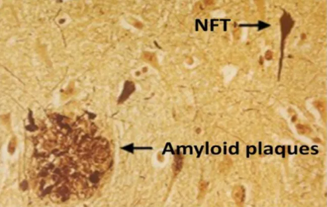
In AD there is neuronal and synaptic loss associated with two histopathological hallmarks: the extracellular amyloid plaques (AP, Fig. 1) and intracellular neurofibrillary tangles (NFTs, Fig. 1), in distinct brain areas including the
16
Figure 1 – Two histopathological hallmarks of AD: Amyloid plaques (AP) and Neurofibrillary tangles (NFTs)20.
Amyloid plaques are extracellular insoluble deposits of a protein fragment, the beta-amyloid (Abeta) generated by proteolytic cleavage of the amyloid precursor protein (APP), surrounded by dystrophic neurites. The Abeta peptide is a physiological soluble cellular metabolite that comprises two predominant forms, the
Abeta1-40 and the Abeta1-42 which differ in their C-terminal15. The Abeta1-40 is the most
predominant and presumably is not neurotoxic while the Abeta1-42 is less prevalent,
more hydrophobic and more toxic. The Abeta1-42, is also proportionally increased in
patients with AD and has a major propensity to aggregate and to form oligomers and
fibrils that ultimately generate amyloid plaques6. The steady state level of Abeta is
controlled by its production, degradation and clearance. In AD it is proposed that the cause of Abeta accumulation is a defect that leads to its over-production or decreased
clearance4. Although, actually, it remains unclear what triggers these alterations in
APP and Abeta metabolism causing this increased production and aggregation of Abeta peptide. However it is known that Abeta accumulation and aggregation results in organelle and membrane damage, which in turn leads to the disruption of cellular
processes and also oxidative stress, inflammation and cell death6.
The NFTs are intracellular aggregates composed of bundles of paired helical filaments (PHF) whose major protein component is the microtubule-associated tau protein. In PHF the tau protein is abnormally hyperphosphorylated and
aggregated14,18,21-22 and so has a reduced ability to bind to microtubules and to
promote their assembly. As a consequence the axonal cytoskeleton is disturbed,
17
example, synapses are very vulnerable to these perturbations in the axonal transport system since it causes dysfunction in neurotransmission and signal propagation
leading to synaptic degeneration.24 Thus, the number of NFT are positively correlated
with the degree of AD, and the same does not occur with amyloid plaques24.
1.1.3. Diagnosis and Treatment
Currently, the diagnosis and treatment of AD is limited and insufficient. As such definitive diagnosis of AD is still only possible after death, with an examination and pathological analysis of brain tissue during an autopsy, where the presence of the senile plaques and neurofibrillary tangles is confirmed. However, there are many current tools that are used to diagnose AD patients or even to exclude this pathology. These tools included detailed patient medical history, information obtained from family members, physical and neurological examinations, laboratory tests, neuropsychological tests to measure, for example language and memory skills and a variety of other approaches, including neuroimaging studies such as computerized tomography (CT), magnetic resonance imaging (MRI) and positron emission tomography (PET). The neuroimaging techniques are of great help since they provide regional structural and functional details of the brain, as well as assist in the
identification of the biochemical profile of brain dysfunction25. Although there are
significant advances in these neuroimaging techniques, the use and identification of novel AD biomarkers is necessary since they give more direct and convenient information to detect the preclinical stages of AD, as well as assisting in the study of
disease progression25-26. The most important potential sources of AD biomarkers are
cerebrospinal fluid (CSF), plasma and urine25. Currently the quantification of Abeta
and tau, both total and phosphorylated tau, in the CSF is the more appropriate to
detect early AD patients26. An early diagnosis of AD is beneficial, as it facilitates the
efficient treatment with new generation of disease modifying drugs12.
Regarding the treatment for AD, it is a complex disease and no single treatment is likely to prevent or cure it. Thus current treatments focus on helping patients maintain mental function, managing behavioral symptoms and delaying the
18
disease14. Actually the therapies for AD can be based in symptomatic approaches or
based in neuroprotective approaches. Therefore there are five major categories of drugs used in AD treatment: the acetylcholinesterase inhibitors, antiglutaminergic treatment, vitamins and antioxidants, anti-inflammatory drugs and pharmacological
management of behavioral disturbances27. From these, the most successful AD drugs
to date are the acetylcholinesterase inhibitors since in AD there is a deficiency in cholinergic neurotransmission that plays a major role in the expression of cognitive functional and behavioral symptoms of AD. Thus acetylcholinesterase inhibitors act
by stopping or slowing down the action of acetylcholinesterase, a catabolic enzyme
that breaks down acetylcholine, the neurotransmitter involved in memory formation,
prolonging its action at cholinergic synapses14,27-28. The antiglutaminergic treatment,
for example the memantine, is another therapeutic approach, but in this case it blocks glutamatergic neurotransmission, since it is an uncompetitive antagonist of NMDA receptors avoiding its hyperstimulation which causes neuronal dysfunction and
death14,27. Evidence that free radicals may accumulate in AD brains, due to the
existence of oxidative stress, has led to interest in the use of antioxidants such as vitamin E. Nonsteroidal anti-inflammatory drugs (NSAIDs) may have a protective role against the development of AD but this effect does not extend once AD is
established27. Concerning the management of behavioral disturbances it can be
achieved by using nonpharmacological (music, light exercise, relaxation exercise) or pharmacological approaches using anxiolytic, antidepressive or antipsychotic drugs. The future of therapies in AD will be based on the understanding of AD pathophysiology and could be achieved with anti-amyloid therapies that are being studied. The development of these therapies has two main approaches: reduce the production of Abeta that can be achieved by inhibiting beta- and gamma-secretase, or increase its clearance by anti-amyloid immunotherapy. The main goal of these new approaches is to modify the progression of the disease; these are called
19
1.2. Alzheimer´s amyloid precursor protein (APP)
APP is a ubiquitously expressed membrane-spanning glycoprotein with a large
N-terminal extracellular domain (EC) and a small cytoplasmic domain (IC) 21,29. It is a
member of a conserved family of type I membrane proteins including APP like protein 1 (APLP1) and APP like protein 2 (APLP2) in mammals. While the APP and APLP2 are ubiquitous but with highly expression in neurons, the APLP1 is
brain-specific29-30. APP is encoded by a gene localized on the mid-portion of the long arm of
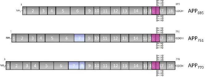
human chromosome 21 (21q21) and contains 18 exons30-31. Alternative splicing of
the exons 7, 8 and 15 generates eight APP isoforms that range from 365-770 amino
acids. The most abundant APP isoforms are APP695, APP751 and APP770 (Fig. 2)15,32-33.
APP751 and APP770 are largely expressed by non-neuronal cells and contain a domain
homologous to the Kunitz-type serine protease inhibitor (KPI), encoded by exon 7,
whereas the APP695 is expressed at higher levels in neurons and does not have the KPI
domain30-31,34. KPI-containing APP isoforms are thought to be more amyloidogenic
and their levels increase in the brains of AD patients15. However the cause and the
functional significance for this tissue-specific isoforms is still not fully understood.
Figure 2 – Schematic representation of the three major APP isoforms in mammalian tissues. Numbers and vertical lines indicate the corresponding exons. The most abundant neuronal isoform, comprising 695 amino acids, is APP695. APP751 and APP770 are alternatively spliced isoforms that differ from APP695 in the expression of
exons 7 and 8, as shown. The sequences encoded by the APP gene exons are indicated approximately to scale. The solid pink region represents the Abeta peptide region, whose sequence is divided between exons 16 and 17. Adapted from Cruz e Silva and Cruz e Silva, 2003.
20
In neuronal and non-neuronal cells, APP is generated in the endoplasmic reticulum (ER) and is known to be transported via the secretory pathway reaching cell surface. During its transit from the ER to the plasma membrane, APP undergoes post-translational modifications that include N- and O- glycosylation and tyrosine
sulfation30,33. Therefore mature APP is located in compartments from the trans-Golgi
to the plasma membrane, being that, only a small fraction is present at the plasma membrane. At the cell surface, APP can be cleaved or rapidly internalized via endocytosis to be recycled back to the membrane, retrogradely delivered to the trans-Golgi-network (TGN) or incorporated into secondary endosomes. The majority of mature APP is proteolytically cleaved either via the alpha-secretase or beta-secretase pathway. It is thought that this proteolytic processing occurs through the secretory
pathway, on the plasma membrane and/or in the endocytic cycle 30,35-36. Moreover, in
neurons, APP is rapidly and anterogradely transported along peripheral and central axons36.
Another important aspect is the fact that the trafficking, metabolism and even the functions of APP are regulated through interactions with several cytoplasmic proteins, for example, the relatively well analyzed Fe65, X11 and X11L, JIP1b
(JNK-interacting protein 1) and KLC (kinesin light chain) proteins15,37 . All these proteins
bind to the APP intracellular domain (AICD) at specific binding domains.
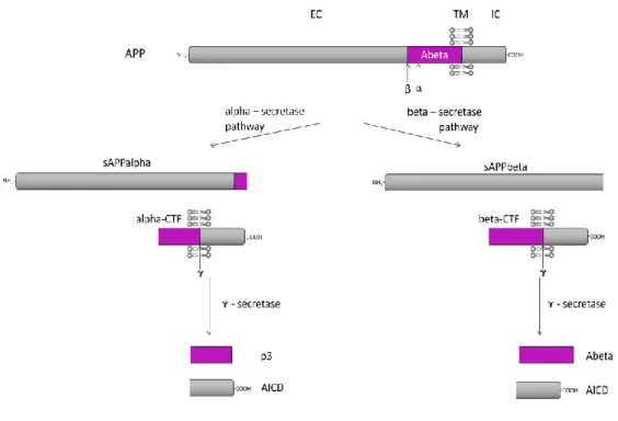
1.2.1. APP Proteolytic processing
APP can be cleaved by two major proteolytic processing pathways: the beta-secretase and alpha-beta-secretase pathway, also called amyloidogenic and non-amyloidogenic, respectively. In the beta-secretase pathway the APP is first cleaved by the beta-secretase, releasing the ectodomain (sAPPbeta) while a C-terminal fragment with 99 amino acids (C99 or beta-CTF) remains membrane bound. Then C99 is cleaved by the gamma-secretase complex to produce Abeta peptide and the AICD. Alternatively, in the alpha-secretase pathway, alpha-secretase primarily cleaves APP releasing the ectodomain (sAPPalpha) and a membrane bound C-terminal fragment with 83 amino acids (C83 or alpha-CTF), which then is also cleaved by
21
processed by the alpha-secretase pathway and so there is a balance between these
two proteolytic pathways21.
Figure 3 – Proteolytic processing of APP30. EC: extracellular domain; TM: transmembrane domain; IC:
intracellular domain. The scheme is not to scale.
Thus, there are three proteases involved in the cleavages of APP: the alpha-, beta- and gamma-secretases. The candidates for the alpha-secretase activity are members of the ADAM family of desintegrin and metalloproteases. BACE (beta-site APP-cleaving enzyme) is a type I transmembrane aspartic protease, has two homologues, BACE1 and BACE2 that are the major beta-secretase in neurons. The gamma-secretase is a multimeric complex with proteolytic activity formed at least by four
22
1.2.2. APP phosphorylation
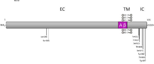
Protein phosphorylation is an important cellular regulatory mechanism that is increased in AD. APP can be phosphorylated at multiple sites in both extracellular and intracellular domains. In the intracellular domains 8 putative phosphorylation residues are described: Tyr653, Thr654, Ser655, Thr668, Ser675, Tyr682, Thr686
and Tyr68729,34,38-39. Since these residues are located in specific protein interacting
sites its phosphorylation may interfere with protein binding and thus interfere with
APP and AICD function.29 Previous studies point to an important role for the Thr668
and Tyr682. The phosphorylation of both these residues is increased in AD brains: the Tyr682 is important for APP interactions with the cytosolic proteins and can promote or abolish them; the Thr668 phosphorylation allows Pin1 (a prolyl isomerase) binding and reduces Fe65 binding to APP and thus it alters APP
processing and Abeta production40. In the APP ectodomain, phosphorylation at
Ser198 and Ser206 residues are present, and occurs in a post-Golgi secretory
compartment and at the cell surface.41 All of these APP phosphorylation sites are
represented in Fig. 4.
Figure 4 – Schematic representation of the phosphorylation residues present in the APP695 isoform
protein: the two phosphorylated Ser residues present in the APP ectodomain, and the eight putative phosphorylated residues in the APP intracellular domain. EC: extracellular domain; TM: transmembrane domain; IC: intracellular domain29,41.
23
1.2.3. APP and APP fragments functions
The precise roles of APP are unknown, although the overall structure of the
protein suggests a role as a receptor or growth factor5. However some studies
describe more putative roles for APP and its fragments in development, cell growth, intercellular communication, signal transduction, nuclear signaling and structural and
functional plasticity29-30,42. Table 1 summarizes some of these putative roles for APP
and its fragments that are produced during APP metabolism.
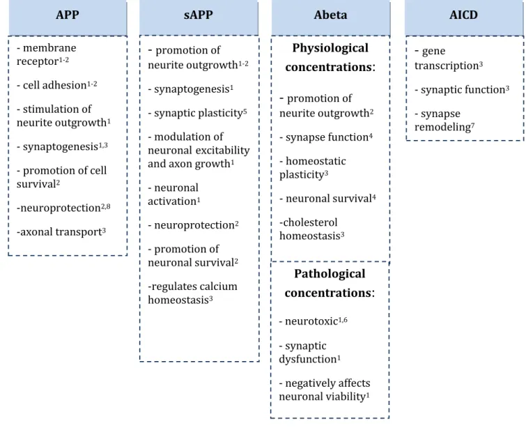
Table 1 - APP and APP fragments (sAPP, Abeta and AICD) putative functions.
APP sAPP Abeta AICD
- membrane receptor1-2 - cell adhesion1-2 - stimulation of neurite outgrowth1 - synaptogenesis1,3 - promotion of cell survival2 -neuroprotection2,8 -axonal transport3
-
promotion of neurite outgrowth1-2 - synaptogenesis1 - synaptic plasticity5 - modulation of neuronalexcitability and axon growth1- neuronal activation1 - neuroprotection2 - promotion of neuronal survival2 -regulates calcium homeostasis3 Physiological concentrations
:
-
promotion of neurite outgrowth2 - synapse function4 - homeostatic plasticity3 - neuronal survival4 -cholesterol homeostasis3-
gene transcription3 - synaptic function3 - synapse remodeling7 Pathological concentrations:
- neurotoxic1,6 - synaptic dysfunction1 - negatively affects neuronal viability124
1.3. Microtubule-associated tau protein
Tau protein belongs to the microtubule-associated protein (MAP) family and was first isolated in 1975 as a protein that co-purifies with tubulin and has the ability
to promote microtubule assembly in vitro24,43. Tau is mainly a neuronal protein,
although it can be expressed in non-neuronal cells43. The human tau gene, MAPT, is
located on the long arm of chromosome 17 at band position 17q21 where it occupies
over 100 kb 43-44. The tau primary transcript contains 16 exons but three of them
(exons 4A, 6 and 8) are never present in any mRNA of the human brain (Fig. 5)44.
Exon 1 is part of the promoter and is transcribed but not translated, as is the case for exon 14. Exons 1, 4, 5, 7, 9, 11, 12 and 13 are constitutive, but exons 2, 3 and 10 are
alternatively spliced, and exon 3 never appears in the absence of exon 2 43-44.
Therefore, the transcript produced by alternative splicing of these three exons yields six different mRNA species that are then translated in six different isoforms of tau
25
Figure 5 – Schematic representation of the human tau gene, mRNA and different protein isoforms24. The
human tau gene is located over 100kb on the long arm of chromosome 17 at position 17q21. It contains 16 exons; exon -1 is a part of the promoter. The tau primary transcript contains 13 exons since exons 4A, 6 and 8 are not transcribed in human. Exons -1 and 14 are transcribed but not translated. Exons 1, 4, 5 ,7, 9, 11, 12, 13 are constitutive, and exons 2, 3 and 10 are alternatively spliced, giving rise to six different mRNAs, translated in six different tau isoforms. These isoforms differ by the absence or presence of one or two 29 amino acids inserts encoded by exon 2 and 3 in the amino-terminal part, in combination with either three (R1, R3 and R4) or four (R1, R2, R3 and R4) repeat-regions in the carboxyl-terminal part.
The isoforms differ by the absence or presence of one or two acidic inserts (0N, 1N or 2N, respectively) at the amino-terminal (N-terminal) part of the molecule and whether they contain three or four repeats of a conserved tubulin binding motif (3R or 4R) at the carboxyl-terminal (C-terminal) region, and they can be designated
as 3R0N, 3R1N, 3R2N, 4R0N, 4R1N and 4R2N44-48. Thus, the longest isoforms in the
CNS has four repeats and two insert (441 residues), and the shortest isoforms has three repeats and no inserts (352 residues) (Fig. 5). The later isoform (3R0N), the smallest form of tau protein, is the only one expressed in fetal tissue while the six
isoforms are expressed in adult brain46. It is thought that tau isoforms have specific
physiological roles since they are differentially expressed during development43.
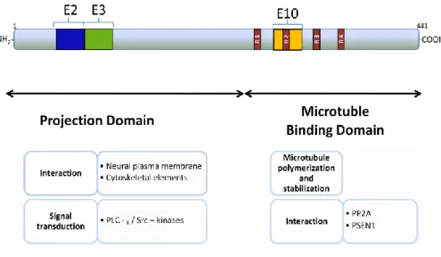
Tau isoforms have two domains: the projection domain and the microtubule-binding domain, that have been proposed to have distinct roles. The projection
26
domain contains the N-terminal two-thirds of the molecule and can be further subdivided into the acidic N-terminal region and a basic proline-rich region. The projection domain of tau determines spacings between axonal microtubules, interacts with other cytoskeletal proteins, for example, spectrin and actin filaments which allow microtubules to interconnect with other cytoskeletal components (Fig. 6). This domain may also allow interaction of tau with proteins associated with the neural plasma membrane and cytoplasmatic organelles, such as mitochondria, and there is some data indicating that tau proteins may interact with src-family non-receptor tyrosine kinases and phospholipase C-ɤ (PLC-ɤ), which suggests that tau may have a role on signal transduction pathways involving these two proteins. Moreover, the interaction of this domain with cytoskeletal and plasma membrane elements is only
possible because this part of themolecule projects from the microtubule surface.
Figure 6 – Summary of biological functions of tau associated with respective functional domain. E: exon; R: repeat domains. Adapted from Buée et.al, 2000.
27
The microtubule-binding domain contains the C-terminal one-third of the molecule and, likewise the projection domain, has been subdivided into the basic tubulin-binding domain region and the acidic C-terminal region (Fig. 6). As its name suggests, this domain is responsible for the binding of tau to microtubules and more specifically, tau binds microtubules through repetitive regions present in this domain. The repetitive regions are the repeat domains (R1, R2, R3 and R4) encoded by exons 9, 10, 11 e 12 and with sequences of 31 or 32 residues very similar but not identical since they are composed by an 18 amino-acid sequence highly conserved and a less conserved sequence composed by 13 or 14 amino-acid sequence. The 18 amino-acid sequence is responsible for binding to microtubules, promoting microtubule polymerization and stabilization. For this reason tau isoforms with 4R (R1, R2, R3 and R4) binds more efficiently to microtubules than the isoforms with 3R (R1, R2 and R3). Besides microtubule assembly recent data suggests that the microtubule-binding domain can also modulate the phosphorylation state of tau proteins since it can bind directly with the protein phosphatase 2A (PP2A) and in consequence microtubules
can inhibit PP2A activity by competing for binding to tau at this domain 43-44,46,49.
1.3.1. Posttranslational modifications of tau protein
Like many other proteins that are implicated in human disease, tau protein is posttranslationally modified. Several modifications have been described for tau protein such as phosphorylation, glycosylation, ubiquitination, glycation, truncation and deamination. Of all posttranslational modifications the most important is phosphorylation because it is an important cellular regulatory mechanism.
28
1.3.1.1 Tau phosphorylation
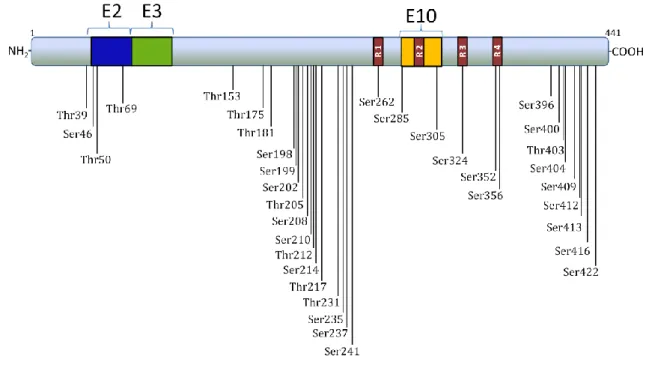
Tau is a phosphoprotein that possesses a large number of potential
phosphorylation sites mainly serine, threonine and tyrosine residues45,49. On the
longest brain tau isoform (441 amino-acids) there are 45 serine and 35 threonine putative phosphorylation sites, and at least 35 phosphorylation sites have already
been described (Fig. 7)43.
Figure 7 – Representation of phosphorylation sites already described on the longest brain tau isoform using phosphorylation-dependent monoclonal antibodies against tau, mass spectrometry and sequencing. All of these sites are localized outside microtubule-binding domains with the exception of Ser262, Ser285, Ser305, Ser324, Ser352 and Ser35643. E: exon; R: repeat domain.
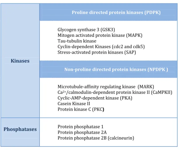
The level of tau phosphorylation is a dynamic process controlled by several protein kinases and protein phosphatases (summarized in table 2). Interestingly, tau phosphorylation is developmentally regulated as fetal isoforms are more phosphorylated during neurogenesis and synaptogenesis and then phosphorylation
29
Table 2- Protein kinases and protein phosphatases most probably involved in tau protein phosphorylation and dephosphorylation, respectively 13,43-44,47-50.
Kinases
Proline directed protein kinases (PDPK)
Glycogen synthase 3 (GSK3) Mitogen activated protein kinase (MAPK) Tau-tubulin kinase Cyclin-dependent Kinases (cdc2 and cdk5) Stress-activated protein kinases (SAP)
Non-proline directed protein kinases (NPDPK )
Microtubule-affinity regulating kinase (MARK)
Ca2+/calmodulin-dependent protein kinase II (CaMPKII)
Cyclic-AMP-dependent kinase (PKA) Casein Kinase II Protein kinase C (PKC)
Phosphatases Protein phosphatase 1 Protein phosphatase 2A
Protein phosphatase 2B (calcineurin)
The majority of the kinases involved in tau phosphorylation belong to the Proline-directed protein kinases (PDPK) which comprise glycogen synthase 3 (GSK3), mitogen activated protein kinase (MAPK), tau-tubulin kinase, cyclin-dependent kinases such as cdk2 and cdk5, and stress-activated kinases (SAP kinases). Another group named non-PDPK or NPDPK comprises microtubule-affinity regulating kinase
(MARK), Ca2+/calmodulin-dependent protein kinase II (CaMPKII),
cyclic-AMP-dependent kinase (PKA), casein kinase II and protein kinase C (PKC)13,43,47-49. GSK3 is
one of the protein kinases that has gained significant attention as a tau kinase and
comprises the highly homologous proteins GSK-3α and GSK-3β46. GSK-3β is highly
expressed in brain and associates with microtubules47. Some studies in which tau and
GSK-3β are co-transfected into non-neuronal cells showed an increase in tau
phosphorylation and impairment in the binding of tau to microtubules46,51-52.
30
having an impact on tau function51. Other important tau kinases are the MAPKs in
which some of its members can phosphorylate tau: p42mapk, p44mapk, pk40erk2 and
p493F12. It was shown in several studies that these MAPKs phosphorylate tau protein
in cultured neurons mainly via activation of tyrosine kinase receptors and protein
kinase cascades46. Cdk5 is a member of cyclin-dependent kinase family and its activity
is highest in neurons due to selective expression of its regulator p35 in these cells51.
This kinase induces phosphorylation of tau protein in vitro, maybe not directly but
instead by regulating the kinases and phosphatases that act on tau47. The NPDPK
cyclic-AMP-dependent kinase (PKA) is shown to phosphorylate tau in vivo47. Since
many kinases are likely to be involved in tau phosphorylation one possibility is that tau might be primed by one kinase before subsequent phosphorylation by a second
kinase that recognizes a nearby phosphorylated residue53.
Regarding protein phosphatases (PP) several studies have shown that three major PPs: PP1, PP2A and PP2B (calcineurin), but not PP2C can dephosphorylate tau
in vitro44,49-50. All of these PPs are present in the brain and are developmentally
regulated43. The PP2A is the most probable phosphatase that acts on most
phosphorylation sites and it is also associated with microtubules46,49-50,54. It was
shown that, in cultured neurons treated with okadaic acid and calyculinA (phosphatase inhibitors) at concentrations sufficient to inhibit PP2A,
phosphorylation of tau was increased54. Similar studies using PP2B inhibitors also
suggest that PP2B is involved in the dephosphorylation of tau protein but at different
sites of the PP2A43. PP1 involvement in tau phosphorylation was also demonstrated
by inhibition of phosphatases in neuronal cell lines. Although, PP2C has been reported to dephosphorylate tau phosphorylated by PKA in vitro, these
dephosphorylations did not affect PHF tau46. Table 3 summarizes the
phosphorylation residues in human tau associated with its tau-directed protein kinases and phosphatases.
31
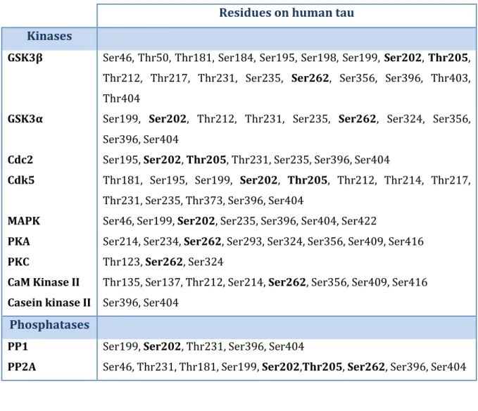
Table 3 – Tau-directed protein kinases and phosphatases and respective phosphorylation residues in human tau 44,46. In bold are the residues addressed in this study.
Residues on human tau Kinases
GSK3β Ser46, Thr50, Thr181, Ser184, Ser195, Ser198, Ser199, Ser202, Thr205, Thr212, Thr217, Thr231, Ser235, Ser262, Ser356, Ser396, Thr403, Thr404
GSK3α Ser199, Ser202, Thr212, Thr231, Ser235, Ser262, Ser324, Ser356, Ser396, Ser404
Cdc2 Ser195, Ser202, Thr205, Thr231, Ser235, Ser396, Ser404
Cdk5 Thr181, Ser195, Ser199, Ser202, Thr205, Thr212, Thr214, Thr217, Thr231, Ser235, Thr373, Ser396, Ser404
MAPK Ser46, Ser199, Ser202, Ser235, Ser396, Ser404, Ser422
PKA Ser214, Ser234, Ser262, Ser293, Ser324, Ser356, Ser409, Ser416
PKC Thr123, Ser262, Ser324
CaM Kinase II Thr135, Ser137, Thr212, Ser214, Ser262, Ser356, Ser409, Ser416
Casein kinase II Ser396, Ser404
Phosphatases
PP1 Ser199, Ser202, Thr231, Ser396, Ser404
PP2A Ser46, Thr231, Thr181, Ser199, Ser202,Thr205, Ser262, Ser396, Ser404
1.3.2.1. Other posttranslational modifications of tau protein
Glycosylation is an enzymatic process through which oligosaccharides are covalently attached to the side chain of polypeptides. There are two types of glycosylation according to the nature of glycosidic bonds: O-glycosylation and N-glycosylation. In tau protein, both types have been reported, but O-glycosylation occurs in unmodified tau whereas N-glycosylation occurs in hyperphosphorylated tau. It was reported that the inhibition of protein phosphatases, which induces tau
hyperphosphorylation, also decreased O-glycosylations49. Thus, later, a reciprocal
relationship between the O-glycosylation and phosphorylation was established, in
32
Ubiquitination consists in the association of ubiquitin, a stress protein, with misfolded or damaged proteins to be degraded in an ATP-dependent manner. The tau protein can be ubiquitinated, however it has only been thus found when in NFTs. Despite the PHF-tau being highly ubiquitinated, it is not degraded and instead it is
deposited as NFTs in AD brain44,49.
In tau isolated from PHF the glycation was present which refers to a non-enzymatic linkage of a reducing sugar to a polypeptide. This glycation might be involved in the insolubility/aggregation of PHFs into NFTs since a cross-linking reaction leading to the formation of insoluble aggregates of proteins is often described as a consequence of proteins glycation. It was also found that glycated tau
can also induce neuronal oxidative stress by generating oxygen free radicals43,49.
Truncation in PHF-tau which consists in the cleavage of tau at the glutaminic acid residue 391 has also been observed. This modification could facilitate aberrant
tau aggregation44,49.
Lastly, the deamination is a chemical reaction in which an amide functional group is removed, that in tau protein is at aspargine or glutamine residues. This tau
modification can also have a role in tau aggregation49.
1.3.2. The physiological role and the pathological effects of tau phosphorylation
Phosphorylation at specific sites and when it is properly coordinated is the predominant mechanism that regulates the different roles of tau protein both in physiological and pathological conditions (Fig. 8).
33
Figure 8 - Physiological and pathological roles of tau phosphorylation in the cell. Taken from
Johnson et al. 2004.
Therefore, tau phosphorylation at normal physiological conditions controls a
variety of processes such as microtubule binding and microtubule assembly47,55,
neurite outgrowth56, axonal transport57 and cell sorting43. As already mentioned tau
protein binds to microtubules through the microtubule-binding domains. However the phosphorylation within this microtubule-binding domain, at the KXGS motifs, has been shown to reduce the binding of tau to microtubules which in physiological
conditions facilitates the formation of cellular processes47. More specifically is the
phosphorylation at residues Ser262 and Ser356 that is required for ‘breaking’ the
binding between tau and microtubules58. Additionally, phosphorylation at Thr231 by
GSK-3beta also plays a role in diminishing the ability of tau to bind to microtubules55.
Tau phosphorylation, probably by GSK-3beta, controls the axonal transport given that when tau is phosphorylated the affinity to microtubules decreases which makes it less effective at competing with kinesin (a protein belonging to a class of motor proteins) for binding sites at microtubules and results in a proper anterogradely
34
organelle transport in neurons49,57. It is also thought that tau is involved in the
regulation of neurite outgrowth and neuronal polarization47. This tau function is also
controlled by phosphorylation, in the KXGS motifs, since phosphorylation at the
proximo-distal gradient in neurons56 can be verified. Tau is present in all cell
compartments, but depending on the cell compartment the levels of tau
phosphorylation also vary, thus contributing to the cell sorting43. It is also important
that tau phosphorylation might have a developmental-specific role since it is more
heavily phosphorylated in fetal than in adult brain52.
However, under pathological conditions, tau is hyperphosphorylated, meaning that it is phosphorylated to a higher degree at normal, physiological sites, and at additional “pathological” sites which can affect its physiological role. Probably this hyperphosphorylation is due to an increase in kinase activity and/or a decrease in
phosphatase activity that causes an imbalance in the
phosphorylation/dephosphorylation of tau 43. Actually, tau obtained from the brain of
Alzheimer patients has 40 phosphorylation sites, 28 serines, 10 threonines and 2
tyrosines, the majority of which can be modified by GSK353. This
hyperphosphorylation seems to occur in a sequential manner in AD brain. Indeed, the phosphorylation of determined sites such as Ser262, Ser202, Thr205 and Thr231 was
frequently observed in the brain of patients at an early stage of the disease59-61.
Hyperphosphorylation of tau, in addition to facilitating tau assembly into PHF, causes a change in the stabilization of microtubules due to decreased microtubule binding which affects the overall organization leading to its dysfunction. Thus the localization and organization of other subcellular structures are affected, and
ultimately increase cell death22.
1.3.3. Tau binding proteins
Another important aspect of tau metabolism is its interacting partners, and actually many proteins have already been described as interacting with tau both in
vitro and in vivo (Table 4). These include proteins such as tubulin62, spectrin63,
35
PP1 and PP2A49, protein interacting with NIMA 1 (Pin1)24,66, PSEN167-68,
alpha-synuclein69-71, fyn tyrosine kinase72, 14-3-3ξ73
,
the heat shock proteins HSP70 andHSP9074-75 and ferritin76.
The most well known tau binding protein is the tubulin as already mentioned above. When tau is phosphorylated its affinity to tubulin is reduced which contributes
to self association and the formation of NFT62. Calmodulin is another tau binding
protein that only binds tau in the presence of Ca2+ which prevents tau from
interacting with tubulin leading to an inhibition of microtubule assembly. Tau also binds to spectrin, an important protein in the maintenance of plasma membrane integrity, but more studies are needed to clarify the physiological relevance of this interaction. Interestingly it has also been demonstrated that spectrin binds calmodulin, however a possible formation of a complex between these three proteins
(spectrin-calmodulin-tau) still remains to be elucidated49,63. The interaction between
tau protein and actin was demonstrated and this interaction of actin occurs through
the tubulin-binding motif of tau77. This interaction might affect actin polymerization
and modulation of its dynamics. It is also possible that this interaction helps in the organization of the cytoskeletal network through the interaction of microtubules to
actin78. Another important interaction of tau protein is with Pin1, which is a member
of the peptidyl-prolyl cis-trans isomerase group of proteins, and can regulate tau phosphorylation and facilitates its dephosphorylation by PP2A. In this case there is a particularity, since the interaction between these two proteins depends on the
phosphorylation of tau: Pin1 only binds tau when phosphorylated at Thr23124,49.
Besides Pin1, the PS1 can also regulate tau phosphorylation. So, PS1 binds directly to
tau and also to the GSK-3beta, and both bind PS1 in the same domain68. It was
observed that in PS1 mutants there is an increase in the association with the
GSK-3beta that leads to increased phosphorylation of tau65. The alpha-synuclein is another
known tau binding protein that stimulates tau phosphorylation through protein kinase A (PKA) and more specifically the interaction is between the C-terminal of alpha-synuclein and the microtubule-binding domain of tau. Since this interaction
36
Table 4 – Tau binding proteins and remarks of these interactions.
Protein Remarks Reference
Tubulin - Promotes microtubule assembly Tseng et al., 1999 Calmodulin - Block tau and tubulin interaction
- Inhibition of microtubule assembly
Carlier et al., 1984 Avila et al., 2004 Spectrin - Physiological relevance of this interaction
is unknown Carlier et al., 1984
Actin
- Interaction affects actin polymerization and modulates its dynamics
- Organization of cytoskeleton network
Yu et al., 2006 Correas et al., 1990
Pin-1
- Regulates tau phosphorylation
- Facilitates dephosphorylation of tau by PP2A
- Pin-1 binds to p-tau at Thr231
Gendron et al., A Agarwal-mawal et al., 2003
Avila et al., 2004
PS1 - Regulates tau phosphorylation - Binds to GSK-3β
Ramirez et al., 2001 Shepherd et al., 2004 Takashina et al., 1993
α-synuclein
- Stimulates PKA tau phosphorylation - Indirectly affects microtubules stability
Shepherd et al., 2004 Jellinger et al., 2011
Jensen et al., 1999
Fyn tyrosine kinase
- Induces tyrosine phosphorylation of tau - Allows signals transduced through fyn to
alter microtubule cytoskeleton
Lee et al., 1998 Klein et al., 2002
14-3-3ξ
- Stimulates tau phosphorylation through
cAMP-dependent protein kinase Hashiguchi et al., 2000
HSP70 and HSP90
- Promotes tau solubility
- Promotes tau binding to microtubules - Prevents tau aggregation
Dou et al., 2003 Jinwal et al., 2009
37
The fyn tyrosine kinase, a src-family non-receptor tyrosine kinase, interacts with a PXXP motif in the proline rich region of tau through its SH3 domains. The interaction results in the tyrosine phosphorylation of tau which results in the
alterations on microtubules by signals transduced through fyn64,79. 14-3-3ξ is another
protein involved in the abnormal phosphorylation in AD since it is an effector of tau protein phosphorylation. More specifically, this protein binds to microtubule-binding domain of tau (phosphorylated and nonphosphorylated) and stimulates tau
phosphorylation through cAMP-dependent protein kinase73. Increasing levels of heat
shock proteins (HSP70 and HSP90), which interact with tau, promote tau solubility
and binding to microtubules preventing aggregation74.
Currently, all the tau interacting proteins and all the role of these interactions have not been described.
1.4. Relationship between Abeta peptide and tau phosphorylation
An important issue in the pathogenesis of AD that is not clear is the association between the two histopathological hallmarks of the disease: amyloid plaques and neurofibrillary tangles. Currently, the most accepted hypothesis is the Amyloid hypothesis in which the accumulation of the Abeta peptide is a central event in the
pathogenesis of AD (Fig. 9)80-82. According to this hypothesis the pathological
processing of APP leads to an increased Abeta concentration in brain that is the main component of the amyloid plaques. The plaques lead to neuronal death and
38
Figure 9 – Pathological cascades of AD. Primarily APP is cleaved by beta-secretase followed by gamma-secretase to produce Abeta1-42 and other shorter fragments. Subsequently Abeta1-42 aggregates resulting in
oligomers and amyloid fibrils that eventually are deposited as amyloid plaques. The toxicity of oligomers and amyloid plaques can lead to the cascade of tau hyperphosphorylation. Tau normally binds to microtubules promoting stability. Following phosphorylation, tau dissociates from microtubules and instead aggregates into NFT which in turn can eventually cause increased cytoskeleton flexibility and neuronal death. Taken from Anoop et al., 2010.
Indeed, an earlier study from Takashima et al. described that 20µM of Abeta1-43
and Abeta25-35 are toxic for rat primary hippocampal cultures and induces an increase
on tau phosphorylation mediated by the activation of the GSK-3beta and tau tubulin
kinase65. In 2002, Zheng et al. reported that aggregated Abeta25-35 induces tau
phosphorylation at Thr181, Ser202 and Thr205 residues in a time and concentration dependent manner in rat septal cultured neurons and activated the MAPK and
GSK-3beta82. Furthermore, Sul et al. showed that Abeta25-35 increased tau phosphorylation
at disease-relevant sites, such as Ser202, and then induced aggregation of tau proteins into NFTs, mediated by GSK-3beta. In this study, PC12 cells were exposed to
10µM of Abeta25-35 for 24 hours83. In marked contrast to these findings, Davis et al.
39
cultures induced no obvious changes in the phosphorylation state of tau at Ser202
and Thr205 residue even if there is an evident toxic effect 84.
A more recent study, revealed that 10µM of Abeta1-42 can also potentiate
hyperphosphorylation of tau at Ser202 in differentiated PC12 cells in a
time-dependent manner being that maximal increase could be achieved within 24 hours85.
Another report from Bulbarelli et al. also showed that in hippocampal neurons the tau
phosphorylation at Ser262 residue progressively increased during Abeta1-42 (2,5µM)
treatment and death significantly increased in a time-dependent manner reaching
60% in 24hours86.
Moreover, it was shown that 5µM of Abeta1-40 activates c-Abl tyrosine kinase
for 30 minutes and 3 hours of exposure in rat hippocampal neurons, with a higher fold increase at the 30 minutes time point. Then c-Abl participates in Abeta-induced
tau phosphorylation through cdk5 activation, by its Tyr15 phosphorylation 87.
Besides the effects of Abeta in mediating the activation of kinases involved in tau
phosphorylation, it has also been described by Vintém et al. that Abeta (Abeta1-40,
Abeta1-42 and Abeta25-35) specifically inhibits different PP1 isoforms at low
micromolar (20 and 50µM) concentrations both in vitro and ex vivo88.
Thus, it is thought that Abeta binds to certain cell receptors and interacts with the signaling pathways that regulate the phosphorylation of tau protein, and multiple
kinases and phosphatases are likely to be involved (Fig. 10) 89. Furthermore,
degradation of hyperphosphorylated tau by proteasome is inhibited by the actions of Abeta 87.
40
Figure 10 – Involvement of multiple interacting candidate tau kinases and phosphatases in Abeta-induced neurodegeneration. Extracellular Abeta activates candidate protein kinases through several different mechanisms, including those represented in this summary. Numerous interactions between protein serine/threonine (pink) and tyrosine (Abl and Fyn, pale blue) kinases as well as phosphatases (PP1 and PP2A, yellow) have been reported. Dashed and solid lines indicate indirect and direct interactions, respectively, and red lines indicate inhibitory relationships between enzymes. CaMKII: calcium-calmodulin kinase II; MARK: microtubule affinity-regulating kinase; MEK: mitogen-activated protein kinase; SAPK: stress-activated protein kinase. Taken from Hanger et al. (2009)53.
Thus although several phosphorylation relevant events have been studied, the cross talk between signaling cascades and all the phosphatase and kinases involved have not been fully elucidated. Clarification of these aspects would undoubtedly be an important step towards developing novel effective therapeutic strategies.
41
42
Alzheimer´s disease is a neurodegenerative disorder characterized by two major histopathological hallmarks: extracellular AP and intracellular NFTs. The latter is primarily composed of hyperphosphorylated tau protein. According to the amyloid cascade hypothesis, the formation of AP precedes tau pathology, which in turn is induced by Abeta oligomers.
However some questions remain regarding the pathological
hyperphosphorylation of tau protein. Anotherimportant feature of tau metabolism is
its interacting proteins; more importantly the proteins that interact specifically with phosphorylated tau (p-tau). The different tau interacting proteins provide relevant information with respect to pathological and protective pathways that are active at different stages of the disease process. These pathways are attractive targets for therapeutical intervention.
Thus the specific aims of this dissertation are to:
Determine the role of Abeta on tau phosphorylation;
Establish the protein phosphatases involved in tau
dephosphorylation.
Identify the proteins that interact with the tau protein and with
phosphorylated tau (p-tau) and evaluate the effects of Abeta on
43
44
3.1. Antibodies
The following primary antibodies were used: rabbit polyclonal p-tau Ser262 antibody (Santa Cruz Biotechnology, Inc) directed against the phosphorylated tau at Ser262; mouse monoclonal anti-phosphorylated tau antibody, clone AT8 (Pierce) which specifically recognizes phosphorylated tau at Ser202 and Thr205; mouse monoclonal anti-tau antibody, clone Tau-5 (Millipore) to detect all phosphorylated and non-phosphorylated isoforms of tau; and mouse monoclonal anti-β-Tubulin antibody (Invitrogen) directed against β-Tubulin (table 5).
Horseradish peroxidase-conjugated anti-mouse (1:5000) and anti-rabbit (1:5000) IgGs were used as secondary antibodies (Amersham Pharmacia) for immunoblotting (table 5).
Table 5- Summary of the antibodies used to detect target proteins and specific dilutions used for the different assays. The specific dilutions used for the different assays are also indicated. IB: Immunoblotting; IP: immunoprecipitation.
Antibody Target Protein/Epitope Dilution Expected bands
site (KDa)
p-tau Ser262 p-tau at Ser 262 IB dilution: 1:1000 IP dilution: 1:150
46-68
AT8 p-tau at Ser202 and
Thr205 IB dilution: 1:1000 46-68
Tau-5 total tau IB dilution: 1:500 IP dilution: 1:100
46-68
45
3.2. Cell culture
3.2.1. Primary Neuronal Cultures
Rat cortical and hippocampal cultures were established from Wistar Hannover 18 days rat embryos whose mother was euthanized by rapid cervical dislocation. After cortex and hippocampus dissection, tissues were dissociated with trypsin (0.23 or 2.25 mg/ml for cortical or hippocampal cultures, respectively) and deoxyribonuclease I (0.15 or 1.5 mg/ml for cortical or hippocampal cultures,
respectively) in Hank’s balanced salt solution (HBSS) for 5 minutes at 37oC. Cells
were washed with HBSS supplemented with 10% FBS to stop trypsinization, centrifuged at 1000 rpm for 2 minutes, and further washed and centrifuged with HBSS for serum withdraw. Cells pellet was ressuspended in complete Neurobasal medium (Gibco), a serum-free medium combination, which is supplemented with 2% NB27 (Gibco). The medium was also supplemented with glutamine (0.5 mM), gentamicin (60µg/ml) and with or without glutamate (25µM) for hippocampal or cortical cultures, respectively. Viability and cellular concentration were assessed by using the Trypan Blue excluding dye (Sigma). For immunoblotting analysis cortical and hippocampal primary neuronal cultures were plated on poly-D-lysine-coated
six-well plates at a density of 0.8x10⁶ cells per well. For immunoprecipitation analysis,
cortical primary neuronal cultures were plated on poly-D-lysine-coated 100 mm
plates at a density of 6.0x10⁶ cells per plate. Cells were maintained in 12 ml of
Neurobasal medium in 100 mm plates and 2 ml of Neurobasal medium in six-well
plates in an atmosphere of 5% CO2 at 37oC for 10 days before being used for
experimental procedures. Five days after plating, ¼ of medium was replaced with glutamate-free complete Neurobasal medium for both cortical and hippocampal cultures.
46
3.3. Cortical and hippocampal neurons treatment with Abeta
To evaluate the effects of Abeta on tau phosphorylation at residues Ser202, Thr205 and Ser262, 10 days cortical and hippocampal neurons were incubated with different concentrations of Abeta peptides for different periods of time.
Synthetic Abeta1-42 , Abeta42-1 and Abeta25-35 peptides(American Peptide) were
dissolved in water to prepare 1mM stock solutions. Exposure of cells to Abeta was preceded by an aggregation step, which was achieved by incubating the different
peptides for 48 hours at 37oC with PBS at concentration of 100µM. 10 days cortical
and hippocampal neurons were used and washed twice with PBS before Abeta treatments. Cells were then incubated for 30 minutes, 3 hours and 24 hours in
Neurobasal medium free of B27 with different Abeta concentrations: 0,5µM Abeta1-42,
2µM Abeta1-42, 10µM Abeta1-42 and 20µM Abeta1-42 for immunoblotting analysis.
After the specific treatments, media and cells were collected. The media were centrifuged at 300 g for 5 minutes, the supernatant transferred to a new microtube and then made up to 1% SDS and boiled for 10 minutes. The cells were collected with RIPA buffer and sonicated twice during 5 seconds. RIPA buffer was used because enables an efficient cell lysis and protein solubilization while avoiding protein degradation and interference with the protein´s immunoreactivity and biological activity.
3.4. Cortical and hippocampal neurons treatment with protein phosphatase inhibitors
In order to establish the protein phosphatases (PPs) involved on tau dephosphorylation at residues Ser202, Thr205 and Ser202, rat primary neuronal cultures were incubated with a PPs inhibitor: okadaic acid..
Stock solution of okadaic acid (0.5µM, Calbiochem) was prepared and used for the following incubations. Rat primary cortical neuronal cultures were plated as described above (section 3.2.1.) and washed twice with PBS before okadaic acid treatments. 10 days cortical and hippocampal neurons were incubated with okadaic
47
acid in Neurobasal medium free of B27 for 30 minutes and 3 hours at different concentrations in order to inhibit specifically different PPs (table 6).
After the appropriate treatments, media and cells were collected as described in section 3.3.
Table 6 – Range of IC50 values of protein phosphatase inhibition. All Values expressed as nanomolar (nM). PP, protein phosphatase; IC50, 50% inhibition concentration. Adapted from Swingle et al., 2007 90.
Inhibition of Ser/Thr Protein Phosphatase activity (IC50)
Drug PP1 PP2A PP2B PP4 PP5 PP7
Okadaic acid (OA)
15-50 0.1 – 0.3 4000 0,1 3.5 >1000
3.5. BCA protein quantification assay
Protein content determination of the cellular lysates was carried out using the BCA Protein Assay (Pierce). This assay is a detergent-compatible formulation based on bicinchoninic acid (BCA) for colorimetric detection and quantification of total
protein. The method combines the reduction of Cu2+ to Cu+ by protein in an alkaline
medium (the biuret reaction) with high sensitivity and selective colorimetric
detection of the cuprous cation (Cu+) using a unique reagent containing bicinchoninic
acid. The purple-coloured reaction product of this assay is formed by chelation of two molecules of BCA with one cuprous ion. This water soluble complex exhibits a strong absorbance at 562 nm that is linear with increasing protein concentration over a working range of 20µg/ml to 2000µg/ml. The standards were prepared as described in table 7, and final volume of each was equal to 50µL.