Developing
Synthetic Tools to
Image and Modulate
the Activity of
Carboxyl terminus
of Hsc70-Interacting
Protein (CHIP)
Mariana Cristina De Sá Cardoso Dos Santos
Mestrado em Bioquímica
Departamento de Química e Bioquímica 2015
Orientadores
Ted Hupp, Chair in Experimental Cancer Research,
University of Edinburgh
Kathryn Ball, Personal Chair of Biochemistry and Cell Signalling,
University of Edinburgh
Coorientador
Todas as correções determinadas pelo júri, e só essas, foram efetuadas. O Presidente do Júri,
The experimental work described in this thesis has been carried out at the Edinburgh Cancer Research Centre (ECRC) of the Institute of Genetics and Molecular Medicine at
the University of Edinburgh in the framework of the ERASMUS+ programme.
The image presented in the cover includes the crystal structure of the CHIP U-box E3 ubiquitin ligase (PDB ID:2C2L) (Zhang et al., 2005) and the structures of the scFv 7A, 7G and 11F predicted by homology from the primary structure sequence using the Raptor X web server (Kallberg et al., 2012). The image was compiled and coloured using the graphic software Visual Molecular
ACCORDING TO THE LEGISLATION, THE REPRODUCTION OF ANY PART OF THIS DISSERTATION IS NOT AUTHORIZED.
ACKNOWLEDGMENTS
This thesis would not have been possible without the help and support of a lot of people. Near or far all the people I will mention next played a part on getting me through the amazing year that made this work come to life.
First of all I would like to thank my supervisors in Edinburgh, Ted Hupp and Kathryn Ball. I cannot thank you enough for accepting me in your group and giving me an opportunity, when I was just a student with very little lab experience; for supporting me and my work throughout the amazing nine months I spent in Edinburgh. I want to thank Ted for your inspirational and never ending enthusiasm for science and your ideas and advice. To Kathryn, a simple thank you would be too little to express how grateful I am for all that you’ve taught me, for all your invaluable help, guidance and support in this amazing journey. You always had a kind word and a hundred new ideas to make up for the ones that didn’t work.
I would also like to thank everyone in the TRH/KLB lab; your support, friendship and help made the time I spent in the lab so fun and enjoyable that it didn’t even feel like work. You all made feel welcome and helped me grown as a researcher and as a person. It was an honour to meet and work with such a special and brilliant group of people. I’d like to direct a special mention to Euan Murray for all his help with the Phage Display technology. And a very big thank you to Jia and Jonas, my partners on ‘Team CHIP’ and ‘Team α-synuclein’ respectively; thank you for your patience and all you’ve taught me, for always being willing to discuss my results and sharing your time, knowledge, opinions (and snacks) with me and for being such amazing friends.
My next thank you goes to my friend Lutske. You were my biggest supporter and friend in my first few months in Edinburgh and there is no way I’ll ever be able to thank you for your support and incredible colourfulness that made my days so much better, when our situation was not ideal.
I’d like to dedicate a special mention to all my Erasmus friends, particularly Beccy, Marta, Francesca and Massimo, with whom I learned so much and had so much fun.
Now it’s time to thank all those who helped me, even though they were far away. My first thank you is for my supervisor in Portugal, Professor Lucília Saraiva for all her help and support and for always believing in me and my work and also her research group from the Faculty of Pharmacy at the University of Porto who always made me feel welcome and part of the fold whenever I went back to Portugal.
Next, I want to thank my friends that I missed so much but from whom I never felt distant because as promised you always kept in touch and kept me updated on all that was going on back home. Ivânia, Paivinha, Fátima, Mariana, Ana Rita, Tânia, Abigail,
Sofia and my fellow Erasmus adventurers Fernando and Pedro, thank you all for your support and friendship that from so many miles away carried me through these months I spent in Edinburgh. You were always willing to congratulate me on my victories and comfort me on my setbacks and share yours with me and for that I am so very thankful. I’d like to direct a special thank you to Marina who besides all that was mentioned above also helped me through the writing of this work, pushing me to write, even when I didn’t feel like it, and always do my best.
Last but definitely not less important I would like to thank my family. You were and always are my biggest supporters. Your love and the confidence you have in me always makes me want to fight and work harder to be great and one day make a difference. Everything I am, I owe it to you for all the great experiences that you provided me with and everything you taught me. I would like to direct a special thank you to my parents and my brother who were the ones who truly made this possible, for always being there, for loving me and always lifting me up to help me overcome all the obstacles that came my way during this last year and always.
ABSTRACT
The C-terminus of the Hsc70 Interacting Protein (CHIP) is a 35kDa homodimeric quality control E3 Ubiquitin ligase that occupies a very central position in the maintenance of cell homeostasis due to its role in the preservation of the integrity of the proteome. CHIP acts as the bridge between the chaperones network and the ubiquitin/proteasome pathway due to its functions as both a co-chaperone for heat shock proteins and as an E3 ligase. Although CHIP’s role in several pathologies such as Alzheimer’s, Parkinson and Cancer has been established little is still known about this protein and the mechanisms that regulate it are not yet well understood.
In this study, a canine scFv antibody library was screened against wild type CHIP and the CHIP TPR mutant K30A with the goal of selecting scFvs that could then be used to image and modulate CHIP’s activity.
Four different clones were selected by Phage Display technology, purified and characterized according to their affinity for the targets and their activity was tested in in vitro ubiquitination assays. The scFv fragments were shown to inhibit CHIP’s ubiquitination of α-synuclein but not CHIP’s autoubiquitination or p53 ubiquitination, suggesting that different intrinsic mechanics may be at play. Additionally, CHIP appeared to induce the formation of synuclein gel-excluding bands which have been predicted to be either α-synuclein aggregates or refolded α-α-synuclein.
This project has shown that scFvs can be an interesting biological tool that can not only be used to design new detection strategies or for possible future therapeutic applications but also to produce a better understanding of the molecular dynamics and mechanisms that regulate CHIP through the modulation of its activity.
RESUMO
C-terminus of the Hsc70 Interacting Protein (CHIP) é uma enzima E3 homodimérica de 35kDa com uma função relevante na manutenção da homeostasia celular devido ao seu envolvimento na preservação da integridade do proteassoma. A CHIP funciona como ponte entre a rede de chaperones e a via proteolítica dependente de ubiquitina/proteassoma devido às suas funções como co-chaperone de proteinas de choque térmico (Hsp) e como ligase de ubiquitina E3.
Apesar do envolvimento da CHIP em várias patologias como a Doença de Alzheimer, Parkinson ou cancro já ser bem reconhecido, ainda são desconhecidos muitos dos mecanismos moleculares que determinam e regulam a atividade desta proteína.
Neste estudo, uma biblioteca canina de anticorpos scFv foi rastreada contra a CHIP e a sua mutante K30A, com o objetivo de selecionar scFvs que pudessem ser usados para a deteção e modulação da atividade da CHIP.
Quatro scFv clones diferentes foram selecionados através da tecnologia de Phage Display, purificados e caracterizados de acordo com a sua afinidade para os alvos. Adicionalmente a sua atividade in vitro foi testada recorrendo-se a ensaios de ubiquitinação.
Os resultados obtidos demonstraram que estes fragmentos scFv eram capazes de inibir a ubiquitinação da α-sinucleina pela CHIP, mas não a auto-ubiquitinação da CHIP ou a ubiquitinção da p53. Estes resultados sugerem o envolvimento de diferentes mecanismos nestes processos de ubiquitinação pela CHIP.
Adicionalmente demonstrou-se também que a CHIP parece induzir a formação de bandas de α-sinucleína que ficam retidas no gel de concentração, as quais poderão ser agregados ou ‘refolded’ α-sinucleína.
Neste projeto foi possivel mostrar que os anticorpos scFv podem ser uma ferramenta biológica relevante não só para desenhar novas estratégias de deteção ou possíveis terapias, mas também para estudar as dinâmicas moleculares e os mecanismos que regulam a CHIP através da modulação da sua atividade.
CONTENTS
TABLES………..………..……….xv
FIGURES ... xvii
ABBREVIATIONS ... xix
I. INTRODUCTION ... 1
The Rise of Recombinant Antibodies ... 1
1.1. General Antibody Structure ... 1
1.2. ScFvs and derived multimeric complexes ... 2
1.3. From the bench to the frontline: current applications of scFvs ... 4
Engineering scFv by Phage Display Technology ... 8
2.1. Molecular Display Libraries ... 8
2.2. Phage Display ... 10
2.3. Alternative Molecular Display Systems ... 13
CHIPping Away at the Unknown ... 14
3.1. Unravelling CHIP’s Structure and Activity ... 14
3.2. Regulation... 18
3.3. Two ends of the same CHIP: physiology and disease ... 20
3.4. Targeting CHIP ... 24
II. AIM ... 25
III. MATERIAL AND METHODS ... 27
1. Material ... 27
1.1. Proteins and Reaction kits ... 27
1.2. Antibodies, Conjugates and Substrates ... 27
1.3. Bacterial Strains ... 28
1.4. Equipment and Applications ... 28
2. Methods ... 28
2.1. Transformation of E. coli by heat shock ... 28
2.2. His-tagged CHIP Production and Purification ... 29
2.3. SDS-PAGE ... 29
2.4. Coomassie Staining ... 29
2.5. Biopanning ... 29
2.6. Polyclonal phage-ELISA ... 30
2.7. Monoclonal scFv Isolation ... 30
2.8. Soluble ScFv Binding Assay ... 30
2.10. ScFv clones Sequencing ... 31
2.11. Medium Scale scFv Production and Purification in TG1 cells... 31
2.12. Medium Scale scFv Production and Purification in BL21-DE3 cells ... 32
2.13. Binding Assays ... 32
2.14. Native Gel ... 32
2.15. Ubiquitination Assays ... 33
2.16. Immunoblotting ... 33
IV. RESULTS ... 35
Expression and Purification of His-CHIP ... 35
ScFv development and selection ... 37
Sequencing analysis and validation of scFv ... 40
Soluble Expression and Purification of scFv clones ... 43
Characterization of Purified scFv clones ... 46
CHIP interacts with α-Synuclein in vitro ... 51
Effect of scFvs in CHIP’s ubiquitination activity in vitro ... 56
CHIP appears to promote in vitro formation of gel-excluding α-synuclein bands . 61 V. DISCUSSION ... 65
TABLES
Table 1. Antibodies and conjugates. ... 27 Table 2. Determination of the Concentration of CHIP after purification by NanoDrop .... 36 Table 3. Comparison of the Primary Structure of the purified scFv Antibodies ... 47 Table 4. Comparative summary of CDRs’ sequence from the purified scFv... 49
FIGURES
Figure 1. Basic Structure of the IgG molecule. ... 1
Figure 2. Schematic representation of proteolysis and engineered rAb fragments. ... 2
Figure 3. Multimeric formats of scFv. ... 4
Figure 4. BiTE. ... 6
Figure 5. Phage Display Cycle. ... 12
Figure 6. Alternative Molecular Display Systems. ... 13
Figure 7. Ubiquitination Pathway ... 15
Figure 8. Representation of CHIP’s structure. ... 17
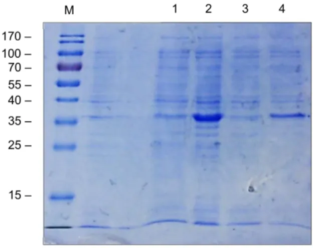
Figure 9. SDS-PAGE analysis of expression trail for CHIP protein. ... 35
Figure 10. SDS-PAGE analysis of CHIP's affinity purification. ... 36
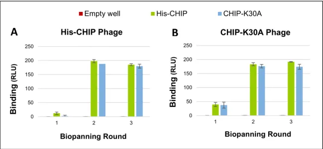
Figure 11. Enrichment of CHIP and CHIP-K30A binding phages through biopanning. ... 37
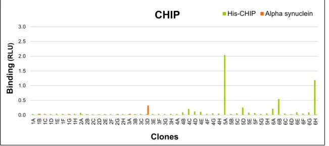
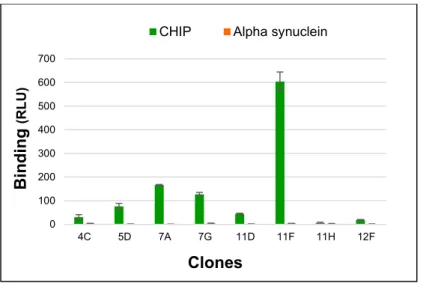
Figure 12. Screening of CHIP library selected clones in soluble scFv binding assay. .... 38
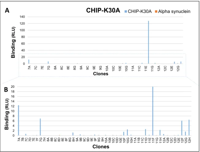
Figure 13. Screening of CHIP-K30A library selected clones in soluble binding assay. ... 39
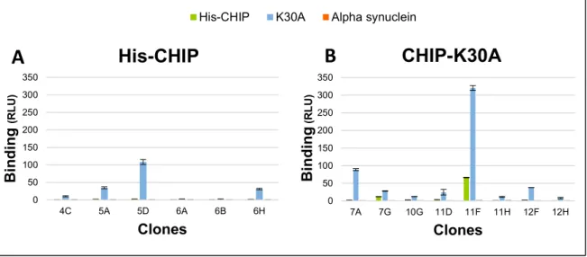
Figure 14. Validation of the scFv clones with highest affinity. ... 40
Figure 15. Alignment of the assumed amino acid sequences for the selected scFv clones with specificity for CHIP. ... 41
Figure 16. Reactivity of selected scFv clones against untagged CHIP. ... 42
Figure 17. Soluble Expression, purification and quantification of scFv 11F in E. coli TG1. ... 43
Figure 18. Transformation of E.coli BL21-DE3 with scFv plasmids. ... 44
Figure 19. SDS-PAGE analysis of affinity purified scFvs 4C, 7A, 7G, 11F. ... 45
Figure 20. Quantification of purified scFv fractions. ... 46
Figure 21. Binding of scFv purified from E.coli ... 48
Figure 22. Reactivity of purified scFvs with CHIP Titration ... 49
Figure 23. Native gel analysis of CHIP bound to scFv. ... 50
Figure 24. CHIP binds to α-synuclein in vitro. ... 51
Figure 25. In vitro ubiquitination of α-synuclein by CHIP. ... 52
Figure 26. In vitro ubiquitination of α-synuclein by CHIP with PFA. ... 53
Figure 27. Optimization of in vitro CHIP/α-synuclein ubiquitination assay. ... 53
Figure 28. Time course ubiquitination assay with modified ubiquitin. ... 54
Figure 29. CHIP autoubiquitination in the presence and absence of α-synuclein. ... 55
Figure 30. ScFv 11F (TG1) interferes with α-synuclein ubiquitination by CHIP. ... 56
Figure 31. ScFv 11F (BL21-DE3) interferes with α-synuclein ubiquitination by CHIP. .... 57
Figure 32. ScFv 4C interferes with α-synuclein ubiquitination by CHIP. ... 58
Figure 34. ScFvs effect in p53 in vitro ubiquitination by CHIP. ... 60 Figure 35. Gel-excluding bands dependence on α-synuclein. ... 61 Figure 36. Identification of gel-excluding bands in the presence and absence of CHIP. . 62 Figure 37. Association of gel-excluding bands to the ubiquitination reaction. ... 63 Figure 38. Dependence of gel-excluding bands on the presence of the ubiquitination assay components. ... 63
ABBREVIATIONS
AAV - Adeno-associated virus
ABL - Abelson murine leukemia viral oncogene homolog 1 BAG - BCL2-Associated Athanogene
BiTE - Bispecific T-cell Engager BSA - Bovine Serum Albumine CD19 - Cluster of Differentiation 19 Cdk5 - Cell division protein kinase 5 CDR Complementary Determining Region
CFTR - Cystic fibrosis transmembrane conductance regulator CH - Constant Variable Chain
CHIP - Carboxyl-terminus of Hsc70-Interacting Protein CL – Constant Light Chain
CXCR2 - C-X-C chemokine receptor type 2 ECL - Enhanced chemiluminescence EGFR – Epidermal Growth Factor Receptor ELISA - Enzyme Linked Imunnosorbent Assay
EMMPRIN - Extracellular Matrix Metalloproteinase Inducer ER – Endoplasmic Reticulum
ERAD – ER Associated Degradation
ERK – Extracellular-signal Regulated Kinases Fab - Fragment Antigen Binding
Fc - fragment crystallisable Fv - Fragment Variable
GRP78 – Glucose Regulated Protein 78
HECT - homologous to E6-associated protein carboxyl terminus HER2 - human epidermal growth factor receptor 2
HIF-1 - Hypoxia-Inducible Factor-1 HRP - Horseradish Peroxidase Hsc – Heat Shock cognate Hsp - Heat shock protein
IPTG - Isopropyl β-D-1-thiogalactopyranoside IRF 1 - Interferon Regulatory factor 1
LB - Lysogeny broth
mAbs - Monoclonal Antibodies
MT1-MMP - Membrane-Type 1 Matrix Metalloproteinase Pael-R - Parkin-associated endothelin receptor-like receptor
PBS - Phosphate-Buffered Saline
PBST - Phosphate-Buffered Saline/Tween PCR - Polymerase Chain Reaction
PEG – Polyethylene Glycol
PTEN - Phosphatase and Tensin homolog rAb – Recombinant Antibody
RING – Really Interesting New Protein RIT - Recombinant Immunotoxin RNAi - Interference RNA
SCF - S-phase kinase-associated protein 1, Cullin, F-box containing complex scFv - Single Chain Fragment Variable
ScFv - single chain variable fragment siRNA – silencing RNA
tAIF - Truncated Apoptosis-inducing factor TPR - tetratricopeptide repeat
VH - Variable Heavy Chain
VHH - camelid heavy-chain antibody
VL - Variable Light Chain
V-NAR - variable region of new or nurse shark antigen receptor WT – Wild Type
I. INTRODUCTION
The Rise of Recombinant Antibodies
As a fundamental part of the immune system, antibodies, also known as immunoglobulins (Igs), are an effective security system against pathogenic organisms or toxins that threaten our body due to their ability to correctly find almost any target antigen and elicit a neutralization response from the host organism (Murphy et al., 2012). As such they are the best “search engines” to detect very small amounts of target molecules, which makes them one of the most used tools in research laboratories all over the world. Besides their broad use in research techniques, new recombinant technologies have facilitated antibody engineering, potentiating their use in diagnostics and as therapeutics for cancer, infectious and inflammatory diseases (Chames et al., 2009).
1.1. General Antibody Structure
IgG is a bivalent, Y-shaped antibody, with a well-established structure (Figure 1) and the most abundant antibody in human serum. It is also the most common format for antibodies in diagnostics and therapeutics. The variable regions determine the specificity, diversity and affinity of the antigen binding while the constant domains mediate the antibody structure, half-life and effector functions. Within each variable domain of the light and heavy chains are three hypervariable regions, also known as complementary determining regions (CDRs), which form loops and exhibit high sequence variability, being predominantly responsible for
antigen recognition. The rest of the VL and VH domains, denominated framework regions,
are less variable and act as a platform to support the CDR loops (Murphy et al., 2012). However this structure is sometimes unattractive for certain applications, due to the effects induced by the Fc domain. For example, unwanted activation of the effector functions can lead to cytokine release mediated toxicity and a long serum half-life is particularly undesirable in imaging applications, where rapid clearance is required in order to improve contrast (Holliger and Hudson, 2005).
Fab Fc Constant region Variable region (Fv) IgG CH CH CH CH CH CH CL CL VH VH VL VL
Figure 1. Basic Structure of the IgG molecule. IgG structure comprises two large heavy chains and two smaller light chains. Each light chain presents one variable domain (VL) and one constant domain (CL)
while the heavy chains contain one variable domain (VH) and three constant domains (CH). The constant
region is coloured blue and the variable regions are designed with stripes representing the CDRs.
Smaller antibody fragments were then generated to overcome the limitations of monoclonal antibodies (mAbs). Initially, this was accomplished by removing the Fc domain using proteolytic treatments with enzymes such as papain or pepsin, yielding Fab and F(ab’)2 fragments, respectively (Porter, 1959; Nisonoff et al., 1960). Inbar and colleagues
were able to obtain a Fv fragment by peptic digestion of a mouse IgA-myeloma (Inbar et al., 1972) however the development of general procedures for Fv isolation was met with limited success (Kakimoto and Onoue, 1974; Sharon and Givol, 1976; Lin and Putnam, 1978; Reth et al., 1979; Sen and Beychok, 1986) and Fab antibodies persisted as the smallest fragment used for biomedical purposes. Later, advances in recombinant DNA technology and antibody engineering led the way to the development of a large variety of recombinant antibody (rAb) fragments with unlimited potential for research, diagnostics and therapy.
Among these fragments can be included Fab, scFv (single chain variable fragment), V-domain molecules (Figure 2) as well as camelid VHH and shark V-NAR
fragments. Compared to mAbs, these minimized antibodies can retain target specificity while presenting several advantages such as a smaller size and reduced immunogenicity, since they lack the Fc domain; better tissue penetration, rapid blood clearance and lower retention time in nontarget tissue, which can be quite beneficial for the purposes of radiotherapy and diagnostics. These and other properties can be tailored and manipulated to better suit the future application of the fragments (Holliger and Hudson, 2005). The rAb fragments can also be easily and cost-effectively cloned and expressed in large quantities in bacterial (Skerra and Pluckthun, 1988), plant (Galeffi et al., 2006), insect (Choo et al., 2002), mammalian and yeast cells (Ho et al., 2006), which makes them more economically viable.
1.2. ScFvs and derived multimeric complexes
ScFv fragments represent the smallest functional VH-VL domains capable of
high-affinity binding to the antigen and were first developed by Huston and co-workers (Huston
ScFv 5’ 3’ Linker VH Fab CH VL CHCL CL VH VL VH CH VL F(ab’)2 VH VL Figure 2. Schematic representation of proteolysis and engineered rAb fragments.
et al., 1988) and immediately followed by Whitlow and his team (Bird et al., 1988). Nowadays, scFvs are the most popular rAb fragment due to their versatility.
Antibodies in the format of scFv are proteins with a molecular weight varying from 26 to 30 kDa that have the ability to bind to the target with identical affinity to that of the parental mAb (Bird et al., 1988) and consist of one VH and one VL chain connected by a
flexible peptide linker (Maynard and Georgiou, 2000; Monnier et al., 2013).
Currently, the most used linkers include sections of glycine and serine residues for flexibility and Glutamic acid and lysine charged residues to improve solubility (Whitlow et al., 1993). Generally, these sequences are 15 to 20 amino acids long, as scFvs joined by a linker with less than 12 amino acids cannot fold into a functional Fv domain (Holliger et al., 1993) and should maintain an hydrophilic nature to keep them from interposing within or between the variable domains during folding of the scFv (Argos, 1990). The variable regions can be associated in either VL-linker-VH or VH-linker-VL orientation, but the latter is
the most common. This factor deserves some attention as the orientation can impact scFv stability, binding to the antigen (Desplancq et al., 1994) and expression efficiency (Merk et al., 1999).
As scFv fragments are small and bind monovalently to their target they often present low functional affinity (also termed avidity) and a short in vivo half-life (Fitch et al., 1999; Mayer et al., 1999). While these properties can be useful for some imaging diagnostic techniques, for example, they can compromise the success of these molecules as therapeutics agents as these may require higher retention times on the target antigen or engagement of multiple receptors in order to activate signal transduction and/or apoptosis (Teeling et al., 2004; Linsley, 2005). In order to overcome this problem while maintaining optimal size for tissue penetration, scFv antibodies are engineered into different types of multimeric complexes for greater binding avidities and better pharmacokinetic properties (Goel et al., 2000; May et al., 2012).
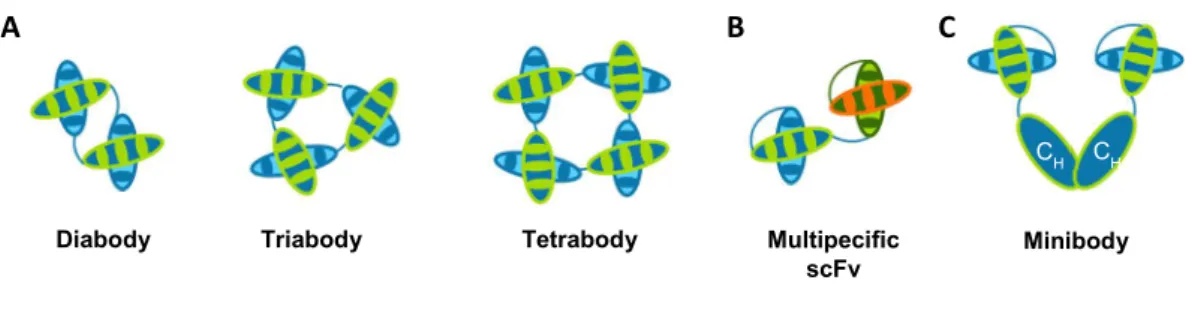
Among the above mentioned complexes it is possible to find minibodies, diabodies, triabodies, tetrabodies and bispecific scFv fragments (Figure 3).
Diabodies, triabodies and tetrabodies are noncovalent molecules that assemble due to short linker lengths. ScFv fragments with linkers of three to twelve amino acids will have the tendency to dimerize, as the VH chain will associate with the VL chain of another
scFv forming a diabody (~60kDa) (Holliger et al., 1993). Decreasing the linker length to three or less amino acids will induce the scFv association into triabodies (~90kDa) (Iliades et al., 1997) or tetrabodies (~120kDa) (Dolezal et al., 2003).
Multispecific scFv antibodies can be developed by combining two or more scFvs with different antigenic targets, enhancing target selectivity and allowing the interaction with multiple epitopes within the same molecule or in different targets (Neri et al., 1995; Coloma and Morrison, 1997).
Minibodies consist of multivalent antibody fragments that have been covalently linked to self-assembling proteins, for example, amphiphilic helix bundles and leucine zippers (Pack and Pluckthun, 1992), IgG constant domains (Hu et al., 1996) or Fc-regions (Li et al., 2000); tetravalent molecules were achieved by covalent linkage with homomultimers such as streptavidin (Kipriyanov et al., 1995; Kipriyanov et al., 1996; Cloutier et al., 2000) or the multimerization domain of p53 (Rheinnecker et al., 1996; Liu et al., 2007) and assessed for pretargeted immunotherapy (Schultz et al., 2000; Goshorn et al., 2001; Lin et al., 2006) and biotinylated drug delivery (Wang et al., 2007).
ScFvs were also engineered into bifunctional fragments through conjugation or attachment to toxins (Chaudhary et al., 1989) and radionuclides (Kuan et al., 1999) mostly for cancer therapy, liposomes (Laukkanen et al., 1994) and enzymes (Sharma et al., 2005) for improved drug delivery, quantum dots (Wang et al., 2008) for imaging, viruses (Nakamura et al., 2004) for gene therapy, and cytokines (Halin et al., 2002) or chemokines (Guo et al., 2004) for immunotherapy.
1.3. From the bench to the frontline: current applications of scFvs
The improvement of methodologies that allow for the development of scFv antibodies and the advantages they present over conventional mAbs has potentiated the use of these fragments in very different, and sometimes complimentary, applications that
Diabody Triabody Tetrabody Multipecific
scFv Minibody
CH CH
A B C
Figure 3. Multimeric formats of scFv.
Several complexes with tailored valences and specificities can be engineered using scFvs as building blocks. Here are represented examples of several categories of multimeric scFv formats. A. Diabodies, triabodies and tetrabodies can be obtained by assembling two, three or four, respectively, scFvs, for an increase in valency. There is also the possibility of engineering these antibody formats for increased specificity (bispecific, trispecific and tetraspecific) by combining scFvs selected for different antigens. B. Multi- or bispecif scFvs combine scFvs with different antigenic targets. C. Minibody assembled by combining scFvs with two IgG’s constant domains.
range from research to therapeutics and in vivo imaging. The contribution of scFvs and its development to these areas and recent examples will be detailed below.
ScFv fragments have become an increasingly useful tool in the study of protein functions and their molecular mechanisms, particularly when only a domain of the protein needs to be studied.
RNAi technology has been used routinely for the purpose of studying protein function but as it down regulates the expression of the whole protein it is unhelpful when the aim is to focus on specific domains. Moreover, the use of this technology in vivo has elicited several problems regarding delivery to the target tissue and consequently off-target toxicity issues (Aagaard and Rossi, 2007). ScFvs, on the other hand, can be selected specifically for different protein domains and for different epitopes in those domains as was the case of a study that reported the development of scFv fragments that targeted different epitopes of the G-protein coupled receptor CXCR2 as allosteric antagonists and showed ligand-dependent differences in functional assays (Rossant et al., 2014).
These antibody fragments are also becoming a strategic tool to study the importance of individual domains in the understanding of the general mechanism of action of a protein and its loss of function. Murphy and colleagues engineered a scFv capable of inhibiting the activity of MT1-MMP, a pericellular protease involved in tumour cell invasion and angiogenesis, by binding to a non-catalytic domain. Also this study presents a potential novel approach to inhibit proteinases by targeting sites outside the catalytic domain (Basu et al., 2012). Another study dissected the activity of Pax6, a homeodomain transcription factor, in the migration of oligodendrocyte precursor cells, using a scFv against the extracellular domain of Pax6 that lead to loss of function (Di Lullo et al., 2011). A plasmid carrying the scFv was electroporated in the neural tube and the scFv was able to neutralize the extracellular domain emphasizing its involvement in the process being studied.
A recent report elucidated the role of EMMPRIN (extracellular matrix metalloproteinase inducer) down-regulation in the promotion of apoptosis through the mitochondrial pathway by intracellular acidosis via intracellular expression of a scFv using a chimeric adenoviral vector (Thammasit et al., 2015). This highlights another advantage of scFvs against RNAi technology as scFvs can be delivered by a viral vector, avoiding the need for multiple administrations and ensuring maintenance of antibody concentration to sustain silencing.
As has been shown scFvs can be used to study protein and domain functions in both in vitro and in vivo studies. Moreover studies of this nature can also lead to new therapeutic targets and reveal molecules with therapeutic potential.
Therapy is perhaps the leading driver for the great improvements registered in scFv development, in the last few years, especially cancer therapy.
ScFvs present several properties that are important in cancer therapy such as the ability to specifically recognize markers expressed in tumour cells and a reduced size for better and more even tissue penetration. A smaller size also means faster clearance rate and while that can be beneficial when scFvs are coupled with drugs or toxins, allowing for a lower exposure for healthy tissue, it also compromises the ability of the scFv to concentrate in the tumour when they’re not conjugated. As was already discussed above, this problem was overcome by engineering multimeric scFv complexes.
ScFv-based tumour therapy involves targeting specific markers in cancer cells and neutralizing the protein, delivering a therapeutic entity such as a toxin, a drug or siRNA, or initiate an immune response.
A scFv derived format that has seen great approval and development in the area of cancer immunotherapy are BiTEs (Bispecific T-cell Engagers) (Figure 4). Recently the first BiTE fragment, Blinatumomab (Amgen), was approved by the FDA for the treatment of refractory Philadelphia chromosome–negative acute lymphoblastic leukemia. This antibody redirects unstimulated primary human T cells towards CD19-positive lymphoma cells but presents the disadvantage of requiring continuous IV infusion due to its low molecular weight. Concurrently some more bispecific antibodies are now
undergoing clinical trials, several are also BiTEs, and the group also includes tetravalent bispecific antibodies that should not require such a frequent administration as blinatumomab (Sheridan, 2015; Wu et al., 2015).
Another area being developed in scFv-mediated cancer therapy are recombinant immunotoxins (RITs) in which scFv are used to direct cytotoxic drugs to cancer cells. This is achieved by replacing the cell binding domain of Pseudomonas exotoxin A with a scFv specific for the desired antigen (Liu et al., 2012). Several immunotoxins are currently undergoing clinical trials such as SSP1 which is being evaluated in combination with chemotherapy for treatment of malignant pleural mesothelioma (Hassan et al., 2014) and moxetumomab pasudotox for advanced hairy cell leukemia (Kreitman et al., 2012) which is in a phase III clinical trial. At the same time more immunotoxins are being investigated
Target cell T cell Antigen BiTE CD3 Figure 4. BiTE.
BiTEs are bispecific diabodies, in which one of the scFv fragments is specific for CD3, the signal transduction element of the T-cell receptor (TCR), while the other engages with a protein found at the surface of target cells. This forms a link between the target cell and the T cell which will release cytotoxic proteins, despite the absence of MHC I or co-stimulatory molecules, triggering apoptosis of the target cell. This imitates physiological events registered during T cell attacks (Wu et al., 2015).
as novel EGFR-specific immunotoxins using scFvs derived from already approved mAb therapies (panitumumab and cetuximab) (Niesen et al., 2015) .
Neurodegenerative diseases have also applied scFvs to attempt to generate therapies which was possible because scFvs are capable of crossing the blood brain barrier, even when they are administered peripherally, and can be overexpressed in vivo, allowing for prolonged therapy without repeated injections (Robert and Wark, 2012).
For example, in Alzheimer’s disease, anti-Aβ scFv, overexpressed via AAV (adeno-associated virus) delivered by intracranial injection, have been shown to reduce amyloid plaques in mice and were still detectable in the brain without causing neurotoxicity (Ryan et al., 2010). Less invasive administration routes were also studied, namely intramuscular (Wang et al., 2010; Yang et al., 2013) and intranasal (Cattepoel et al., 2011); both were successful and lead to reduction of Aβ accumulation. A more recent study reported scFvs that bind toxic oligomeric but not monomeric or fibrillar tau and were capable of detecting it in earlier stages than usual, demonstrating potential for biomarker development (Tian et al., 2015). Another study demonstrated the in vivo effects of a pan-amyloid specific scFv antibody that mitigated memory deficits and brain pan-amyloid load in mice with Alzheimer’ Disease (Zhao et al., 2014).
In vivo imaging is another area that was has been improved by the use of scFvs especially regarding diagnostic applications such as tumour detection. The low molecular weight of scFvs allows them to be coupled to radionuclei, quantum dots and nanoparticles while maintaining the necessary properties for in vivo imaging. Among these properties are high affinity for the target, deep tissue penetration and a fast clearance rate. As such the scFv format provides an ideal non-invasive tool to detect and analyse the expression of a specific target in vivo.
For example, a scFv fragment specific for GRP78, a protein important for cell proliferation and angiogenesis, was linked to quantum dots and delivered in a xenograft mouse model. The complex quantum dot-scFv allowed for easy visualization of the target in vivo. Additionally it was also able to inhibit breast tumour growth (Xu et al., 2012).
Quantum dots were also conjugated to anti-tumour scFvs specific for the oncomarkers, HER1/EGFR and HER2/neu, forming self-assembling fluorescent complexes with the target that enabled visualization of cancer cells in vitro (Zdobnova et al., 2012).
Magnetic resonance imaging is an emerging field in the application of scFv fragments as it sensitivity has been shown to improve by linking scFv antibodies with supramagnetic iron oxide nanoparticles (SPIONs). In tumour imaging, these particles
provide a clearer contrast between healthy and cancer cells due to different uptake levels and adding scFv allows targeting of specific cells (Vigor et al., 2010).
Another promising imaging modality that has already adopted the scFv format is Photoaccoustic which also employs iron oxide nanoparticles. In this case, the nanoparticles were conjugated with an anti-HER2 scFv and used to image HER2-positive tumours (Kanazaki et al., 2015).
Since they were introduced, scFvs have become progressively more applicable in research laboratories, diagnostic and therapeutic applications. As this format of fragments continues to evolve and its uses become more widely acknowledged it is expected that it will also become more relevant in the progress of all the areas mentioned above, but more particularly in therapy.
Engineering scFv by Phage Display Technology
Conventionally, scFv antibodies were produced from hybridoma cells acquired from immunized animals by amplification of the VH and VL domains from mRNA and connecting them by a polylinker followed by insertion in the choice vector (Huston et al., 1988). However, lately, this technology has been surpassed by in vitro molecular displaying technologies due to their adaptability to high throughput formats not to mention the ease of manipulation to optimize scFv properties in order to produce a pool of varied and highly functional antibodies (Bradbury et al., 2011).
Three different molecular displays from which scFv can be selected and affinity matured have been reported, namely phage-display (McCafferty et al., 1990), ribosome display (Hanes and Pluckthun, 1997) and cell surface display (Francisco et al., 1993; Boder and Wittrup, 1997). All three formats share the same basic principle as molecular display libraries, created by cloning a diverse collection of rAb genes, are screened for target antigen binding and the resulting enriched pools are amplified after each round of selection with the target. After a few selection rounds the polyclonal pools are screened for antigen reactivity.
2.1. Molecular Display Libraries
Antibody diversity results from somatic recombination in B lymphocytes through a combination of three different gene segments, V, D and J. Depending on the source of the variable region genes, four different types of molecular display libraries can be built.
Immune libraries derive from immunoglobulin genes of lymphocytes from immunized animals (Clackson et al., 1991) or naturally immunized (Jacobin et al., 2002) or infected, humans (Burton et al., 1991). These libraries have been constructed from various species, besides humans, including mouse, chicken, rabbit and camel (Hoogenboom et al., 1998). Despite being antigen-specific and enriched in affinity matured clones they also present several disadvantages such as the need to create a new library for each antigen, the time required for immunizations and the unpredictability of the immune response (Azzazy and Highsmith, 2002).
Naïve, synthetic and semi-synthetic libraries are ‘single pot’ repertories, resulting from non-immunized donors and are antigen independent, consequently they represent a source of antibodies against a diverse assortment of antigens including self, toxic and non-immunogenic antigens (Marks et al., 1991; Griffiths et al., 1993; Vaughan et al., 1996).
Naïve libraries offer the possibility to select antibodies of desired specificity and high affinity without immunization being necessary (Burton et al., 1991). Genes responsible for the variable domain are obtained through B-cell mRNA amplification using oligonucleotides specific for the VL and VH families. The heavy and light chains are then
randomly combined and cloned to create a combinatorial scFv library. This procedure gives access to germline antibodies, this is, antibodies that have yet to be exposed to antigens, however the frequency of those antibodies is dependent on the source of the B-cells (Marks et al., 1991).
Synthetic libraries result from in vitro assembly of V, D and J gene segments. Assembly of V-genes artificially allows for introduction of diversity using PCR techniques and degenerate primers to randomize CDR regions (Hoogenboom and Winter, 1992). The heavy chain CDR3 has been the main target of these modifications in synthetic repertoires. Examples of synthetic libraries include the Tomlinson I and J libraries (de Wildt et al., 2000; Goletz et al., 2002) and the human combinatorial antibody libraries (HuCAL) for scFv (HuCAL-scFv) (Knappik et al., 2000) and Fab fragments (HuCAL-Fab1, HuCAL GOLD, HuCAL PLATINUM) (Rauchenberger et al., 2003; Rothe et al., 2008; Prassler et al., 2011) Semi-synthetic libraries combine elements of both natural and synthetic origin in order to increase natural diversity while introducing synthetic functional diversity. These libraries are typically created by amplifying natural naïve CDR1 and CD2 regions and synthetically randomizing CD3 regions, by shuffling natural CDR regions or by combinatorial mutation of certain amino acids in the VH and VL chains of the CDR3 region
(Barbas et al., 1992). It has also been reported a synthetic library that combined synthetic CDR1 and CDR2 regions and a natural CDR3 (Hoet et al., 2005).
All these three libraries are amenable to high throughput screening and capable of generating and selecting thousands of antibodies specific to different epitopes on the same target.
2.2. Phage Display
Phage-display is the oldest and most frequently used system for molecular display. This technique was first implemented in 1985, when Smith and colleagues showed that foreign DNA fragments could be fused to the gene encoding the phage minor coat protein, pIII, of a nonlytic filamentous bacteriophage and consequently expressed in the form of a fusion protein at the surface of the phage without affecting the its infectivity (Smith, 1985). Later, a team of researchers based in Cambridge successfully applied this technology to the production of scFv antibodies by showing that these fragments could be displayed at the surface of the virion as functional proteins capable of binding to antigens (McCafferty et al., 1990).
The phage of choice is M13, a flexible rod like shaped filamentous phage with a 6000 to 8000 bases circular genome surrounded by a coat of five different proteins. One end of the phage displays by five copies of each of the two minor coat proteins, pIII and pVI while the other end presents three to five copies of pVII and pIX; the rest of the phage particle is covered with several thousand copies of the coat protein pVIII. (Smith and Petrenko, 1997).
The most commonly used coat proteins for phage display are pIII and pVIII. The pIII protein is involved in phage-host interactions during infection and is 460 amino acids long while pVIII protein has only 50 amino acids (Crissman and Smith, 1984; Smith, 1985). As these proteins have an important role in viral packaging and infectivity displaying large foreign peptides on every copy of selected coat protein could interfere with these functions limiting the performance of the phage display technology.
This obstacle could be overcome by expressing the foreign fragment only in a portion of the coat protein and different solutions were engineered to achieve this goal. In some phage display systems instead of introducing the DNA encoding the scFv library directly into the phage genome, the library is inserted as a gene cassette encoding scFv-pIII fusion proteins. This way the phage will retain the wild type scFv-pIII protein copy ensuring that only some pIII coat proteins display scFvs (Smith and Petrenko, 1997).
Another option is to insert the fusion coat protein in a phagemid (a plasmid with both phage and E. coli derived origins of replication), while the WT coat protein and the remaining genes essential for phage assembly are delivered by a helper phage with a deficient packaging signal. Co-infection of the bacteria by the phagemid and the helper
phage produces hybrid virions with the phagemid genome and displaying only limited copies of the fusion coat protein (Vieira and Messing, 1987; Mead and Kemper, 1988). During phage assembly WT pIII competes with the fusion protein for integration into the phage particle however since in a wild type phage there is only three to five copies of pIII coat protein, the majority of phagemid particles will only display zero or one copy of the fusion protein per phage. This is the reason why higher affinity antibodies are more often selected from phagemid than phage libraries, as there is no avidity (O'Connell et al., 2002).
The selection of scFv from phage display libraries is achieved by multiple rounds of probing against the desired antigen interspaced with washing steps to remove nonspecific phage clones. Typically, several rounds of panning with progressively increased washing stringency in between are performed to enrich the resulting phage pools with the highest affinity binders (Figure 5). This process is denominated biopanning and it is generally performed against a target immobilized onto a solid support as microtiter plate wells (Schofield et al., 2007), immunotubes (Weisser et al., 2007) or columns (McWhirter et al., 2006). Additionally, the phage library can be incubated with biotinylated antigen and then the antigen-phage complex is captured using a streptavidin surface (Winter et al., 1994). This approach could be beneficial for proteins that suffer conformational changes due to immobilization or so as not to limit the epitopes exposed to the panning.
In order to retrieve the phage particles that express scFv fragments capable of binding to the target protein an elution step is performed. Several elution strategies have been used including low (Smith, 1985) or high (Parmley and Smith, 1988) pH buffers, proteolytic cleavage (Ward et al., 1996), competition with free antigen (Oldenburg et al., 1992) and even ultrasound (Lunder et al., 2008).
The eluted phages retain their infective capacity and can therefore be propagated by infecting new bacterial hosts, to yield an amplified library, already more specific for the target protein, that can serve as input for the next affinity panning round. Usually only two to three rounds of panning are necessary however this can vary depending on the target protein. The eluate from the final round of panning are propagated and subjected to further analysis to isolate clones of interest.
Phage display of scFvs presents several advantages over other techniques. High stability of the phages allows storage at 4ºC for several years (Burritt et al., 1996) and makes phage display amenable to obtain binders for a specific conformation, structure, folding or enzymatic activity of the target protein (Forrer et al., 1999). Additionally, scFv production can be achieved swiftly and economically just by infecting E.coli.
Recent improvements in this technique include the development of a high-throughput phage-display screening in array format using a protein chip carrying recombinant phage particles that aims to facilitate antibody identification and characterization (Diez et al., 2015); an industrialized platform to generate high affinity antibodies for transcription factors and epigenetic antigens through an optimized automated phage display and antigen expression pipeline capable of producing a great number of sequenced Fabs with high affinity stability and good expression in E. coli (Hornsby et al., 2015); and generation of scFv fragments capable of revealing structural differences of amyloid-β fibrils resulting from variations in the acidity of the environment during the fibrillogenesis process (Droste et al., 2015).
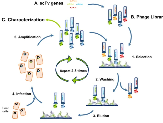
In summary, phage display technology has seen great improvements since it was first developed in 1985 in order to accommodate the new applications and demands of today’s science. This technique has been particularly useful in the high throughput development of new scFv fragments which in turn have been widely used in basic research Figure 5. Phage Display Cycle.
A library of gene variants of antibody fragments (A) is expressed on the surface of M13 phage (B) and incubated with an immobilized antigen to select based on the expressed epitopes antigen binding scFv (1). After extensive washing to remove unbound and low affinity phage particles (2), the remaining bound phage particles are eluted (3). The selected phage are used to infect host cells (4) which are then grown (5) originating a “new” library enriched with variants of antibody fragments capable of high affinity interaction with the desired antigen (polyclonal pool). This cycle is usually repeated from two or three times to increase the pool of high affinity antibodies. The use of phage display results in selection of high affinity antibodies, that can be expressed and characterized (C) (Smith, 1985).
A. scFv genes Repeat 2-3 times B. Phage Library 2. Washing 3. Elution 4. Infection 5. Amplification Host cells C. Characterization 1. Selection
and immunotherapy, as has been described before. Considering the evolution this technique has achieved and seeing as it is still extensively employed, new developments are to be expected and new applications unveiled.
2.3. Alternative Molecular Display Systems
Even though phage display is the most commonly used molecular display system, Ribosome and Cell Surface display also have to be considered as two well-established techniques which also have their advantages.
Ribosome display doesn’t require cell growth or transformation as it is a fully in vitro transcription/translation system. The concept involves the translation of the scFv encoding nucleic acids to form a complex that is then incubated with the target protein to select specific binders (Figure 6). This system can overcome the limitations of cell-based displays such as the expression bias and generate larger libraries than phage or cell surface display constituting a powerful alternative to the remaining molecular display systems (Hanes and Pluckthun, 1997).
In the cell surface display system, thousands of copies of the scFv are anchored to proteins such as the Lpp-OmpA chimera on the membrane of E.coli (Francisco et al., 1993) or the α-agglutinin adhesion receptor on the cell wall of yeast (Boder and Wittrup, 1997) and consequently displayed on the surface of the cells and selected with Fluorescent Activated Cell Sorting (FACS) technology. This selection technique allows for Figure 6. Alternative Molecular Display Systems.
A. Ribosome Display. mRNA molecules are incubated with a stoichiometric amount of ribosome and once the translated antibody fragment emerges from the ribosome, the lack the stop codons will lead to the formation of a protein-ribosome-mRNA complex. The presence of a spacer region at the 3’ terminal of the DNA library ensures the fusion to the ribosome while allowing the protein to fold correctly. The complexes are then incubated with the immobilized target to probe for binding. Elution is achieved by destruction of the complex or competitive elution with free ligands. The eluted mRNA is subjected to reverse transcription-PCR in order to obtain DNA for the next round of panning (Hanes and Pluckthun, 1997). B. Cell Surface Display. The protein of interest is expressed at the surface of the cell fused with one of its surface proteins and high-affinity binders can be selected and quantified, according to scFv expression and antigen binding, by flow cytometry using both fluorescently labelled antigens and anti-epitope tagged reagents (Boder and Wittrup, 1997).
scFv
Cell Surface Display Ribosome Display
scFv
Nucleic acid Spacer
a swift and quantitative screening and ensures that tightly bound clones will be recovered. The most popular format is yeast display but E. coli and Bacillus thuringiensis (Du et al., 2005).
CHIPping Away at the Unknown
The Carboxy-terminus of Hsc-70 Interacting Protein (CHIP) is a 35kDa homodimeric quality control E3 ligase. CHIP is highly expressed in tissues with high metabolic rates such as the heart, the adult striated muscle and the brain. Intracellularly, it is known for being present in both the cytoplasm (Ballinger et al., 1999) and the nucleus under different conditions (Meacham et al., 2001)
Evolutionarily, CHIP’s amino acid sequence is well-conserved across several species, sharing a particularly high similarity with that of mouse (~98%) and its ubiquitination domain, the U-box, is the least altered region of the sequence. This protein was first described in 1999 when it was discovered during an assay that aimed to identify TPR-containing proteins in the heart by screening a phage library of human heart cDNA against a fragment of cytochrome 40 (Ballinger et al., 1999).
Fifteen years later, even though several CHIP interactors and substrates have been identified and its relevance in physiology and disease has been widely studied, little is still known about its molecular mechanistics and regulation.
3.1. Unravelling CHIP’s Structure and Activity
The maintenance of normal cellular functions rests heavily on the integrity of the cell’s proteome; to this end, the cell possesses a set of pathways responsible for monitoring and maintaining the health of its proteins. Central to these pathways are the molecular chaperones that can promote the folding of misfolded protein, and if that’s not possible target them to the ubiquitin-proteasome system (UPS) which is responsible for the degradation of proteins usually marked with ubiquitin tags.
The addition of ubiquitin to the target proteins is accomplish through a process known as ubiquitination, which requires a succession of biochemical reactions catalysed by three different groups of enzymes (Figure 7). CHIP belongs to the group of the E3 ubiquitin ligases.
Fi gu re 7 . U bi qu iti na tio n Pa th w ay Pr ot ei n ub iq ui tin at io n in vo lve s th re e cl as se s of e nz ym es . F irs t, th e C-te rm in us o f u bi qu itin (U B) is lin ke d by a th io es te r b on d to th e ac tiv e sit e cy st ei ne o f a u bi qu itin -a ct iv at in g en zy m e E1 , g en er at in g an U b-E1 c om pl ex . T hi s re ac tio n is AT P-de pe nd en t a nd a ct iva te s ub iq ui tin . A fte rw ar ds u bi qu itin is tr an sf er re d to th e ac tiv e si te c ys te in e of a n ub iq ui tin -c on ju ga tin g en zy m e (E 2) , f or m in g an E 2-Ub th io es te r, an d re le as in g E1 . E 3 ub iq ui tin li ga se s in te ra ct b ot h wi th th e co m pl ex E 2-Ub a nd th e su bs tra te b y ca ta lys in g th e fin al tr an sf er o f u bi qu itin b y tw o di ffe re nt m ec ha ni sm s, d ep en di ng o n th e ty pe o f l ig as e an d ar e th e m ai n so ur ce o f s pe ci fic ity in th e ub iq ui tin s ys te m . H EC T-ty pe E 3s a ct a s co va le nt in te rm ed ia te s as u bi qu itin is in iti al ly tra ns fe rre d to th e ac tiv e sit e cy st ei ne o f E 3 an d on ly th en c on ju ga te d to th e su bs tra te . I ns te ad R IN G a nd U -b ox E 3s fa cil ita te th e tra ns fe re nc e of u bi qu iti n di re ct ly fro m th e co m pl ex E 2-Ub to th e su bs tra te . U bi qu iti na tio n re ac tio ns o cc ur m os tly o n pr im ar y am in es in L ys in es , a nd , l es s of te n, a fr ee N -te rm in us , r es ul tin g in s ta bl e pe pt id e bo nd s wi th th e C-te rm in us o f u bi qu itin . T hi s pr oc es s ca n oc cu r o nc e (m on ou bi qu itin at io n) o r m ul tip le ti m es on d iff er en t L ys in e re si du es o f t he p ro te in (m ul ti-m on ou bi qu itin at io n) . A dd itio na lly , u bi qu itin c an a lso b e tra ns fe rre d to o ne o f t he L ys in es o r t he N -te rm in al m et hi on in e of o th er u bi qu iti n m ol ec ul es al re ad y at ta ch ed to th e su bs tra te o rig in at in g po ly-ub iq ui tin c ha in s. T he ty pe o f u bi qu itin at io n in flu en ce s th e fa te a nd fu nc tio n of th e m od ifie d pr ot ei n as m on ou bi qu itin at io n of te n le ad s to in vo lve m en t in p ro ce ss es a s DN A re pa ir, p ro te in tr af fic ki ng , a nd tr an sc rip tio n (R am an at ha n an d Ye , 2 01 2) w hi le m ul ti-m on ou bi qu itin at io n ca n al so re di re ct p ro te in s fo r p ro te as om al d eg ra da tio n (D im ov a et a l., 20 12 ; S ha be k et a l., 2 01 2) . R eg ar di ng p ol yu bi qu itin at io n, K 11 -li nk ed c ha in s ar e in te gr al to p ro te as om al ta rg et in g of a na ph as e-pr om ot in g co m pl ex /c yc lo so m e (A PC /C ) s ub st ra te s (W ick liff e et a l., 20 11 )m ea nw hi le li nk ag e th ro ug h K4 8 an d ot he r l ys in es c ha in s of fo ur o r m or e ub iq ui tin m ol ec ul es e ffi ci en tly ta rg et p ro te in s fo r p ro te as om al d eg ra da tio n (X u et a l., 2 00 9; K im e t a l., 2 01 1) . L in ea r an d K6 3-lin ke d ub iq ui tin c ha in s ar e as so ci at ed w ith n on -d eg ra da tio n ev en ts o f N F-κB s ig na lin g (S ch m uk le a nd W al cz ak , 2 01 2) a nd a re a ls o im pl ica te d in D NA re pa ir an d ta rg et in g of e nd oc yt ic pr ot ei ns fo r l ys os om al d eg ra da tio n (R am ae ke rs a nd W ou te rs , 2 01 1; C la gu e et a l., 2 01 2) .
In fact, CHIP plays an important role in the ubiquitin-proteasome scheme as a bridge between the molecular chaperone system and the degradation pathway due to its functions as both a quality control E3 ubiquitin ligase and a co-chaperone of several heat shock proteins (Hsp) (McDonough and Patterson, 2003).
CHIP’s structure is intimately associated with its activity. Each monomer (34.5kDa) displays two specialized domains, which impact CHIP’s activity in a different but complementary manner, joined by a central coiled-coil region. The amino terminus contains three tetratricopeptide repeats (TPR), responsible for the interactions with chaperones, and the carboxyl terminus displays a U-box domain that grants CHIP its E3 ubiquitin ligase activity (Figure 8) (Zhang et al., 2005).
The U-box, positioned at the C-terminus, is structurally similar to RING finger domains, the main difference resting on the fact that U-boxes are stabilized by hydrogen bonds instead of zinc binding (Aravind and Koonin, 2000). CHIP’s U-box (residues 232-298) contains a pair of β-hairpins running into a short α-helix followed by a third hairpin and concluding in in a C-terminal α-helix (Zhang et al., 2005). This region acts as a scaffold or an adaptor, positioning the substrate in proximity with the E2-ubiquitin complex, as opposed to the HECT ubiquitin ligases, that form transient thioester links with the ubiquitin molecule and transfer it to the substrate (Passmore and Barford, 2004).
The TPR domain (residues 26-131) includes three TPRs, each of which consists of two antiparallel α-helices separated by a turn to form a ‘knob and hole’ structure (Zhang et al., 2005) with a hydrophobic surface that facilitates protein:protein interactions (Das et al., 1998). This domain is primarily responsible for CHIP’s interactions with the Hsc/Hsp proteins.
Initially it was believed that TPR domains were mostly rigid, invariable structures, even upon ligand binding. Nevertheless recent structural and dynamic studies have suggested otherwise and added that binding to the TPR domain can actually lead to large conformation changes in the protein as a whole (Parashar et al., 2013).
This possibility has already started to be investigated for CHIP. A study conducted using Hydrogen/Deuterium Exchange coupled with Mass Spectrometry (HDX-MS) demonstrated that the TPR domain in CHIP is not only loosely folded but also that the first sixty amino acids are intrinsically disordered. Also it was revealed a high degree of flexibility in the TPR domain which decreased upon Hsp70 or Hsp70 peptide binding (Graf et al., 2010).
These results above were supported by a recent publication that additionally looked at a CHIP TPR mutant (CHIP-K30A), in which the lysine in position 30 was replaced by an alanine, a residue that encourages helix formation. This protein had only been studied as a non-chaperone binding mutant of CHIP with decreased catalytic activity. However it was revealed that, when compared with wild type CHIP, the mutant presents a TPR domain with reduced flexibility, equivalent to the decrease seen for the ligand-bound TPR domain. Additionally molecular dynamics studies were conducted and it was shown that the mutation affected not only TPR movements but also the U-box’s. This observation lead the way to a new mechanism of regulation for CHIP and established the K30A mutant as a useful tool to study the effect of TPR stabilization in the absence of a ligand (Narayan et al., 2015).
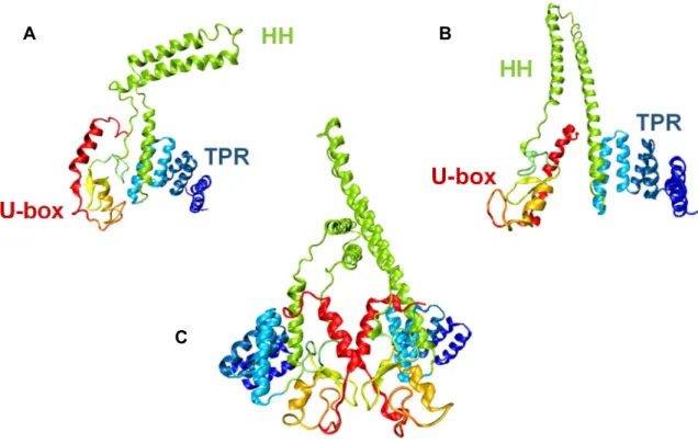
The TPR and the U-box domains are brought together by a charged coiled-coil domain (helix 7 and 8), essential for the coupling of CHIP’s inactive monomers and consequently its function. as enzymatic activity of CHIP is dependent on its dimerization (Nikolay et al., 2004). When CHIP dimerizes, the two protomers adopt significantly different conformations forming an asymmetric dimer. The assembly of CHIP’s two monomers involves the interaction of the U-box domain and the central helical domain of Figure 8. Representation of CHIP’s structure.
(A) Structure of the CHIP protomer that presents the ‘broken’ coiled coil domain (CC) indicating the tetratricopeptide repeat domain (TPR) and the U-box domain. (B) As in (A) but for the protomer that maintains a straight coiled coil domain. (C) The two protomer assembled. These images were obtained from PDB file 2C2L (Zhang et al., 2005) using the graphic software Visual Molecular Dynamics (VMD) (Humphrey et al., 1996) coloured according to the different secondary structure motifs present.
HH
TPR
TPR
U-box
U-box
HH
A B Ceach monomer. However helix 7 adopts distinct conformations in the two monomers; in one remains a straight helix while in the other it breaks forming two perpendicular α-helices. This, along with a dislocation in the U-boxes and the helical domains symmetry axes leads to structural arrangements that positions the TPR domain of one of the protomers in front of its U-box domain, blocking it. For this reason CHIP displays a “half-of-sites” activity (Zhang et al., 2005).
CHIP is predominantly responsible for the ubiquitination of chaperone-bound substrates by binding to C-terminal of Hsp/c70 and Hsp 90 with its TPR domain in order to facilitate ubiquitination via its catalytic U-box by the 26S proteasome. Chaperone clients usually include chaperone activated signalling proteins and proteins prone to aggregation that are subjected to chaperone assisted quality control (Connell et al., 2001; Jiang et al., 2001).
In order to perform its role as ubiquitin ligase, CHIP depends on the interaction with E2 proteins. CHIP has been shown to interact with UbcH5 to produce Lys-48-linked polyubiquitination and with E2 complex Ubc13-Uev1A to generate Lys-63-linked polyubiquitination. This suggests the product formed in CHIP-mediated ubiquitination reactions is dependent on which E2 is involved (Xu et al., 2008). Another study identified a set of other seven other E2 enzymes that bind and function with CHIP in vitro to produce all types of ubiquitination events. This study also confirmed that CHIP requires the SPA motif in loop 7 of E2 for recognition and binding (Soss et al., 2011). Different ubiquitination patterns have also been described depending on the E2 present but also due to small changes in the substrate, as ubiquitination assays showed that the distribution of multiple ubiquitination chain types is different for Hsp70 versus Hsc70, even though these proteins present highly sequence homology and similar structure (Soss et al., 2015).
Additionally, CHIP has also been shown to interact with other E3 ligases to facilitate their ubiquitination activity, functioning as an E4 ligase. So far this function of CHIP has been demonstrated for Parkin, considered the culprit for a juvenile form of Parkinson (Imai et al., 2002), and for the complex SCFSkp2 (Nie et al., 2008).
3.2. Regulation
CHIP is regulated at different levels and new insights about possible mechanisms have been in study lately.
Few studies have been dedicated to studying CHIP’s transcriptional regulation under physiological and pathological contexts. Still, it is to be expected that in case of a massive accumulation of misfolded proteins, quick adjustments of the levels of Hsp70
chaperones and its co chaperones would be required so as to maintain homeostasis. The levels of CHIP and/or Hsp70 mRNA are in fact upregulated and have, in vivo and in vitro, protective effect under stress conditions such as heat shock, pathological polyQ overexpression (Miller et al., 2005; Dikshit and Jana, 2007) and oxidative stress (Stankowski et al., 2011). A decrease in CHIP’s mRNA and protein levels has been observed in breast cancer (Kajiro et al., 2009; Patani et al., 2010), colorectal (Ruckova et al., 2012; Wang et al., 2014b) and gastric cancer (Gan et al., 2012) and correlated greatly with prognosis. These results are in agreement with evidence showing that CHIP acts as tumoral suppressor.
The one example of CHIP posttranscriptional regulation that has been reported so far is in the context of bone morphogenesis as translational repression of CHIP by miR-764-5p was deemed essential for adequate osteoblast differentiation (Guo et al., 2012).
Posttranslational modifications (PTM) of CHIP have also been investigated however only ubiquitin modifications have been reported. CHIP undergoes a regulatory ubiquitination in cells and in vitro which doesn’t promote its turnover but instead facilitates substrate targeting for proteasomal degradation (Jiang et al., 2001; McDonough and Patterson, 2003). For instance, Ataxin 3, an ubiquitin-interacting motif containing deubiquitinase, provides chain editing activity for CHIP by binding and deubiquitinating CHIP upon completion of substrate ubiquitination however this activity is dependent on E2 Ube2w ubiquitination of CHIP (Scaglione et al., 2011). Additionally it has also been reported that CHIP undergoes extensive autoubiquitination however the extension of this process seems to be dependent on the E2 enzyme present in the reaction (Soss et al., 2011; Soss et al., 2015). Until recently no direct evidence of other PTMs had been found. It had only been proposed that CHIP had functional phosphorylation sites (Dephoure et al., 2008) and interacted with protein kinases such as ERK5 and Lim Kinase 1 (LIMK1) (Lim et al., 2007; Woo et al., 2010). However recently a study reporter that Cdk5 phosphorylates CHIP at Ser20, promoting tAIF-mediated neuronal death (Kim et al., 2015).
Moreover substrate PTM and conformation changes also have roles in functional regulation of CHIP. For instance, under stress conditions, Abl phosphorylation of MST1 kinase inhibits its degradation by CHIP allowing it to bind to FOXO3 and trigger neuronal cell death (Xiao et al., 2011). Also Landré and colleagues reported that IRF-1 (interferon regulatory factor-1) ubiquitination by CHIP was inhibited when IRF-1 adopted a DNA bound conformation as it obstructed the E3 docking site (Landre et al., 2013).
The activity of CHIP is also regulated by its interactions with other proteins. This regulation occurs by varied mechanisms including: competition with substrate binding (eg. S100 proteins) (Shimamoto et al., 2013); competition with chaperone binding (eg. Xap2)
(Lees et al., 2003); conformational modification of the chaperone complex (eg.HspBP1) (Alberti et al., 2004); interference with CHIP:E2 interaction (eg. BAG2) (Arndt et al., 2005); facilitation of chaperone binding (eg. BAG1/3) (Demand et al., 2001; Dai et al., 2005) and of the interaction with the E2 (eg. S5a) (Kim et al., 2009).
It has also been proposed a chaperone-mediated allosteric model of CHIP regulation. An in vitro study, carried out using wild type and a U-box mutant (P269A) CHIP without E3 ligase activity, suggested that CHIP-Hsc70 binding is dependent on allosteric interactions between the U-box and the TPR domains. The increased binding efficiency of TPR to Hsc70 by the U-box mutation raised the possibility that the U-box contributes to TPR domain folding, induced during binding to Hsc70 (Matsumura et al., 2013). In line with this allosteric model, recent publication reported that changes in the TPR domain flexibility, secondary structure and motion also impact the U-box, as a TPR mutant with a less flexible conformation also showed decreased catalytic activity. Furthermore this study suggested that Hsp70 can modulate CHIP’s ubiquitination activity on native proteins, through the TPR domain, on top of its function as a targeting signal for CHIP in the chaperones and protein control pathways (Narayan et al., 2015).
3.3. Two ends of the same CHIP: physiology and disease
In view of CHIP’s position as hub between the ubiquitin/proteasome system and the chaperones’ pathway it is not difficult to conceive its involvement in numerous cellular processes and the important role it presents in the regulation of a great number of proteins. Thus CHIP’s significance in several physiological and disease related processes has been avidly studied.
Currently, CHIP is well established as an E3 ubiquitin ligase and as such, one of its main physiological functions is to ensure the maintenance of protein quality under both normal and stress situations. Denatured proteins or nascent polypeptides are recognized by chaperones, due to their exposed hydrophobic surfaces, and failure to refold them triggers degradation. As CHIP associates closely with chaperones it gains access to a whole portfolio of clients.
Several publications have linked CHIP to the ER associated degradation pathway (ERAD). First it was shown that CHIP was necessary for proper biogenesis of CFTR (Cystic fibrosis transmembrane conductance regulator) as it promoted degradation of misfolded receptors. Consequently CHIP became a possible target for strategies that aim to rescue misfolded but potentially functional receptors from ERAD without affecting pro-folding activities (Matsumura et al., 2013). Then Donelly and co-workers identified CHIP