doi: 10.2983/0730-8000(2006)25[857:REDCBO]2.0.CO;2
RESTRICTION ENZYME DIGESTION CHROMOSOME BANDING ON TWO COMMERCIALLY
IMPORTANT VENERID BIVALVE SPECIES: RUDITAPES DECUSSATUS AND CERASTODERMA
EDULE
ALEXANDRA LEITÃO,1,2* RAQUEL CHAVES,1 DOMITÍLIA MATIAS,2 SANDRA JOAQUIM,2 FRANCISCO RUANO3 AND HENRIQUE GUEDES-PINTO1
1
Department of Genetics and Biotechnology, Centre of Genetics and Biotechnology of the University of Trás-os-Montes and Alto Douro CGB/UTAD P-5000-911 Vila Real, Portugal;
2
IPIMAR/CRIPSul Avenida 5 de Outubro 8700-305 Olhão, Portugal;
3IPIMAR, Aquaculture Department, Avenida de Brasília, 1449-006 Lisboa, Portugal
ABSTRACT Reliable banding techniques are a major necessity for the genetic research in marine bivalves. Restriction
enzyme banding (HaeIII) was performed, in this study, on chromosomes of two commercially important species of veneroid bivalves: the clam Ruditapes decussatus (Adams and Reeve) and the cockle Cerastoderma edule. Identification of the nineteen individual chromosome pairs was obtained for both species. The cytogenetic studies made in marine molluscs have recently experienced a very fast devel- opment caused by the introduction of new molecular techniques mainly fluorescence in situ hybridization (FISH). Recently it has been shown in mammalian chromosomes that restriction enzyme banding is compatible with FISH, allowing simultaneous banding, and consequent accurate identification of the localization of the probes and unambiguously identification of the chromosome(s) carrier(s). As far as we know this is the first RE-banding obtained in karyotypes of veneroid species. The application of restriction enzyme chromosome banding in veneroids are diverse and this study can constitute a fundamental step for future gene mapping on this commercially important group of bivalves and could offer a new approach to specific problems in veneroid taxonomy and genetics.
KEY WORDS: Cerastoderma edule, chromosome banding, in situ restriction enzyme banding, Ruditapes decussatus, veneroid
INTRO DUCTION
Cytogenetic investigations in veneroid and marine bivalves in general were first mainly concerned with data on chromosome number and gross morphology. Later, morphometric analysis of karyotypes provided characterization of chromosome morphology based on centromeric position. Afterwards, the application of dif- ferential staining techniques such as Ag-NORs for nucleolar or- ganizer regions, C-banding for heterochromatin or G-banding for individual chromosome identification allowed the identification of specific chromosome pairs in the karyotypes of bivalve species (see Thiriot-Quiévreux 2002, for review).
In the last 20 years, the introduction of new molecular tech- niques essentially fluorescence in situ hybridization (FISH) al- lowed a significant development in the cytogenetic studies made in marine molluscs. However the classical banding techniques such as G-, R- or Q-banding used for individual chromosomal identi- fication, are not compatible with FISH. In fact these bandings are often lost during the in situ hybridization procedure even following refixation using relatively low temperatures and short times for denaturation (Chaves et al. 2002) making difficult the accurate chromosomal localization of the probes and the identification of the chromosome(s) carrier(s).
In several recent studies on the application of the FISH tech- nique to marine bivalves, and although these applications were successful in obtaining positive signal(s) of hybridization, a diffi- culty in the unambiguous identification of the exact chromosomes carrier of the probes was encounter, except on the cases of local- ization of the probes on the largest or smallest chromosome pairs, which can be easily distinguished by their highly differentiated size. Chromosome carriers could only be barely identified based on their size and centromeric index, not by means of individual chromosome banding identification (e.g., Clabby et al. 1996,
*Corresponding author. E-mail: [email protected]
Zhang et al. 1999, Insua et al. 1999, Gonzalez-Tizon et al. 2000, Xu et al. 2001, Wang & Guo, 2004, Hurtado & Pasantes 2005), which unfortunately limits the extent of the potential of this tech- nique.
In situ digestion with restriction endonucleases (REs), which cleave DNA at specific target sequences, has been shown to pro- duce consistent banding patterns in fixed mammalian and insect chromosomes and more recently has been successfully applied to mussels (Martinez-Lage et al. 1994), scallops (Gajardo et al. 2002) and oysters (Leitão et al. 2004, Bouilly et al. 2005, Cross et al. 2005). In all cases, specific longitudinal chromosomal banding patterns were obtained after digestion with REs, allowing the in- dividual identification of all chromosome pairs and the establish- ment of precise karyotypes. This technique has also been applied in a chromosomal evolution study within the Ostreidae family (Leitão et al. 2004).
RE banding presents a major advantage, in fact it has been recently shown in mammals that restriction enzyme banding is compatible with FISH (Chaves et al. 2002). Moreover, the com- bined use of different REs can also be useful in the detection of different classes of heterochromatin not revealed by standard banding techniques.
Genetic studies of commercially important marine bivalves have considerably increased in recent years, becoming crucial for the development in aquaculture. The harvesting of veneroid bi- valve species has been an important component of European and Northern Africa fisheries since ancient times. The clam Ruditapes decussatus and the cockle Cerastoderma edule are of great socio- economic importance in Europe and are widely distributed along the coastline. The culture of these bivalve molluscs, mainly the culture of the clam, R. decussatus, represents a major fraction of the molluscan mariculture in Portugal with over 10,000 people directly or indirectly involved in this activity (Ruano 1997, Ruano & Cachola 1986).
Clam species of the subfamily Tapetinae (see Fischer-Piette & Métivier 1971, and Partridge, 1977 for taxonomic revision) are of considerable commercial importance but very little has been pub- lished on their basic genetics. Only 2 (Wilkins & Mathers 1974, Walne & Wood 1975) of the 225 references cited by Patridge (1977) on R. decussatus, concern genetics. Since 1975, genetic studies on the genus Ruditapes remain scarce (Borsa & Thiriot- Quiévreux 1990). In cytogenetics concerns, the studies consisted only of chromosome number determination of R. phillipinarum and R. decussatus (2n=38) (Gérard 1978), triploidy induction in R. phillipinarum (Beaumont & Contaris 1988, Goslin & Nolan 1989), and only standard karyotype characterization with intrage- nus comparison of R. phillipinarum, R. aureus and R. decussatus (Borsa & Thiriot-Quiévreux 1990).
Very little karyological data have been published until now on Cerastoderma. A chromosome complement of 2n 38 has been reported in C. edule and C. glaucum (Koulman & Wolff 1977). Only standard karyotype has been described in an Atlantic popu- lation of C. edule (Insua & Thiriot-Quiévreux 1992) and standard karyotype, and the location of the nucleolar organizer regions were described from Baltic and Mediterranean populations’ of C. glau- cum (Thiriot-Quiévreux & Wolowicz 1996). FISH was success- fully applied in C. edule for the study of the 5S rDNA repeated unit (Insua et al. 1999).
However, up till now, no banding technique, which allows the individual identification of all chromosome pairs was applied to any of these species. The unambiguous identification of all indi- vidual chromosome pairs is essential for: (1) the establishment of the precise karyotype of these species; (2) chromosome evolution studies (through the study of possible fissions, translocations, and deletions); (3) aneuploidy studies and (4) the precise localization of in situ hybridization probes, because the chromosomes pairs carrying the probes cannot be accurately determined without un- ambiguous identification of all chromosome pairs.
To fulfill this gap in these two veneroid species, we applied in this study the restriction enzyme banding technique to fixed met- aphase chromosomes of R. decussatus (Veneridae) and C. edule (Cardiidae).
MATERIALS AND METHODS
Biological Material
Specimens of R. decussatus were intertidally collected at Lameirão (Ria Formosa lagoon, south of Portugal) and specimens of C. edule were intertidally collected at Almargem (Ria Formosa lagoon, south of Portugal). Before processing, the animals of both species were acclimated at the IPIMAR-Culture Molluscs Experi- mental Station Hatchery for one week.
Chromosome Preparation
Whole juvenile animals (ca. 1.5-cm length) were incubated for 7 h in a 0.005% solution of colchicine in seawater. Then the gills were dissected and treated for 30 min in 0.9% sodium citrate in distilled water. The material was fixed in a freshly prepared mix- ture of absolute alcohol and acetic acid (3:1) with three changes of 20 min each. Fixed pieces of gill from each individual were dis- sociated in 50% acetic acid with distilled water solution. Slides were prepared following an air-drying techn ique (Thiriot- Quiévreux & Ayraud 1982). The slides were kept at −20°C until further used.
In situ Restriction Endonuclease Digestion
Slides were aged during 6 h, in a dry incubator at 65°C, before the restriction endonuclease treatment. The restriction enzyme used: HaeIII (GG/CC) was diluted in the buffer indicated by the manufacturer (Invitrogen, Life Technologies), and final concen- trations of 30 U were obtained per 100 µL (following Leitão et al. 2004). The 100- µL of this solution was placed on each slide and covered with coverslips. These slides were incubated in a humid chamber for 16 h at 37°C. Control slides were submitted to the same treatment as described earlier in this study but incubated only with buffer. The slides were then washed in distilled water, air dried and stained with Giemsa (1% solution, diluted in phosphate buffer at pH 6.8).
Microscopy and Image Processing
Images of metaphases of R. decussatus and C. edule banded with the restriction endonuclease HaeIII were acquired with a CCD camera (Axiocam, ZEISS) coupled to a ZEISS Axioplan 2 Imaging microscope. Digitized photos were printed from Adobe Photoshop (version 5.0) using only contrast optimization functions that affected the whole of the image.
Karyotype Organization
The karyotypes of the banded metaphases were organized based on the length, centromeric position and RE-banding pattern. Because we are working with somatic tissues, we had to use many animals to obtain a sufficient number of mitoses. A total of 38 RE-banded karyotypes were examined for R. decussatus and 42 for C. edule.
RE SULT S
The diploid complement of both R. decussatus and C. edule was 2n=38, and the proportion of the different morphometric types of chromosomes observed in both species was similar to the one observed in previously published results, 6 metacentric, 3 submetacentric and 10 subtelocentric pairs for R. decussatus (Borsa & Thiriot-Quiévreux 1990) and 12 submetacentric, 4 sub- telocentric and 3 telocentric for C. edule (Insua & Thiriot- Quiévreux 1992).
The RE (HaeIII) tested yield specific banding pattern in the 38 RE-banded karyotypes examined of R. decussatus and the 42 of C. edule. Moreover, the banding patterns were consistent between members of homologous chromosome pairs. The in situ RE ex- periment was compared with control treatment on slides from both species. Control slides were tested with the same treatment as the in situ restriction banding slides, but incubated only with buffer. In both cases, there was no banding pattern induced in the chromo- somes, and all chromosomes incubated (from both species), with only buffer from HaeIII showed a Giemsa standard staining.
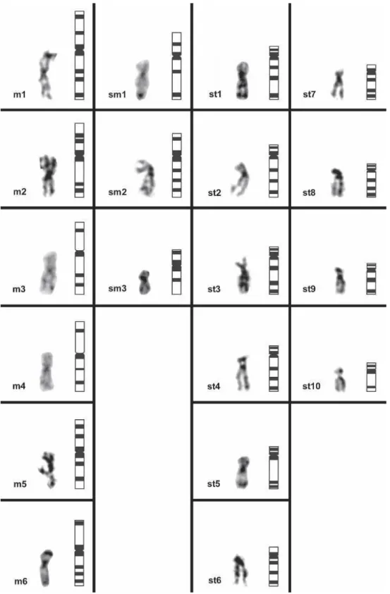
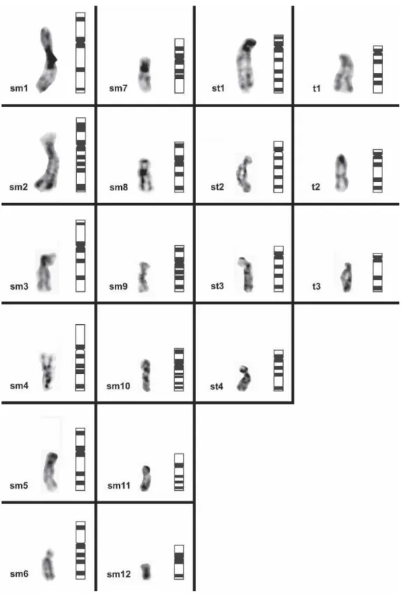
Examples of banded metaphases with HaeIII are present in Figure 1 for R. decussatus (Fig. 1a) and C. edule (Fig. 1b). Karyo- types with consistent banding pattern between homologous pairs are shown in Figure 2 for R. decussatus (Fig. 2a) and C. edule (Fig. 2b). All results are assembled and summarized in Figures 3 and 4, which show the haploid distribution of HaeIII chromosomal bands in the two species. HaeIII produced a banding pattern along the length of each chromosome (Figs. 3 and 4). The restriction band- ing produced was adequate for the single identification of all chro- mosomes for both species and organization of their respective
karyotypes (Figs, 2, 3 and 4). Interstitial, centromeric and telo- meric bands were observed along the chromosomes of both species. In the right side of each column of Figures 3 (for R. decussatus) and 4 (for C. edule) is shown a schematic representation of the in situ restriction banding patterns obtained for each species.
DISCUSSION
The diploid chromosome number of 2n= 38 is confirmed in both R. decussatus and C. edule and appears to be the modal number of the Veneridae and Cardiidae families (Nakamura 1985, Corni & Trentini 1986), and it is also common among the super- order Veneroida (Thiriot-Quiévreux et al. 1987).
The application, for the first time, of the RE HaeIII to the chromosomes of the clam R. decussatus and the cockle C. edule produced specific banding patterns and allowed the unambiguous individual identification of all the chromosome pairs making pos- sible the preparation of accurate karyotypes and their respective ideograms (Figs. 1–4). Therefore, this technique has demonstrated to be a reliable technique and more prompt (compared with con- ventional banding techniques) for veneroid chromosome banding.
For the construction of the ideograms, we only described the presence of the bands and each band’s relative position; the inten- sity of the bands was not considered. The intensity of the bands in the RE treatments seems to be related to the type of counterstain used (e.g., Giemsa or fluorochroms) (Gonsálvez et al. 1991). Sev- eral authors demonstrate that the loss of DNA after a RE digestion can increase the capacity of the stain to bind to a specific chro- mosome region (Gonsálvez et al. 1991, Nieddu et al. 1999). There- fore, it seemed reasonable not to consider the intensity of the bands in the construction of ideograms but only their presence and position.
The in situ restriction banding technique, applied here to both species, presents a major advantage, that can be used simulta- neously with FISH techniques (Chaves et al. 2002), demanding only one round of observation and minimal extra preparation steps. Consequently, the in situ restriction banding technique will facilitate physical mapping in this group of bivalves, besides being compatible with more traditional banding techniques.
Furthermore because tissue culture protocols are not yet avail- able in marine bivalves, the chromosomes are prepared directly from the animals and are of poor morphology. The in situ restric- tion banding technique better preserves the morphology of the chromosomes (compared with other conventional banding meth- ods), representing an additional advantage for the identification of veneroid chromosomes, when using further techniques such as FISH or C-banding (Chaves et al. 2002).
The use of the RE-banding technique can be very useful in several studies of economic or ecological importance within this group of veneroid bivalves. For instance: (a) in evolution of chro- mosome and karyotypes; (b) can also provide a rapid method for the identification of the missing chromosomes in aneuploidy situ- ations, for which a negative correlation with the growth rate was put in evidence in other bivalve species (Leitão et al. 2001) and (c) for the study of the impact of contaminants (anthropogenic com- pounds, and so forth) on the genetic patrimony of veneroids (through the identification of possible neoplasias, missing chro- mosomes, deletions, translocationsand so forth). This last application is far more important because the clam R. decussatus has been recently proposed as a potential bio-indicator species in areas were mussels are not available (Bebianno et al. 2004).
In fact clams are appropriate organisms for monitoring because they are sedentary filter feeders that exhibit a high level of diversity at a large number of loci (Moraga et al. 2002). Moreover, the tissue most currently used for cytogenetic analysis is from the gills, which is one of the most “interesting” tissues from the ecotoxicological point of view (Bebianno et al. 2004).
This study shows that the applications of restriction enzyme chromosome banding in veneroids are diverse and can constitute a fundamental step in gene mapping in this commercially important group of bivalves and could offer a new approach to specific problems in veneroid taxonomy and genetics.
ACKNOWLEDGMENTS
The authors thank C. Thiriot-Quiévreux for constructive com- ments, M. Matias, E. Domingos and M. Teixeira for technical assistance and A. Good for revising the English. This work was partially supported by a Portuguese grant from
the Ministry of Science and Technology (FCT)
(SFRH/BPD/18961/2004).
LITERATURE CITED
Beaumont, A. R. & M. H. Contaris. 1988. Production of triploid embryos of Tapes semidecussatus by the use of cytochalasin B. Aquaculture 73:37–42.
Bebianno, M. J., F. Géret, P. Hoarau, M. A. Serafim, M. R. Coelho, M. Gnassia-Barelli & M. Roméo. 2004. Biomarkers in Ruditapes decus-
satus: a potential bioindicator species. Biomarkers 9:305–330.
Borsa, P. & C. Thiriot-Quiévreux. 1990. Karyological and allozymic char- acterization of Ruditapes philippinarum, R. aureus and R. decussatus (Bivalvia, Veneridae). Aquaculture 90:209–227.
Bouilly, K., A. Leitão, R. Chaves, H. Guedes-Pinto, P. Boudry & S. Lapègue. 2005. Endonuclease banding reveals that atrazine-induced aneuploidy resembles spontaneous chromosome loss in Crassostrea
gigas. Genome 48:177–180.
Chaves, R., F. Adega, S. Santos, J. S. Heslop-Harrison & H. Guedes-Pinto. 2002. In situ hybridization and chromosome banding in mammalian species. Cytogenet. Genome Res. 96:113–116.
Clabby, C., U. Goswami, F. Flavin, N. P. Wilkins, J. A. Houghton & R. Powell. 1996. Cloning, characterization and chromosomal location of a satellite DNA from the Pacific oyster, Crassostrea gigas. Gene 168:205–209.
Corni, M. G. & M. Trentini. 1986. A chromosome study of Chamelae
gallina (L.) (Bivalvia, Veneridae). Boll. Zool. 53:23–24.
Cross, I., E. Díaz, I. Sánchez & L. Rebordinos. 2005. Molecular and cytogenetic characterization of Crassostrea angulata chromosomes.
Aquaculture 247:135–144.
Fischer-Piette, E. & B. Métivier. 1971. Révision des Tapetinae (Mol- lusques Bivalves). Mém. Mus. Natl. Hist. Nat. Paris. 71:1–106. Gajardo, G., M. Parraguez & N. Colihueque. 2002. Karyotype analysis and
chromosome banding of the Chilean-Peruvian Scallop Argopecten pur-
puratus. (Lamarck, 1819). J. Shellfish Res. 21:585–590.
Gérard, A. 1978. Étude des garnitures chromosomiques de deux Veneridae:
Ruditapes decussatus (L.) et Ruditapes philippinarum (Adams et
Reeve). Haliotis 9:69–71.
Gosálvez, J., R. Mezzanotte & C. López-Fernández. 1991. Selective di- gestion of mouse chromosomes with restriction endonucleases. II. X- ray microanalysis of HaeIII-treated chromosomes. Cytogenet. Cell
Genet. 56:82–86.
Gonzalez-Tizon, A., A. Martinez-Lage, L. Rego, J. Ausio & J. Mendez. 2000. DNA content, karyotypes, and chromosomal location for the 18S-58S-28S ribosomal loci in some species of bivalve molluscs from the Pacific Canadian coast. Genome 43:1065–1072.
Gosling, E. M. & A. Nolan. 1989. Triploidy induction by thermal shock in the Manila clam Tapes semidecussatus. Aquaculture 78:223–228.
Hurtado, N. S. & J. J. Pasantes. 2005. Surface spreading of synaptonemal complexes in the clam Dosinia exoleta (Mollusca, Bivalvia).
Chromo-some Res. 13:575–580.
Insua, A. & C. Thiriot-Quiévreux. 1992. Karyotypes of the Cerastoderma
edule, Venerupis pullastra and Venerupis rhomboides (Bivalvia,
Veneroida). Aquat. Living Resour. 5:1–18.
Insua, A., R. Freire & J. Mendez. 1999. The 5S rD NA of the bivalve
Cerastoderma edule: nucleotide sequence of the repeat unit and
chromosomal location relative to 18S-28S rDNA. Genet. Sel. Evol. 31:509–518.
Koulman, J. G. & W. S. Wolff. 1977. The Mollusca of the estuarine regions of the rivers Rhine, Meuse and Schedt in relation to hydrographic of the area. V. The Cardiidae. Basteria 41:21–32.
Leitão, A., P. Boudry & C. Thiriot-Quiévreux. 2001. Negative correlation between aneuploidy and growth in the Pacific oyster, Crassostrea gi-
gas: ten years of evidence. Aquaculture 193:39–48.
Leitão, A., R. Chaves, S. Santos, H. Guedes-Pinto & P. Boudry. 2004. Restriction enzyme digestion chromosome banding in Crassostrea and
Ostrea species: comparative karyological analysis within Ostreidae. Genome 47:781–788.
Martinez-Lage, A., A. Gonzalez-Tizon & J. Mendez. 1994. Characteriza- tion of different chromatin types in Mytilus galloprovincialis L. after C-banding, fluorochrome and restriction endonuclease treatments. He-
redity 72:242–249.
Moraga, D., E. Mdelgi-Lasram, M. S. Romdhane, A. El Abed, I. Boutet & M. Auffret. 2002. Genetic responses to metal contamination in two clams: Ruditapes decussatus and Ruditapes philippinarum. Mar.
Envi ron. Res. 54:521–525.
Nakamura, H. K. 1985. A review of molluscan cytogenetic information based on the CIS-MOCH-computerized system for molluscan chromo- somes. Bivalvia, Polyplacophora and Cephalopoda. Venus, Jap. J. Ma-
lacol. 44:93–225.
Nieddu, M., R. Rossino, G. Pichiri, M. Rocchi, M. D. Setzu & R. Mezza- notte. 1999. The efficiency of in-situ hybridization on human chromo- somes with alphoid DNAs is enhanced by previous digestion with AluI and TaqI. Chromosome Res. 7:593–602.
Partridge, J. K. 1977. Littoral and benthic investigations on the west coast of Ireland. VI. Annotated bibliographies of the genus Tapes (Bivalvia; Veneridae). Part I. Tapes decussatus (L). Part II. Tapes semidecussatus Reeve. Proceedings of the Royal Irish Academy, Section B. Biol. Geol.
Chem. Sci. 77:1–64.
Ruano, F. 1997. Fisheries and farming of important marine bivalves in Portugal. Marine Fisheries Review, NOAA Technical Repport NMFS 129:191–200.
Ruano, F. & R. Cachola. 1986. Outbreak of a severe epizootic of Perkinsus
marinus (Levin-78) at Ria de Faro clam’s culture beds. Proceedings of
2nd International Colloque Pathology Marine Aquatic. pp. 4–42. Thiriot-Quiévreux, C. 2002. Review of the literature on bivalve
cytoge-netics in the last ten years. Cah. Biol. Mar. 43:17–26.
Thiriot-Quiévreux, C. & N. Ayraud. 1982. Les caryotypes de quelques espèces de bivalves et gastéropodes marins. Mar. Biol. 70:165–175. Thiriot-Quiévreux, C. & M. Wolowicz. 1996. Karyotypes of Cerastoderma
glaucum (Bivalvia) from Baltic and Mediterranean populations. Hy- drobiologia 324:149–155.
Thiriot-Quiévreux, C.,J. Soyer & M. Bouvy. 1987. Etude des chromo- somes du bivalve protobranche Malletia sabrina Hedley, 1916. Vie
Milieu 37:175–180.
Walne, P.R. & P.C.Wood. 1975. A review of the shellfish research undertaken at the fisheries laboratories in 1974. Shellfish Information Leaflet, Ministry of Agriculture, Fisheries and Food, U.K, nº34. 43 pp. Wang, Y.& X. Guo. 2004. Chromosome rearrangement in Pectinidae
Revealed by rRNA Loci and implications for bivalve evolution. Biol.
Bull. 207:247–256.
Wilkins, N. P. & N. F. Mathers. 1974. Phenotypes of phosphoglucose isomerase in some bivalve molluscs. Comp. Biochem. Physiol. B Bio-
chem. Mol. Biol. 48B:599–611.
Xu, Z., X. Guo, P. M. Gaffney & J. C. Pierce. 2001. Chromosomal location of the major ribosomal RNA genes in Crassostrea virginica and Cras-
sostrea gigas. Veliger 44:79–83.
Zhang, Q. Y., G. Yu, R. K. Cooper & T. R. Tiersch. 1999. Chromosomal location by fluorescence in situ hybridization of the 28S ribosomal RNA gene of the eastern oyster. J. Shellfish Res. 18:431–435.
Figure 2. Examples of diploid-banded karyotypes. Figure 2a, diploid karyotype of R. decussatus banded with HaeIII, Figure 2b, diploid karyotype of C. edule banded with HaeIII. These examples are presented, to show the banding pattern consistency between homologous pairs. Scale bar = 5 µm
Figure 1. Examples of banded metaphases with the RE HaeIII: Figure 1a, metaphase of R. decussatus; Figure 1b, metaphase of C. edule. Scale bar = 5 µm
Figure 3. Haploid distribution of the chromosomal bands (left side of each column) and schematic representation of the in situ restriction banding patterns (right side of each column) obtained for the RE HaeIII in R. decussatus.
Figure 4. Haploid distribution of the chromosomal bands (left side of each column) and schematic representation of the in situ restriction banding patterns (right side of each column) obtained for the RE Hae III in C. edule.