Universidade de Aveiro Ano 2017
Departamento de Química
Carolina Oliveira
Pandeirada
Caracterização dos
hidratos
de
carbono
de
Nannochloropsis oculata e sua utilização em
microarrays
Characterization
of
carbohydrates
from
Nannochloropsis
oculata
and
their
use
in
Universidade de Aveiro Ano 2017
Departamento de Química
Carolina Oliveira
Pandeirada
Caracterização
dos
hidratos
de
carbono
de
Nannochloropsis oculata e sua utilização em
microarrays
Characterization
of
carbohydrates
from
Nannochloropsis
oculata
and
their
use
in
microarrays
Dissertação apresentada à Universidade de Aveiro para cumprimento dos requisitos necessários à obtenção do grau de Mestre em Bioquímica, Ramo Alimentar, realizada sob a orientação científica da Doutora Cláudia Nunes, investigadora de pós-doutoramento do Departamento de Química da Universidade de Aveiro, e do Doutor Manuel A. Coimbra, Professor associado com agregação do Departamento de Química da Universidade de Aveiro.
Thanks are due to FCT through national founds and FEDER, within the PT2020 Partnership Agreement, for funding QOPNA (FCT UID/QUI/00062/2013)
O júri/ The jury
Presidente/ President Professora Doutora Rita Maria Pinho Ferreira
Professora auxiliar do Departamento de Química da Universidade de Aveiro
Arguente/ Arguent Doutora Maria Angelina de Sá Palma
Ivestigadora da Faculdade de Ciências e Tecnologia da Universidade NOVA de Lisboa
Orientador/ Supervisor Professor Doutor Manuel António Coimbra Rodrigues da Silva
Agradecimentos/ Acknowledgments
Os meus sinceros agradecimentos a/ My sinceriously thanks to:
À Drª. Cláudia Nunes, minha orientadora, por toda a confiança que depositou em mim ao longo deste ano, por toda a disponibilidade, por todas as interrogações e pelos inúmeros conhecimentos transmitidos. A si, Cláudia, agradeço todos os conselhos, o carinho e a força. Um grande Obrigada Ao Prof. Manuel A. Coimbra, meu co-orientador, pela oportunidade que me deu de poder ingressar num desafio que se tornou muito enriquecedor. Agradeço também toda a sabedoria partilhada e todas as interrogações feitas com olhar crítico ao meu trabalho, as quais me fizeram pensar, refletir e aprender. Obrigada!
À Drª. Angelina Palma da Universidade NOVA de Lisboa pela prestabilidade e acompanhamento ao longo dos ensaios de microarrays. Agradeço também a simpatia, os conhecimentos transmitidos e o olhar atento, crítico e preocupado sobre o meu trabalho. Obrigada
À Élia, que em muito me ajudou a progredir e ganhar aptidões a nível laboratorial. Agradeço-te também toda a amizade. Obrigada
À Viviana e à Drª. Benedita da Universidade NOVA de Lisboa por todo o acompanhamento, disponibilidade e conhecimentos que me transmitiram ao longo do tempo que passei na NOVA, foram incansáveis, Obrigada Em particular, quero agradecer aos membros dos grupos GlycoLab e X-tal da NOVA pelo acolhimento, por disponibilizarem os laboratboratórios para realizar os ensaios e pela partilha de conhecimentos. Obrigada!
Aos meus companheiros de laboratório, Joana, Pedro, Sónia, Angélica, Andreia, Guido e restantes membros do grupo ResPoStA, não só pelo companheirismo, diversão e “aturarem a minha criança”, mas acima de tudo pelos conhecimentos e chamadas de atenção. Obrigada
Aos meus amigos mais próximos, Cristiana, Rita, Sofia, Catarina, Catarina, Ana, Marco, Lisandra e Lino pelos momentos de diversão e apoio. Obrigada
À família Calado, pelos conhecimentos transmitidos, pelo carinho e apoio. Obrigada
À minha irmã, Mariana, por ser um exemplo e por me dar não só bons conselhos e visões, mas por toda a amizade e forças ao longo destes anos. Obrigada Á minha família, Pai, Mãe e Avôs, por até aos dias de hoje me acolherem e pela educação que me deram. A vós devo a pessoa que hoje sou! Obrigada E porque, “Cada um é responsável por multiplicar os dons que Deus lhe deu”, eu agradeço ter tido a sorte de ter cada um de vós junto de mim para me ajudar numa parte desta difícil missão! Um grande bem-haja
Palavras-chave Nannochloropsis oculata; Polissacarídeos; Glicolípidos; Microarrays; Interações proteínas-hidratos de carbono.
Resumo As microalgas marinhas são fáceis de cultivar e acumulam compostos, como polissacarídeos (PS) e glicolípidos (GL), aos quais têm sido atribuídas atividades biológicas. O presente trabalho teve como objetivo estudar as características estruturais dos PS e dos hidratos de carbono dos GL da microalga marinha Nannochloropsis oculata. A biomassa foi desengordurada, extraída com água quente (HW) e ultrafiltrada. Isto originou um resíduo rico em (1→4)-Glc, atribuído à presença de celulose na parede celular, um filtrado rico em manitol e um retentato (Mw>10 kDa) que continha os PS solúveis. Os PS solúveis foram fracionados por precipitação em etanol originando três frações, Et50, Et85 e EtSN. Os PS da fração Et85 foram sujeitos a fraccionamento por cromatografia de troca aniónica, obtendo-se as frações #1, #2 e #3. Todas as frações contiveram ácidos urónicos e grupos sulfato, sendo este conteúdo quase negligenciável na fração #1.
As análises de açúcares, juntamente com os microarrays usando proteínas de especificidades conhecidas, permitiram verificar que os PS da fração Et50 são constituídos por (Glc, exibindo também domínios de (β1→3, β1→4)-glucanas. Os PS das frações Et85 e EtSN incluíram maioritariamente Rha e Man, contendo também outros açúcares em menores quantidades, Xyl, Fuc e Gal. As frações Et85, #1, #2 e #3, e EtSN possuiram principalmente (1→2)-Rha, com uma ramificação na posição C-3. As frações Et85, #2 e #3 continham também o resíduo substituído 2,3,4-Rha. Na fração Et85 os grupos sulfato encontraram-se na posição C-3, e simultaneamente nas posições C-3 e C-4 de (1→2)-Rha. As frações #2 e #3, além de resíduos de Rha continham Xyl, Fuc e Gal, sugerindo que estes fazem parte de heteropolissacarídeos aniónicos sulfatados. A Man, nas frações Et85 e EtSN, encontrou-se maioritariamente como (1→3)- e (1→4)-Man, fazendo parte de PS não-aniónicos na fração Et85. A análise por microarrays evidenciou que a Man deve encontrar-se em configuração α devido à interação com a ConA. A proteína BT0996 da bactéria Bacteroides thetaiotaomicron, do sistema digestivo humano, usada nos microarrays, mostrou que os PS nas frações Et50, Et85, #1 e #3 podem conter um epítopo comum. Esta interação evidencia que os PS de N. oculata poderão ser metabolizados no sistema digestivo humano.
Os GL de N. oculata ocorrem principalmente como mono- e digalactolipídos, sendo a sua presença confirmada pelos microarrays por interação com a proteína RCA120. A análise de açúcares evidenciou também que a fração lipídica
deve conter sulfoquinovosil diacilglicerol.
Considerando os resultados obtidos, a combinação das análises de açúcares com os microarrays mostraram ser uma abordagem adequada para identificar as características estruturais dos PS e GL da microalga N. oculata.
Keywords Nannochloropsis oculata; Polysaccharides; Glycolipids; Microarrays; Carbohydrate-protein interactions.
Abstract Marine microalgae are of easy culturing and able to accumulate compounds, such as polysaccharides (PS) and glycolipids (GL), which have been attributed biological activities. The present work aimed to study the structural features of the PS and the carbohydrates of the GL of the marine microalga Nannochloropsis oculata using carbohydrate analyses and microarrays. Biomass was defatted, extracted with hot-water (HW), and ultrafiltered. This yielded a residue rich in (1→4)-Glc that was assigned to the presence of a cellulose cell-wall, one filtrate rich in mannitol, and one rentante (Mw>10 kDa) that comprised the soluble PS. The soluble PS were fractionated through ethanol precipitation giving three fractions, Et50, Et85 and EtSN. The PS in Et85 were further fractionated through an anion-exchange chromatography, yielding fractions #1, #2, and #3. All PS fractions comprised uronic acids and sulfate groups, being this content almost negligible in fraction #1.
Carbohydrate analyses together with microarrays experiments using proteins with known carbohydrate-binding specificities allowed to verify that the PS in Et50 are dominated by (β1→4)-Glc and exhibited also domains of (β1→3, β1→4)-glucans. PS in Et85 and EtSN fractions were composed by Rha and Man, comprising other sugars in lower content, Xyl, Fuc, and Gal. Fractions Et85, #1, #2, #3, and EtSN comprised mainly (1→2)-Rha with a branching point occurring at position C-3. Fractions Et85, #2, and #3 included also the substituted residue 2,3,4-Rha. In fraction Et85, the sulfate group occurred at position C-3, and simultaneously at positions C-3 and C-4 of (1→2)-Rha. Fractions #2 and #3, besides Rha residues, contained Xyl, Fuc, and Gal, suggesting that these are part of anionic sulfated heteropolysaccharides. The Man, in Et85 and EtSN, occurred mainly as (1→3)- and (1→4)-Man residues, being these part of non-anionic PS in fraction Et85. Carbohydrate microarrays highlighted that Man may be present in α-configuration due to the interaction with ConA. The protein BT0996 of the Bacteroides thetaiotaomicron, a human gut bacteria, used in microarrays, displayed that the PS in fractions Et50, Et85, #1, and #3 may share a common epitope. This interaction is remarkable as it shows that these PS are probably metabolized in the human gut.
The GL occur mainly as mono- and digalactolipids, being the presence of galactolipids confirmed through microarrays by interaction with RCA120.
Carbohydrate analyses highlighted that the lipid fraction may comprise sulfoquinovosyl diacylglycerol.
Considering the results obtained, the joint use of carbohydrate analyses with microarrays showed to be a suitable approach to identify the structural features of the PS and the GL of the marine microalga N. oculata.
I
Contents
_______________________________________
List of Figures
_____________________________________________ V
List of Tables
_____________________________________________ IX
List of Abbreviations
_____________________________________ XI
Chapter 1 – Introduction and Objectives
______________ 1
1.1. Carbohydrates in marine microalgae
_____________________ 3
1.1.1. General characteristics of polysaccharides ___________________________________________ 4
1.1.2. General characteristics of glycolipids ________________________________________________ 7
1.1.3. Carbohydrates of Nannochloropsis oculata __________________________________________ 10
1.2. Carbohydrate microarrays
_____________________________ 12
1.2.1. Immobilization methods __________________________________________________________ 13
1.2.1.1. Noncovalent immobilization ____________________________________________________ 13 1.2.1.2. Covalent immobilization _______________________________________________________ 15 - Chemically modified carbohydrates ________________________________________________ 15 - Chemically unmodified carbohydrates ______________________________________________ 16 1.2.2. Carbohydrate-binding proteins ____________________________________________________ 17
1.2.2.1. Lectins _____________________________________________________________________ 17 1.2.2.2. Antibodies __________________________________________________________________ 20 1.2.2.3. Carbohydrate-binding modules __________________________________________________ 22 - Human gut microbiome __________________________________________________________ 24
1.3. Objectives of the thesis
__________________________________ 28
Chapter 2 – Material and Methods
___________________ 31
2.1. Characterization of polysaccharides from N. oculata
_____ 33
2.1.1. Sample ________________________________________________________________________ 33
2.1.2. Sequential extraction of polysaccharides from biomass of N. oculata ____________________ 33
- Biomass defatting _______________________________________________________________ 33 - Extraction of hot-water soluble polysaccharides ______________________________________ 33 - Sequential ethanol precipitation of the HW soluble PS _________________________________ 34 - Anion-exchange chromatography of the fraction Et85 __________________________________ 34 2.1.3. Protein content__________________________________________________________________ 36
II
2.1.4. RNase digestion of the fraction Et50 ________________________________________________ 36
2.1.5. Carbohydrate analyses ___________________________________________________________ 36
2.1.5.1 Neutral sugars, non-free and free alditols analyses __________________________________ 37 2.1.5.2. Uronic acids content __________________________________________________________ 38 2.1.5.3. Determination of sulfate content _________________________________________________ 38 2.1.5.4. Glycosidic linkages analysis ____________________________________________________ 39 Methylation ________________________________________________________________________ 39 Reduction of carboxyl groups of methylated PS ___________________________________________ 40 Hydrolysis, Reduction and Acetylation of methylated PS ____________________________________ 40 2.1.5.5. Desulfation of Et85 fraction ____________________________________________________ 41 2.1.5.6. Determination of molecular weight of the PS _______________________________________ 41 2.1.6. Statistical analysis _______________________________________________________________ 41
2.1.7. Recombinant expression of proteins from Bacteroides thetaiotaomicron VPI-5482: BT3698 and BT0996 _____________________________________________________________________________ 42
2.1.7.1. DNA, bacterial strains and plasmids _____________________________________________ 42 2.1.7.2 Competent cells _______________________________________________________________ 43 2.1.7.3. Transformation ______________________________________________________________ 43 2.1.7.4. Expression of proteins BT3698 and BT0996 _______________________________________ 43 - Protocol for expression of BT3698 with auto-induction _________________________________ 44 - Protocol for expression of BT0996 with IPTG-induction ________________________________ 44 2.1.7.5. Cell harvesting and lysis _______________________________________________________ 44 2.1.7.6. Purification of BT3698 and BT0996 by affinity chromatography _______________________ 45 - Affinity purification with His GravitrapTM columns ___________________________________ 45 - Purification with ÄKTA START ____________________________________________________ 46 2.1.7.7. Protein quantification _________________________________________________________ 46 2.1.7.8. SDS-PAGE analysis ___________________________________________________________ 47 2.1.8. Development of microarrays with the PS extracted from N. oculata _____________________ 47
2.1.8.1. Construction of the carbohydrate microarrays _____________________________________ 47 2.1.8.2. Carbohydrate microarrays binding assay _________________________________________ 50 2.1.8.3. Carbohydrate microarrays data analysis __________________________________________ 52
2.2. Characterization of glycolipids from N. oculata
__________ 54
2.2.1. Extraction of lipids from biomass of N. oculata ______________________________________ 54
2.2.1.1. Carbohydrate analyses ________________________________________________________ 54 2.2.2. Development of microarrays with the dialysed lipid fraction extracted from N. oculata ____ 55
2.2.2.1. Construction of the carbohydrate microarrays _____________________________________ 55 2.2.2.2. Carbohydrate microarrays binding assay _________________________________________ 57
III 2.2.2.3. Carbohydrate microarrays data analysis __________________________________________ 59
Chapter 3 – Results and Discussion
__________________ 61
3.1. Characterization of polysaccharides from N. oculata
_____ 63
3.1.1. Characterization of the fractions derived from sequential extraction of PS from N. oculata _ 63
3.1.1.1. Biomass of Nannochloropsis oculata _____________________________________________ 63 3.1.1.2. Hot-water (HW) extracts _______________________________________________________ 66 3.1.1.3. Fractions of the sequential ethanol (EtOH) precipitation _____________________________ 70 3.1.1.4. Fractions derived from the anion-exchange chromatography of Et85 ___________________ 84 3.1.2. Recombinant expression of proteins from Bacteroides thetaiotaomicron VPI-5482: BT3698 and BT0996 _____________________________________________________________________________ 92
3.1.2.1. Proteins quantification and SDS-PAGE analysis ____________________________________ 92 3.1.3. Carbohydrate microarrays developed with PS from N. oculata _________________________ 94
3.1.3.1. Plant lectins _________________________________________________________________ 96 3.1.3.2. Monoclonal antibodies ________________________________________________________ 99 3.1.3.3. Microbial carbohydrate-binding modules (CBMs) _________________________________ 101 - CmCBM6-2 __________________________________________________________________ 101 - CBMs belonging to B. thetaiotaomicron ____________________________________________ 102
3.2. Characterization of glycolipids from N. oculata
_________ 108
3.2.1. Carbohydrate characterization of the lipid fraction __________________________________ 108
3.2.2. Carbohydrate microarray developed with the dialysed lipid fraction extracted from N. oculata
___________________________________________________________________________________ 112
3.2.2.1. RCA120 ____________________________________________________________________ 114
3.2.2.2. hGalectin-3 ________________________________________________________________ 115
Chapter 4. – Conclusions and future perspectives
_ 117
References
_______________________________________________ 125
Supplementary Material
____________________________________ 137
Supplementary information 1 _________________________________________________________ 139 Supplementary information 2 _________________________________________________________ 141 Supplementary figures _______________________________________________________________ 142 Supplementary tables ________________________________________________________________ 144 Supplementary information 3 _________________________________________________________ 146 Supplementary information 4 _________________________________________________________ 147V
List of Figures____________________________________
Figure 1. Examples of microalgae polysaccharide structures. (A) A model of (β1→3)-D-glucan branched at position O-6 of Glc residue from the diatom P. tricornutum (18). (B) Models a or b for the possible repeating unit in EPS from Porphyridium sp. R = H, SO2O, terminal Gal or terminal Xyl, m = 2 or 3 (20). (C) The highlybranched structure of the (β1→3,1→6)-D-glucan from I. galbana (21). (D) The structure suggested for the arabinomannan extracted from the cell-wall of C. vulgaris (a+b=2, b>a, C=1, d=1) (22). 5
Figure 2. Structures of mono- and digalactosyl diacylglycerols (MGDG and DGDG) and sulfoquinovosyl
diacylglycerol (SQDG) mainly found in photosynthetic membranes of microalgae. R1 and R2 represent the two
fatty acid chains linked to the glycerol backbone. Image adapted from Zhang et al. (35). 8
Figure 3. Illustrations of cells viewed by light microscopy of two strains of N. oculata, CCMP525 (p) and
CCMP533 (q). Black droplet = red body (44). 10
Figure 4. Preparation of neoglycolipids (NGLs) from reducing oligosaccharides by reductive amination or
oxime ligation reactions (63). 14
Figure 5. Immobilization methods for the construction of carbohydrate microarrays. Noncovalent
immobilization: attachment of carbohydrates (A) through hydrophobic interactions on the underivatized surface, and (B) through electrostatic or fluorous-fluorous interactions on the derivatized surface. Covalent immobilization: attachment of (C) chemically modified carbohydrates onto derivatized surface, and (D) unmodified carbohydrates onto derivatized surface. Image adapted from Park et al. (58). 17
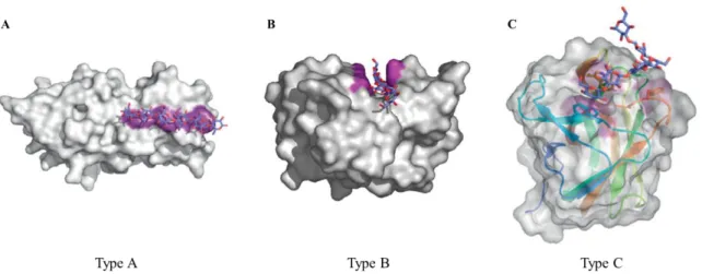
Figure 6. Structural examples of CBMs from Type A, B and C. (A) The crystallographic dimer of CBM63
from Bacillus subtilis (PDB ID 4FER) in complex with cellohexaose (blue sticks). Surface composed by the aromatic amino acids that form the planar hydrophobic surface are shown in purple (106). (B) The crystallographic structure of the CBM4-2 from Celllulomonas fimi (PDB ID 1GU3] in complex with a cellopentaose molecule. The amino acid side chain involved in binding are shown in purple (104). (C) The X-ray crystal structure of the CBM6 from Bacilus halodurans in complex with laminarihexaose (PDB ID 1W9W). The CBM specifically recognizes the non-reducing end of the sugar. The secondary structures are shown as colored ramped cartoons with relevant amino acid side chains involved in carbohydrate binding shown as sticks. Solvent accessible surfaces are shown in gray with the surfaces contributed by the aromatic residues
colored purple (106). 23
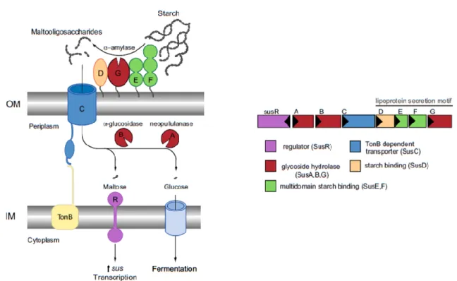
Figure 7. Overview of the starch utilization system (Sus) in B. thetaiotaomicron. The inner
membrane-spanning protein SusR to sense the disaccharide inducer, maltose, in the periplasm and subsequently drives the transcription of the locus susABCDEFG. Starch is bound to the surface of the cell by the starch-binding outer membrane lipoproteins SusDEF. Subsequent hydrolysis by a similarly membrane-tethered α-amylase, SusG, generates oligosaccharides small enough to transit through the TonB-dependent transporter (SusC). Once in the periplasm, SusA and SusB, a neopullulanase and α-glucosidase, respectively, process oligosaccharides into glucose. The monosaccharide is then shuttled into the cytoplasm by an unknown transporter. Image adapted
from Foley et al. (111). 25
Figure 8. Diagram of the procedure used to extract sequentially the fractions enriched in polysaccharides from
N. oculata. 35
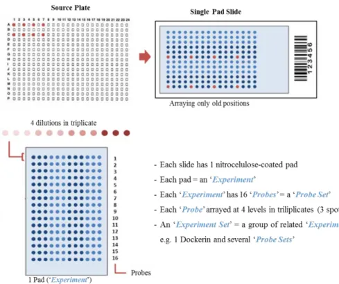
Figure 9. Scheme of the arraying method using single pad slides. An example of a layout of one single pad
slide with 16 probes printed at 4 different dilutions in triplicate each is represented, as well as designations used during a carbohydrate microarray experiment. Image adapted from ISBio training course, Microarray screening analysis supporting material, 2015, Caparica, Portugal (124). 49
Figure 10. Scheme of the carbohydrate microarray binding experiment using slides of 2 pads. Adapted from
ISBio training course, Microarray screening analysis supporting material, 2015, Caparica, Portugal (124). 52
Figure 11. Composition in neutral and acidic sugars (UA), and in sulfate (SO3-) groups in the biomass and in
defatted biomass of N. oculata. Results are expressed in molar percentage (mol %). Asterisks represent values significantly different (p<0.05) between the biomass and defatted biomass. 66
Figure 12. Composition in neutral and acidic sugars (UA), and in sulfate (SO3-) groups present in the Residue,
Filtrate (Mw<10 kDa) and Retentate (Mw>10 kDa) fractions obtained from the hot-water (HW) extraction after ultrafiltration (cut-off 10 kDa). Results are expressed in molar percentage (mol %). 68
VI
Figure 13. Chromatogram from the GPC analysis of the fractions obtained during the sequential EtOH
precipitation (Et50, Et85 and EtSN). Numbers 1 and 2 represent the populations of polymers present in each fraction with different molecular weights (Mw). 72
Figure 14. Polysaccharides (PS) composition of the EtOH fractions (Et50, Et85 and EtSN). (A) Composition
in neutral monosaccharides, uronic acids (UA), and in sulfate (SO3-) groups. (B) Composition in alditols, UA
and SO3- of the PS. Results are expressed in molar percentage (mol %) of the total sugars, alditols, and sulfate
content. 74
Figure 15. Chemical structure of riboflavin (134). 78
Figure 16. Composition in neutral monosaccharides, alditols, uronic acids (UA), and in sulfate (SO3-) groups
of the polysaccharides (PS) in Fraction #1, #2, and #3 obtained from the anion-exchange chromatography of Et85 with 0, 0.125 and 0.250 M NaCl, respectively. Results are expressed in molar percentage (mol %) of the total content in sugars, alditols and sulfate. 86
Figure 17. Chromatogram from the GPC analysis of the fractions #1, #2, and #3 obtained from the
anion-exchange chromatography of the fraction Et85. Numbers 1 and 2 represent the populations of polymers present in each fraction with different molecular weights (Mw). 90
Figure 18. SDS-PAGE (10 % acrylamide) results showing purification of the proteins BT3698 and BT0996. Results of purification with His Gravitrap columns for BT3698 and with ÄKTA START for BT0996.
BT3698 and BT0996 proteins were eluted with 5 ml of 50 mM HEPES at pH 7.5, 1 M NaCl, 5 mM CaCl2 and
300 mM imidazole at a final pH of 7.5, and with 50 mM HEPES at pH 7.5, 1 M NaCl, 5 mM CaCl2 and 500
mM imidazole at a final pH of 7.5, respectively. SDS-PAGE: Gel was performed with a constant amperage of 60 mA. Visualization of proteins molecular weight (Mw) was assed by coloration with NZYTech BlueSafe. For SDS gel was used the respective concentrations: BT3698 33 µg and BT0996 16 µg. A Marker II from NZYTech® was used (Marker) as reference. 93
Figure 19. Screening of the microarrays developed with Microalgae PS set 1 probes by analysis with two plant
lectins: ConA and RCA120. The binding signals are depicted as means of fluorescence intensities of triplicate
spots of probe arrayed (with error bars). Each probe was printed in triplicate at two levels: 0.1 (blue bars) and 0.5 (red bars) mg/mL (30 and 150 pg/spot). Quantified fluorescence intensity is plotted on the x-axis. Carbohydrate probes are plotted on the y-axis. 97
Figure 20. Screening of the microarrays developed with Microalgae PS set 1 probes by analysis with two
antibodies (Abs). Ab names are depicted on top of each graph and both Abs have different specificities (Table 6). Carbohydrate sequence information of the probes is in Table 19. The binding signals are depicted as means of fluorescence intensities of triplicate spots of probe arrayed (with error bars). Each probe was printed in triplicate at two levels: 0.1 (blue bars) and 0.5 (red bars) mg/mL (30 and 150 pg/spot). Quantified fluorescence intensity is plotted on the x-axis. Carbohydrate probes are plotted on the y-axis. Fluorescene intensity signals of the bars marked with an asterisk in the Anti-heteromann graph are not considered due to contamination of this microarray experiment with the mAb anti-(β1→3, β1→4)-D-glucan during the binding assay. 100
Figure 21. Screening of the microarray developed with Microalgae PS set 1 probes by analysis with the family
6 CBM CBM6-2 from Cellvibrio mixtus (CmCBM6-2). Cabohydrate-binding specificity of CmCBM6-2 is in Table 6, and the carbohydrate sequence information of the probes is in Table 19. The binding signals are depicted as means of fluorescence intensities of triplicate spots of probe arrayed (with error bars). Each probe was printed in triplicate at two levels: 0.1 (blue bars) and 0.5 (red bars) mg (dw)/mL (30 and 150 pg/spot). Quantified fluorescence intensity is plotted on the x-axis. Carbohydrate probes are plotted on the y-axis. 101
Figure 22. Analysis of the carbohydrate microarrays developed with Microalgae PS set 1 probes with the CBM
domains of BT3698 and BT0996, from the bacterium Bacteroides thetaiotaomicron. Carbohydrate-binding specificity of each protein is in Table 6, and carbohydrate sequence information of the probes included in the microarray is in Table 19. The binding signals are depicted as means of fluorescence intensities of triplicate spots of probe arrayed (with error bars). Each probe was printed in triplicate at two levels: 0.1 (blue bars) and 0.5 (red bars) mg (dw)/mL (30 and 150 pg/spot). Quantified fluorescence intensity is plotted on the y-axis. Carbohydrate probes are plotted on the x-axis. 103
Figure 23. Comparison of the relative binding intensities of the proteins investigated in carbohydrate
microarrays: ConA, RCA120, Anti-heteromannan (Rat IgM), Anti-(β1→3, β1→4)-D-glucan, CmCBM6-2,
BT3698 and BT0996. The heat map representation shows the different polysaccharides (PS) binding patterns revealed by the microarray analysis containing the Microalgae PS set 1 (Table 19). The relative binding
VII
intensities were calculated as the percentage of the mean fluorescence signal intensity at 0.5 mg/mL PS probe given by the probe most strongly bound by each protein (normalized as 100%). 105
Figure 24. Monosaccharides composition of the lipid fraction extracted with CHCl3:MeOH (2:1, v/v) from N.
oculata before (Lipid fraction) and after dialysis (Dialysed lipid fraction). Results are expressed in molar percentage (mol %) of the total content in sugars. 110
Figure 25. Screening of the carbohydrate microarrays developed with Microalgae GL set 2 probes, which
comprise the dialysed lipid fraction (GL) from N. oculata, by analysis with two lectins, the plant lectin RCA120
and the animal lectin hGalectin-3. Protein names are depicted on top of each graph and the specificity of each protein towards Gal residues is described in table 8. Carbohydrate composition of the liposome probes included in the microarray is in table 22. Binding signals are depicted as means of fluorescence intensities of triplicate spots of probe arrayed (with error bars). Quantified fluorescence intensity is plotted on the x-axis. Liposome probes comprising the GL are plotted on the y-axis. 115
Supplementary Figure 1. Approaches to detect molecular interactions through fluorescence-based methods. (A) Direct fluorophore (FITC) labelling of the protein, (B) use of fluorophore (Alexa Fluor 6747)-labelled
secondary reagent (streptavidin) that binds to biotinylated protein, and (C) use of fluorescent secondary antibody that binds to anti-glycan antibody. (D) The average relative fluorescence units generated during the process of fluorescence scanning of replicate spots are calculated and the data are presented as histograms of fluorescence intensity. Image adapted from Smith and Cummings (153). ... 139
Supplementary Figure 2. Representative scheme of the steps performed to identify the glycosidic linkages
present in the PS of fraction Et85_#1. 1º Separation of the PMAAs by gas chromatography (GC); 2º Identification of each peak in the chromatogram using mass spectrometry (MS); and 3º Integration of the area of each peak on the chromatogram corresponding to sugars and posterior relative quantification. ... 141
Supplementary Figure 3. Native proteins expressed by Bacteroides thetaiotaomicron, BT3698 and BT0996.
Red arrows are the selected domains used for cloning. Key: SP, signal peptide; GH, glycoside hydrolase; CBM, carbohydrate-binding module; N’, N-terminal; C’, C-terminal; GH and CBM alternative numbers indicate different families according to CAZy classification (http://www.cazy.org/, (107) and Correia, V. G. et al. unpublished). ... 142
Supplementary Figure 4. Recombinant proteins overexpressed in Escherichia coli BL21 (DE3) and TUNER
(DE3) expression strains, BT3698 and BT0996, respectively. A pHTP1 vector with kanamycin (Kan) resistance was used for cloning the domains CBM58 and CBM57. The recombinant proteins BT3698 and BT0996 contained an N-terminal (N’) His-tag (His6) comprising the following sequence: MGSSHHHHHHSSGPQQGLR, and the respective CBM module (Correia, V. G. et al. unpublished). ... 142
Supplementary Figure 5. Chemical structures of the carrier lipids used to produce the liposome probes. 1,2-Diacyl-sn-glycero-3-phosphocholine (PC1) from egg yolk, R and R’= Fatty acid chains. Typical egg yolk
phosphatidylcholine have fatty acid contents of approximately 33 % 16:0 (palmitic), 13 % 18:0 (stearic), 31 % 18:1 (oleic), and 15 % 18:2 (linoleic) (other fatty acids being in minor contributors).
1,2-Di-O-hexadecyl-sn-glycero-3-phosphocholine (PC2) and Cholesterol (C). Images adapted from http://www.sigmaaldrich.com.
... 143
Supplementary Figure 6. Top graph: Composition in neutral and acidic sugars (UA) of the five commercial
polysaccharides (PS) (PGA apple, PGA citrus, GHM, GLM, and Oat β-glucan). Results are expressed in molar percentage (mol %) of the total content in sugars (NS + UA). Bottom graph: Composition in NS of the same commercial PS, results are expressed in mol % of the total content in NS. ... 149
IX
List of Tables_____________________________________
Table 1. Typical carbohydrate ligands reported for various plant and animal lectins. 19Table 2. Carbohydrate ligands recognised by various monoclonal antibodies (mAbs) through carbohydrate
microarrays. 21
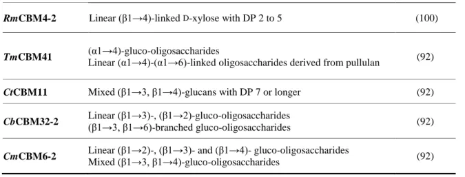
Table 3. Carbohydrate ligands of various microbial carbohydrate-binding modules (CBMs) ascertained
through carbohydrate microarrays. 24
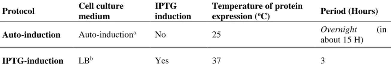
Table 4. Main differences in the auto-induction and IPTG-induction protocols for protein expression. 44
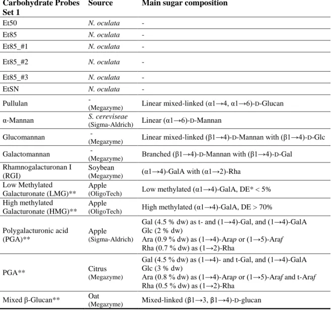
Table 5. List of all carbohydrate probes that composed the Microalgae polysaccharides (PS) set 1, which were
analysed in the binding charts and in the matrix (heat-map). The microarray comprises PS from N. oculata and from commercial sources. The main sugar composition in the commercial polysaccharide probes is described.
48
Table 6. List of the lectins, antibodies and CBMs used in the carbohydrate microarrays developed with
Microalgae PS set 1 and their reported specificities. 50
Table 7. List of all liposome probes that composed the Microalgae glycolipids (GL) set 2, which were analysed
in the binding charts. The microarray comprised liposomes that were composed by the GL present in the dialysed lipid fraction from N. oculata, one phosphatidylcoline (PC), 1,2-Diacyl-sn-glycero-3-phosphocholine (PC1) or 1,2-Di-O-hexadecyl-sn-glycero-3-phosphocholine (PC2), and the cholesterol (C). Composition of the probes differed in the mol ratio PC:C:GL. 56
Table 8. Lectins used in the carbohydrate microarrays developed with Microalgae GL set 2 and their reported
specificities. 57
Table 9. Content in protein, neutral sugars (NS), uronic acids (UA), and sulfate groups (SO3-) present in the
biomass of N. oculata and in their defatted biomass. Results are expressed in percentage of dry weight (dw). 63
Table 10. Yields of extraction with hot-water (HW) after ultrafiltration (cut-off 10 kDa) from the defatted
biomass, results are expressed as weight percentage (%) of defatted biomass. Content in protein, neutral sugars (NS), uronic acids (UA) and sulfate groups (SO3-) present in the Residue (material insoluble in HW), Filtrate
(F Mw<10 kDa) and Retentate (Ret Mw>10 kDa) after ultrafiltration obtained from the HW extraction. Results are expressed as weight % of fraction. 67
Table 11. Yields of sequential EtOH precipitation of the retentate to each fraction (Et50, Et85 and EtSN),
expressed as weight percentage (%) of retentate. Content in protein, and in neutral sugars (NS), uronic acids (UA) and sulfate (SO3-) groups that compose the polysaccharides (PS) present in Et50, Et85 and EtSN. Results
expressed as weight % of fraction. 71
Table 12. Estimation of the weight average molecular weight (Mw) and number average molecular weight
(Mn) present in the EtOH fractions by GPC. Data are expressed in Dalton (Da) unit. 72
Table 13. Glycosyl linkage composition (mol % of the total sugars) of the fractions obtained from sequential
EtOH precipitation (Et50, Et85 and EtSN) and after desulfation of the fraction Et85 (Et85 desulfated). Glycosidic linkages of the PS in Et50 and EtSN were determined from the carboxyl-reduced partially methylated alditol acetates (PMAAs), and the Et85 and Et85 desulfated through PMAAs without reduction of
carboxyl groups. 75
Table 14. Neutral sugars (NS) composition of the fraction Et50 before and after digestion with RNase. Results
are expressed as mean ± SD in molar percentage (%) of the total NS from duplicate analyses. 77
Table 15. Yields of the Fractions #1, #2 and #3 obtained from the anion-exchange chromatography of the
fraction Et85 with 0, 0.125 and 0.250 M NaCl, respectively, results are expressed as weight percentage (%) of Et85. Content in neutral sugars (NS), uronic acids (UA) and sulfate (SO3-) groups that compose the
polysaccharides (PS) present in fractions #1, #2, and #3. Results expressed as weight % of fraction. 85
Table 16. Glycosyl linkage composition (mol % of the total sugars) of the fractions Et85_#1, Et85_#2 and
X
NaCl, respectively, and of the initial Et85 fraction. Glycosidic linkages were determined from the carboxyl-reduced partially methylated alditol acetates (PMAAs), except for Et85. 87
Table 17. Estimation of the weight average molecular weights (Mw) and number average molecular weight
(Mn) present in the fractions obtained from the anion-exchange chromatography (#1, #2 and #3) by GPC. 90
Table 18. Characteristics of the recombinant proteins BT3698 and BT0996. Organism that they belong, cloned
domain and concentration of each protein (µg/mL). 93
Table 19. List of all carbohydrate probes that composed the Microalgae polysaccharides (PS) set 1 that were
printed in microarrays. The main sugar composition is described for each probe. 95
Table 20. Composition in neutral sugars (NS), alditols and sulfate (SO3-) groups of the lipid fraction extracted
with CHCl3:MeOH (2:1, v/v) from N. oculata, and the lipid fraction after being dialysed. Results are expressed
in weight percentage (%) of the lipid fraction. 109
Table 21. Glycosyl linkage composition (mol % of the total neutral sugars) of the lipid fraction extracted with
CHCl3:MeOH (2:1, v/v) from the biomass of N. oculata. 111 Table 22. List of all liposome probes that composed the Microalgae glycolipids (GL) set 2. The microarray
comprised liposomes composed of the GL present in the dialysed lipid fraction from N. oculata, one phosphatidylcholine (PC), 1,2-Diacyl-sn-glycero-3-phosphocholine (PC1) or 1,2-Di-O-hexadecyl-sn-glycero-3-phosphocholine (PC2), and the cholesterol (C). Composition of the probes differed in the mol ratio PC:C:GL. The main sugar composition is depicted for each probe. 113
Supplementary Table 1. Carbohydrate-binding modules (CBMs) expressed by Bacteroides thetaiotaomicron
produced through recombinant expression, CBM57 and CBM58. The protein parameters of each protein are depicted, which include the length in number of amino acids (aa), the molecular weight (Mw), the extinction coefficient and the isoelectric point (parameters were calculated using the tool online bioninformatic tool http://web.expasy.org/protparam). 144
Supplementary Table 2. Conditions of the proteins analysed in the carbohydrate microarrays with Microalgae
PS set 1. BI, biotin. 144
Supplementary Table 3. Conditions of the proteins analysed in the carbohydrate microarrays with Microalgae
GL set 2. BI, biotin. 145
Supplementary Table 4. Composition in neutral sugars (NS) and content in acidic sugars (UA) present in the
following commercial polysaccharides: PGA apple (purchased from SIGMA), PGA citrus (purchased from Megazyme), High Methylated Galacturonate (HMG) and Low Methylated Galacturonate (LMG) purchased from OligoTech, and Oat β-glucan (purchased from Megazyme). Results are expressed in weight percentage
(% w/w). 148
Supplementary Table 5. Glycosyl linkage composition (mol % of the total sugars) of the five commercial
polysaccharides (PS) (PGA apple, PGA citrus, HMG, LMG, and Oat β-glucan). Glycosidic linkages were determined by methylation analysis with reduction of the carboxyl groups in PGA apple, PGA citrus, HMG and LMG, and in Oat β-glucan by methylation analysis without reduction of carboxyl groups. 149
XI
List of Abbreviations______________________________
Ab (or Ig) – Antibody
ADHP -
N-aminoacetyl-N-(9-
anthracenylmethyl)-1,2-dihexadecyl-sn-glycero-3-phosphoethanolamine
AF – Alexa-Fluor
AnGal – 3,6-anhydro-Gal
AOPE – Aminooxy-functionalized DHPE AraOH – Arabinitol
BSA – Bovine serum albumin C – Cholesterol CBM – Carbohydrate-binding module CBPs – Carbohydrate-binding proteins CE – Carbohydrate esterase ConA – Concanavalin A CRD – Carbohydrate-recognition domain C-type – Ca2+-dependent Cy – Cyanine DAG – Diacylglycerol DB – Degree of branching
DC-SIGN – Dendritic cell-specific ICAM-3 grabbing nonintegrin DE – Degree of esterification
DGDG – Digalactosyl diacylglycerols DHPE -
1,2-dihexadecyl-sn-glycero-3-phosphoethanolamine dH2O – Destilled water
DMSO – Dimethyl sulfoxide DP – Degree of polymerization dw – Dry weight
EBV-EA – Epstein-Barr virus-associated early antigen
EPA – Eicosapentaenoic acid EPS – Exopolysaccharides ER – Endoplasmic reticulum
Et50 – Precipitate with 50 % EtOH (v/v) Et85 – Precipitate with 85 % EtOH (v/v) EtSN – Supernatant of the 85 % EtOH
(v/v)
FA – Fatty acid
FI – Fluorescence intensity FID – Flame ionization detector FITC – Fluorescein isothiocyanate Fraction #1 (Et85_#1) – Fraction eluted
with 0 M NaCl from the anion-exchange chromatography of Et85 Fraction #2 (Et85_#2) – Fraction eluted
with 0.125 M NaCl from the anion-exchange chromatography of Et85 Fraction #3 (Et85_#3) – Fraction eluted
with 0.250 M NaCl from the anion-exchange chromatography of Et85 FucOH – Fucitol
GalA – Galacturonic acid GalOH - Galactitol GlcA – Glucuronic acid GC – Gas chromatography GH – Glycoside hydrolase GIT – Gastrointestinal tract GL – Glycolipids
GlcOH – Glucitol
GPC – Gel permeation chromatography GT – Glycosyltransferase
XII
HG – Homogalacturonnan
HMG – High methylated galacturonate HW – Hot-water
IL – Interleukin Ig – Immunoglobuline
IMAC – Immobilized-metal affinity IPTG – β-D-1-thiogalactopyranoside Kan – Kanamycin
LA – LB-agar
LacNAc – N-Acetyllactosamine LB – Luria-Bertani
LC-PUFA – Long-chain polyunsaturated fatty acid
LMG – Low methylated galacturonate LPS – Lipopolysaccharide
mAb – Monoclonal antibody ManOH – Mannitol
MFF – m-phenylphenol MGDG – Monogalactosyl
diacylglycerols
Microbiota – Microorganisms in the large intestine
MS – Mass spectrometry
Mw – Molecular weight
N. oculata – Nannochloropsis oculata
Neu5Ac – N-acetylneuramic acid (sialic acid)
NGL – Neoglycolipid
NHS – N-hydroxysuccinimide NMR – Nuclear magnetic resonance NO – Nitric oxid
N-Prot factor – Nitrogen-to-nitrogen
protein conversion NS – Neutral sugars OS – Oligosaccharides PAGE – Polyacrylamide gel
electrophoresis PC – Phosphatidylcholine PC1 - 1,2-Diacyl-sn-glycero-3-phosphocholine PC2 - 1,2-Di-O-hexadecyl-sn-glycero-3-phosphocholine PFPA – Perfluorophenylazide PGA – Polygalacturonic acid pI – Isoelectric point
PL – Polysaccharide lyase PMII – Protein Marker II
PMAA – Permethylated alditol-acetate PMT – Photomultiplier
PO – Polysaccharide oxidase PS- Polysaccharides
PUL – Polysaccharide utilization loci RCA-I – Ricinus communis agglutinin I RCA120 – R. communis agglutinin 120
RGI – Rhamnogalacturonan I RGII – Rhamnogalacturonan II RhaOH – Rhamnitol
RibOH – Ribitol
SDS – Sodium-dodecyl sulfate
SEC – Size exclusion chromatography Speedvac – Centrifuge concentrator at
reduced pressure
XIII
Sus – Starch-utilization system t – terminal
TCA – Trichloroacetic acid TFA – Trifluoroacetic acid
TPA – 12-O-tetradecanoylphorbol-13-acetate
Troom – Room temperature
UA – Uronic acids
UEA-I – Ulex europaeus agglutinin I WGA – Wheat germ agglutinin XylOH - Xylitol
1
3
Introduction_____________________________________
1.1. Carbohydrates in marine microalgae
Marine microalgae are unicellular photosynthetic organisms of easy growing and culturing found in aquatic habitats. These organisms have also an interesting chemical composition that have been promoting their cultivation in large scale for biotechnological exploitation (1,2). In this field, microalgae may work as a source of biological/bioactive natural compounds with potential applications in various industries, such as food, biomedical and cosmetics (1–3). In food, these compounds are able to enhance its nutritional content and contribute positively to human health. One type of these compounds that have already proved to show various biological activities are carbohydrates (3,4).
Carbohydrates are a class of biological molecules composed of monosaccharide residues with highly diverse structures. Monosaccharides are linked to each other by glycosidic linkages and, depending on the number of residues that form carbohydrates, they are nominated as oligosaccharides (OS) or polysaccharides (PS) (large number of monosaccharide residues). In addition, carbohydrates can be incorporated into glycoconjugates, such as glycoproteins, proteoglycans, and glycolipids. The glycosyl portion of glycoconjugates, which are found largely on the cell surfaces, can mediate many biological processes, often participating as recognition elements (ligands) in cellular recognition systems through molecular interactions (binding events), like protein-carbohydrate interactions (5,6).
Regarding to carbohydrates in marine microalgae, they are found mainly as polysaccharides (PS) (7) and a minority amount can be found incorporated into glycolipids (GL) (8). Both PS and GL have been associated with various biological activities, such as immunomodulatory ability, antifungal, antiviral, anticoagulant, antitumor properties, among others (4,8). The biological activities of these compounds are mainly due to their chemical structures and physicochemical characteristics. However, establishing the relationship between the structures of these compounds and their bioactivities is difficult due to its high complexity and diversity among and inside microalgae species. As a result, the structure of these compounds and its relation in molecular interactions remains understudied in many species, like those that belong to the class Eustigmatophyceae. Thus, considering the potential biological roles of the carbohydrates in marine microalgae, it is important to
4
unravel the structural features of PS and GL in each species and understand how their structures can be involved in molecular interactions.
This chapter describes briefly the general structural characteristics of PS and GL present in marine microalgae, and an insight about the carbohydrates of Nannochloropsis
oculata, a species of the class Eustigmatophyceae, it will be discussed in order to understand
their properties (section 1.1.). An overview about a technology that allows to study interactions between carbohydrates and various molecules, the carbohydrate microarrays technology, will be also presented (setion 1.2.).
1.1.1. General characteristics of polysaccharides
The carbohydrate fraction in microalgae is present in a range of 5-30 %, and 80 to 95 % of the total carbohydrate fraction is composed of polysaccharides (PS) (7,9–12). Regarding to the characteristics of microalgae PS, including its sugars composition and chemical structure, some studies have been performed in order to unravel their features. The microalgae PS are variable in sugar composition but glucose tends to be the predominant sugar (20-90 % of total sugars). This is consistent with the fact that glucans are the major reserve PS in microalgae (7,13). Besides Gal and Man are also common sugars, and Rha, Fuc, Rib, Ara, Xyl, and uronic acids (UA) can be found in variable proportions. In addition, it should be referred that many of the PS in marine species can be found substituted with sulfate groups, which have been associated with several biological activities, such as immunomodulatory and antimicrobial properties (14–16). Therefore, this high diversity in sugars composition of microalgae PS suggests the presence of complex hetero-polymers.
The characterization of PS, in terms of composition and glycosidic linkages, has been described only for few PS. One example is the PS soluble in hot-water (HW) from microalgae that belong to the diatoms class. These PS were composed mainly of Glc, Gal, Man, Xyl and Rha as neutral sugars, with 3-Glcp residue as dominant glycosidic linkage, being found also other Glc residues, such as 3,6- and 2,3-Glcp residues. This indicates the presence of (β1→3)-linked glucan with low levels of branching at positions O-6 and O-2. The (β1→3)-glucans with a small degree of branching (DB) at C-6, (β1→3, β1→6)-branched glucans, is normally referred as chrysolaminarin (Fig. 1 A) (17–19).
5
In the diatom Phaeodactylum tricornutum other residues were also detected, such as t-Glc, t- and 6-Gal, being the terminal residues in high content. This suggests the presence of HW-polymers with low molecular weight (Mw), since the ratio of terminal residues to 3-linked Glcp residues and 6-3-linked Gal was relatively high (17). Moreover, the PS in the HW soluble extracts from P. tricornutum contained sulfate groups and high levels of UA, in addition to the neutral sugar residues mentioned above for diatoms. The UA were found mostly as glucuronic acids (GlcA), which occurred as terminal (t-) and 2,4-glucuronosyl residues (19). This highlights that these PS are probably anionic sulfated PS, being the negative character conferred by the presence of UA and sulfate groups. These structures are possibly responsible for various biological activities. As example, Guzmán et al. (16) reported that the crude PS extracts with HW from P. tricornutum, which comprised UA and sulfate groups, showed anti-inflammatory effects in the paw edema test. This activity was related to the presence of sulfated PS, which probably were determined by its structural conformation.
Figure 1. Examples of microalgae polysaccharide structures. (A) A model of (β1→3)-D-glucan branched at position O-6 of Glc residue from the diatom P. tricornutum (18). (B) Models a or b for the possible repeating unit in EPS from Porphyridium sp. R = H, SO2O, terminal Gal or terminal Xyl, m = 2 or 3 (20). (C) The highly
branched structure of the (β1→3,1→6)-D-glucan from I. galbana (21). (D) The structure suggested for the arabinomannan extracted from the cell-wall of C. vulgaris (a+b=2, b>a, C=1, d=1) (22).
6
Besides t- and 2,4-glucuronosyl residues and sulfate groups, it was verified that the PS from P. tricornutum frustule are formed mainly by Man, which occurs predominantly as 2,3- and 3-Man linkages (19,23). This suggests the presence of a branched sulfated glucuronomannan, with a backbone composed of (β1→3)-mannan branched at C-2 with sulfate or OS chains with terminal glucuronosyl residues.
Another interesting characteristic of microalgae is that some species contain cells encapsulated within a complex cell-wall polysaccharide (20). The external part of this complex, designated as exopolysaccharides (EPS), may dissolves into the medium. This allows an easy recollection of PS, which represents an advantage for further application. Studies performed about the structure of Porphyridium sp. EPS, red microalgae of the Rhodophyta division, highlighted a high complexity of these PS. The EPS from
Porphyridium sp. are normally sulfated polyanionic hetero-polymers. These tend to be
formed by D- and L-Gal, D-Glc, D-Xyl, and D-GlcA, and some of these residues can be
sulfated (20,24–26). They may also contain a few amount of methylated sugars, such as
2-O-Me-D-GlcA (24). In terms of glycosidic linkages, the methylation analysis revealed the
presence mainly of t-, 2- or 4-Xylp, t- and 3-Galp, and 3- and 3,6-Glcp residues (20). This probably indicates that the linear backbone is composed of 2- or 4-Xylp, 3-Galp and 3-Glcp residues, with a branching point at the O-6 position of Glc residue, which may be ramified with t-Xylp or t-Galp (Fig. 1 B). Furthermore, Geresh et al. (20) verified the presence of 3-GlcA as a backbone component. In addition, after desulfation it was verified an increase of 3-Glcp and a decrease in 3,6-Glcp, proposing that sulfate groups are linked at the O-6 position of Glc residue. However, the NMR spectroscopy of EPS from Porphyridium sp. revealed that these PS were ramified at O-2 position of Gal with Xyl (25), instead of O-6 position of Glc as proposed before. These results display that the EPS structure is not invariable and may appear some differences even though inside the same microalgae species, which was explained as a consequence of the difference in the methodologies used during the analysis, or in the different origin of alga strains (20).
Remarkably, the EPS from P. cruentum showed antiviral activity especially against the Vesicular stomatitis virus (14). The antiviral activity was explained due to the anionic properties of the EPS conferred by the presence of sulfate groups along with carboxyl groups, making the EPS a good choice to protect against viruses infections. A possible mechanism proposed to explain this result is that the anionic polymers can interact with the
7
positive charges of the amino acids on the envelope surface of the viruses, inhibiting the virus penetration into the host cells (14,27). As a result, unravel the structural characteristics of the microalgae PS is important in order to understand/determine the biological processes that they can manage.
Other interesting PS have been recently described for various marine microalgae species (21,22,28,29). Some of these examples include the presence of highly branched (β1→3, 1→6)-D-glucans in Isochrysis galbana (21), which exhibited a potential anti-tumor
activity (Fig. 1 C); the presence of one arabinomannan (Fig. 1 D) from the cell-wall of
Chlorella vulgaris that was determined to possess a highly branched structure composed of
6- and 2-O-linked mannan as main chain, comprising also 5-Araf residues. The last residues were organized as Araf-[(β1→2)-Araf-(α1→]n, being side chains with an average chain
length of two linked to the mannan main chain (22). A water-soluble α-glucan also present in C. vulgaris contains a backbone composed of (α1→6)-D-glucan branched at C-3 position
with α-D-Glcp (29).
In addition, marine microalgae are emerging as a potential source of dietary fibres due to the presence of PS like chrysolaminarin and (β1→3, 1→6)-D-glucans (β-glucans), among
others (30). This is particularly important due to the large number of beneficial effects that dietary fibres can display in the human health. This cames from the fact that these compounds are not able to be hydrolysed by the human enzymatic machinery, crossing along the gastrointestinal tract (GIT) without being metabolized and digested. These PS are not also fermented, at least completely, by the microorganisms (microbiota) in the large intestine. However, some of these PS can be selectively fermented by the colonic microbiota, working as prebiotics, to short-chain fatty acids which may promote various beneficial effects to the host’s health. Although some chemical structures of the PS have come to light, the polysaccharide characterization of many species still remains unknown, especially due to high complexity of these polymers and the high diversity of microalgae.
1.1.2. General characteristics of glycolipids
Glycolipids (GL) are a class of lipids that comprise on its composition carbohydrates (6). In photosynthetic organisms, like microalgae, these compounds are found in photosynthetic organelle membranes such as in chloroplast and thylakoids (31). GL
8
comprise mostly galactolipids, such as monogalactosyl diacylglycerols (MGDGs) and digalactosyl diacylglyrcerols (DGDGs), and the sulfolipid sulfoquinovosyl diacylglycerol (SQDG), which play an important role in the membrane characteristics. Galactolipids are able to influence the ratio of bilayer to non-bilayer forming lipids, which determines the biophysical characteristics of membranes and it is essential for functional activity of membrane proteins (32). This is an important aspect in the photosynthetic process, since membrane lipids interact with the protein complexes of photosynthesis, like those present in (light harvesting complexes) photosystem I and photosystem II (33,34).
Relatively to the structure of these GL, they are composed of a glycerol backbone with two acyl chains, (R1 and R2) esterified at positions sn-1 and sn-2, and a sugar moiety,
comprising the polar head group of the GL, located at position sn-3 (8,35) (Fig. 2). The acyl chains can accommodate fatty acids (FAs) with different chain lengths and degrees of unsaturation, being the FA composition highly influenced by the growth phase and the environmental conditions, like salinity, light and nutrient availability. In addition, this may not only influence the FA profile of the GL, but also alter the content in MGDG, DGDG and SQDG (36–38).
Figure 2. Structures of mono- and digalactosyl diacylglycerols (MGDG and DGDG) and sulfoquinovosyl
diacylglycerol (SQDG) mainly found in photosynthetic membranes of microalgae. R1 and R2 represent the two
fatty acid chains linked to the glycerol backbone. Image adapted from Zhang et al. (35).
The MGDG contains one Gal residue bound by a glycosidic linkage to the sn-3 position of the glycerol backbone in β-configuration, referred as 1,2-diacyl-3-O-(β-D
9
diacylglycerol (DAG). The disaccharide is composed of a terminal α-Gal residue (1→6) linked to one β-Gal residue that binds the sn-3 of the glycerol backbone, forming the 1,2-diacyl-3-O-(Gal-(α1→6)-O-(β-D-Galp)-sn-glycerol (DGDG) (Fig. 2). Unlike to MGDG and DGDG, SQDG has a negatively charged character. SQDG comprises one 6-deoxy-6-sulfo-α-D-Glcp (sulfoquinovose) as a polar head group linked to the DAG instead of Gal, referred
as 1,2-diacyl-3-O-(6-sulfo-6-deoxy-α-D-Glc)-sn-glycerol (Fig. 2). The presence of the sulfonic acid linked at the position 6 of the Glc residue is responsible for the negative character of the SQDG (8,31,32,35).
Noteworthy, these type of GL extracted from various marine microalgae showed being able to mediate various biological activities, which are dependent not only on the glysosyl portion but also in their acyl chains (35). As example, the MGDG and DGDG extracted from
Chlorella vulgaris exhibited antitumor activity through inhibition of the tumor promoting
stage of Epstein-Barr virus-associated early antigen (EBV-EA) activation on Raji cells induced by 12-O-tetradecanoylphorbol-13-acetate (TPA) (39). Furthermore, MGDG and DGDG extracted from Nannochloropsis granulata (40) and Pavlova lutheri (41) exhibited anti-inflammatory properties. These were demonstrated through inhibition of nitric oxide (NO) production in RAW264.7 macrophage cells through the downregulation of iNOS expression, and through downregulation of the production of the pro-inflammatory interleukin (IL)-6 in response of inflammation stimulated with lipopolysaccharide (LPS) in human THP-1 macrophages, respectively.
Moreover, the SQDG extracted from the Porphyridium sp. showed anti-inflammatory, antioxidant and anti-proliferative effects (42). The anti-inflammatory and antioxidant activities were observed in in vitro assays through inhibition the production of the superoxide anion generated by phorbol myristate acetate in rat peritoneal leukocytes, which have a role in allergy and inflammation processes. The anti-proliferative effect was showed through the growth inhibition of human colon adenocarcinoma DLD-1 cells.
As a result, these biological activities assigned to GL various beneficial health effects with important nutritional significance. Therefore, detect and evaluate the structural characteristics of GL in each microalgae is important in order to understand the mechanisms in that they can be involved and try to establish its structure-activity relationship.
10
1.1.3. Carbohydrates of Nannochloropsis oculata
Nannochloropsis oculata is a unicellular marine microalga that belongs to the class
Eustigmatophyceae. Their cells are spherical, green, and the size range from 1-9 µm (Fig. 3) (43,44).This microalga is considered one of the most promising feedstock for developing new food products, due to their high content in long-chain polyunsaturated fatty acids (LC-PUFAs) ω-3, particularly in eicosapentaenoic acid (C20:5, EPA) (12,43,45). Although their lipid fraction has been much studied, only few studies about the carbohydrate fraction of N.
oculata have been done.
Figure 3. Illustrations of cells viewed by light microscopy of two strains of N. oculata, CCMP525 (p) and
CCMP533 (q). Black droplet = red body (44).
The carbohydrates fraction represents 4 to 17 % of dry weight (dw) in N. oculata (9– 12). However, this composition can change when the microalga are cultured under different conditions or harvested at different growth phases (46). N. oculata in stationary phase batch cultures seems to contain 2-4 times more carbohydrates than in logarithmic phase batch cultures. This result occurs due to nitrogen (N) limitation at the beginning of stationary phase, which should be responsible for the accumulation of carbohydrates and/or lipids at the expense of protein, being also recently verified for N. oceanica (47). So, this suggests that it is possible to influence the content in carbohydrates depending on the culture phase, which may represent an advantage to obtain carbohydrates for further exploration.
PS comprise the major part of carbohydrates in N. oculata, ranging from 74 to 88 % of total carbohydrates (10,11). In terms of sugar composition, the PS are composed mainly of Glc (45-68 weight % of PS), followed by Rha, Man, Gal, Rib, Fuc, and Xyl in about 4 to 10 % for each residue (9–11), suggesting a diversified composition of the PS and possible the presence of hetero-PS, as showed above for other marine microalgae. Moreover, recently Hafsa et al. (48) found that the total sugar content in the aqueous extract of N. oculata was
11
59 %, corresponding to 4 % of total dried matter. This extract was composed mainly of Glc (68 %) followed by inositol (20 %), containing also mannitol (6 % of extract). In addition, these authors verified the presence of Xyl, Man, and Gal in minor amounts and, notable, they verified the presence of sulfate groups in the N. oculata aqueous extract in about 6 %. As a result, this points out that the water soluble PS of N. oculata besides being hetero-PS, they are also probably sulfated and consequently negatively charged. In addition, these authors verified that the PS in this aqueous extract possessed antioxidant, antimicrobial, anticholinesterase and antiproliferation activities.
Regarding to chemical structure of PS from N. oculata, in terms of glycosidic linkages, to our knowledge, none, detailed study has been done. Therefore, despite being superficially known their sugar composition, the way that these residues are organised into the PS remains unclear. However, a recent study regarding to the carbohydrate constituents of N. oculata by
13C NMR revealed the presence of resonances whose chemical shifts correspond to cellulose
and both (β1→3)- and (β1→6)-glucans, which were assigned to the presence of chrysolaminarin (49). Additionally, in N. oceanica, a species that also belongs to the Eustigmatophyceae class, has been proposed the presence of (β1→3)-glucans with occasional (β1→6)-linked branches with some of the chains containing mannitol residues at the reducing end. These kind of polymers are storage PS normally designated as laminarin (47,50). Laminarin differs from chrysolaminarin since contains some of the chains with 1-linked mannitol residue at the reducing end, whereas chrysolaminarin does not contain mannitol (51). Furthermore, in both microalgae N. oculata and N. oceanica was highlighted the presence of one (β1→3)-glucan synthase gene (52). Therefore, the chemical shifts corresponding to (β1→3)- and (β1→6)-glucans in N. oculata might indicates the presence of chrysolaminarin or laminarin as storage polysaccharide.
Various studies propose that N. oculata contains GL comprising mostly galactolipids (about 25% of the total lipids (53)) and sulfolipid SQDG (45,49,53–55). The galactolipids were found mainly as MGDG and DGDG, in agreement with the reported previous for marine microalgae. Particularly, the presence of galactolipids are specially interesting since Banskota et al. (40)found that the MGDG and DGDG isolated from N. granulata, a species of the Eustigmatophyceae class, may work as anti-inflammatory agents. It should be mentioned that the content in GL depends on the growth and environmental conditions, similarly to PS. At the onset of stationary phase, MGDG, DGDG and SQDG account to 17,