1
Fundação Oswaldo Cruz, Instituto Nacional de Saúde da Mulher, da Criança e do Adolescente Fernandes Figueira, Ambulatório de Nutrição/Pediatria. Av. Rui Barbosa, 716, Flamengo, 22250-020, Rio de Janeiro, RJ, Brasil. Correspondência para/Correspondence to: ALP CUNHA. E-mail: <analuc.cunha@gmail.com>.
2
Fundação Oswaldo Cruz, Instituto Nacional de Saúde da Mulher, da Criança e do Adolescente Fernandes Figueira, Departamento de Pesquisa Clínica. Rio de Janeiro, RJ, Brasil.
3
Universidade Federal do Rio de Janeiro, Centro de Ciências da Saúde, Instituto de Nutrição Josué de Castro, Laboratório de Bioquímica Nutricional. Rio de Janeiro, RJ, Brasil.
Support: Fundação de Amparo à Pesquisa do Estado do Rio de Janeiro (processo no23848) e Conselho Nacional de Desenvolvimento Científi co e Tecnológico (processo nº305329/2016-2).
Article based on the thesis by ALP CUNHA entitled “Impacto da suplementação com óleo de peixe na concentração de ácidos graxos em eritrócitos, nos marcadores infl amatórios e no estado clínico e nutricional em crianças e adolescentes com fi brose cística”. Universidade Federal do Rio de Janeiro; 2013.
Como citar este artigo/How to cite this article
Cunha ALP, Costa ACC, Vasconcelos Z, Tavares do Carmo MA, Chaves CRMM. Fatty acid profi le in erythrocytes associated with serum cytokines in pediatric cystic fi brosis patients. Rev Nutr. 2018;31(5):455-66. http://dx.doi.org/10. 1590/1678-98652018000500003
Fatty acid profi le in erythrocytes
associated with serum cytokines in
pediatric cystic fi brosis patients
Perfi l de ácidos graxos nos eritrócitos associados
com citocinas séricas em pacientes pediátricos
com Fibrose Cística
Ana Lucia Pereira da CUNHA1 0000-0002-0970-0180
Ana Carolina Carioca da COSTA2 0000-0002-9456-3319
Zilton VASCONCELOS2 0000-0002-2193-2224
Maria das Graças TAVARES DO CARMO3 0000-0003-1376-1098
Celia Regina Moutinho de Miranda CHAVES1 0000-0002-6727-188X
A B S T R A C T
Objective
Methods
A cross-sectional study was performed at a reference center in Rio de Janeiro, Brazil. We have included all pediatric patients aged 5-19 years with confirmed cystic fibrosis diagnosis. Erythrocyte fatty acid composition and serum cytokine (TNF-α, IL-1β, IL-6 and IL-8) and C-reactive protein levels were measured. The cut-off point to determine essential fatty acids deficiency was the linoleic acid concentration of <21%.
Results
Twenty-six children (<10 years old) and thirty-one adolescents were studied. Most patients were female and heterozygous for DF508 mutation and suffered from exocrine pancreatic insufficiency. Both children and adolescents had lower linoleic acid concentration (<21%). TNF-α was the only pro-inflammatory marker whose levels were increased; the increase was greater in children. An association between fatty acid composition in erythrocytes and cytokines IL-1β and IL-6 was observed (p<0.05).
Conclusion
The pediatric cystic fibrosis patients studied presented a deficiency of essential fatty acids, and an association between fatty acid profile in erythrocytes and serum pro-inflammatory cytokines was observed. These findings highlight the importance of this type of assessment that may open new possibilities for studying pathophysiology and treating cystic fibrosis patients, such as the dietary supplementation with n-3 fatty acids (eicosapentaenoic and docosahexaenoic acids). However, further longitudinal studies are needed for better clarification of the imbalance in lipid metabolism and inflammation in cystic fibrosis
Keywords: Adolescent. Child. Cystic fibrosis. Cytokines. Erythrocytes. Fatty acids.
R E S U M O
Objetivo
Analisar a composição dos ácidos graxos nos eritrócitos e sua associação com os níveis séricos de citocinas em pacientes pediátricos com fibrose cística.
Métodos
Estudo transversal, realizado em um centro de referência no Rio de Janeiro/Brasil. Foram incluídos todos os pacientes pediátricos com diagnóstico confirmado de fibrose cística, entre cinco e 19 anos de idade. Foram analisadas a composição de ácidos graxos nos eritrócitos, as citocinas séricas (TNFα, IL-1, IL-6 e IL-8) e a proteína C reativa. O ponto de corte para determinar a deficiência de ácidos graxos essenciais foi a concentração de ácido linoleico <21%.
Resultados
Foram estudadas 26 crianças (<10 anos) e 31 adolescentes, sendo a maioria do sexo feminino, heterozigoto para a mutação DF508 e com insuficiência pancreática exócrina. Nas crianças e nos adolescentes as concentrações de ácido graxo linoleico eram baixas (<21%). O TNF-α foi o único marcador pró-inflamatório cujas concentrações estavam aumentadas, principalmente nas crianças. Uma associação entre a composição de ácidos graxos nos eritrócitos e as citocinas IL-1β e IL-6 foi observada (p<0,05).
Conclusão
Os pacientes pediátricos estudados apresentaram deficiência de ácidos graxos essenciais e foi observada associação entre o perfil de ácidos graxos nos eritrócitos com as citocinas pró-inflamatórias séricas. Os achados destacam a importância deste tipo de avaliação sobre a fisiopatologia e o tratamento de pacientes com fibrose cística, como a suplementação com ácidos graxos n-3 (eicosapentaenoico e docosahexaenoico). No entanto são necessários mais estudos longitudinais no esclarecimento entre o desequilíbrio do metabolismo lipídico e a inflamação na fibrose cística.
Palavras-chave: Adolescente. Criança. Fibrose cística. Citocinas. Eritrócitos. Ácidos graxos.
I N T R O D U C T I O N
Airway disease in Cystic Fibrosis (CF) is generally characterized by a continuous cycle of
cycle of inflammation and infection has yet to be elucidated [1].
Chronic inflammation in CF may also contribute to the deficiency in Essential Fatty Acids (EFA). A possible mechanism to explain this association involves increased eicosanoid synthesis due to increased EFA oxidation [2]. At the same time, the imbalance in the metabolism of n-3 and n-6 Polyunsaturated Fatty Acids (PUFA) can result in competition for desaturase and elongase enzymes, which are involved in eicosanoid synthesis, and may promote the formation of pro-inflammatory lipids. Cells involved in the inflammatory response are typically rich in n-6 fatty acids and Arachidonic Acid-derived (AA-derived) eicosanoids, which play an important role in inflammation [3]. Changes in the conversion of Linoleic Acid (LA) to AA trigger an imbalance between AA synthesis and degradation, modulating Cystic Fibrosis Transmembrane Conductance Regulator (CFTR) activity [4,5].
Defects in CFTR activity have been associated with fatty acid disturbances and increased production of pro-inflammatory mediators [2]. Alterations in fatty acid metabolism are therefore considered a primary CF feature rather than a secondary one due to deficient nutrient absorption in these patients. Understanding the mechanisms of fatty acid abnormalities and their association with CF pathophysiology may also be relevant for dietary therapy [6]. In addition, as chronic inflammatory insults are common in CF patients, these individuals have increased levels of inflammatory cytokines and acute phase proteins, including C-Reactive Protein (CRP) [1].
Despite advances in the study of CFTR defects and lung inflammation in CF, the role of fatty acid imbalance in the CF pathophysiology is not fully understood [1,2], and few studies have attempted to correlate the fatty acid profile and inflammatory markers in pediatric CF patients. In the present study, we conducted a cross-sectional study to analyze erythrocyte fatty
acid composition and its association with serum pro-inflammatory cytokines in Brazilian pediatric CF patients.
M E T H O D S
This cross-sectional study was conducted at a reference center for pediatric CF treatment in Rio de Janeiro, Brazil. All patients aged 5-19 years with confirmed CF diagnosis, treated between January 2009 and December 2010, were included. The medical records were examined, and results of exocrine pancreatic function and pulmonary function were obtained.
Exocrine pancreatic function was assessed by measuring fat or pancreatic elastase-1 content in feces. Patients with more than 5g of fecal fat per day or less than 200mg of elastase-1g of feces were considered to suffer from Exocrine Pancreatic Insufficiency (EPI) [7].
Lung function was assessed based on the Forced Expiratory Volume in the first second (FEV1). The techniques and reference values followed the recommendations of the American Thoracic Society [8].
Fatty acids in erythrocytes
Blood samples were taken by venipuncture using BD Vacutainer® tubes (Beckton and
Saturated Fatty Acids (SFA), Monounsaturated Fatty Acids (MUFA), n-6 PUFA, n-3 PUFA and their long-chain derivatives were analyzed by gas chromatography (Agilent Technologies 7890A gas chromatograph) in a standard SP2330 column (60mx320uMx0.2µm) equipped with a split injector and a Flame Ionization Detector (FID). Analysis time was 50 minutes. Initial temperature was 150°C and final temperature 200°C; the carrier gas was hydrogen and the makeup gas was ultrapure nitrogen.
Fatty Acid Methyl Esters (FAME) were identified by comparing their relative retention times with a known standard (GLC-463 reference standard; Nu-Check Prep Inc.) and quantified by comparing the peak area with that of a known internal standard. Results were expressed as mean±Standard Deviation (SD) of weight percentage (g/100g of total fatty acids). The percentage of LA<21% was used as cut-off point to determine EFA deficiency [10].
Inflammatory markers
Concentrations of Tumor Necrosis Factor Alpha (TNF-α), Interleukin-1β (IL-1β), Interleukin-6 (IL-6), Interleukin-8 (IL-8), Prostaglandin E2 (PGE2) and Leukotriene B4 (LTB4) were determined by Enzyme-Linked Immunosorbent Assay (ELISA) using the eBioscience® ELISA kit (Thermo Fisher Scientific, Massachusetts, United States). Plasma samples for ELISA assays were frozen in Eppendorf tubes at -80°C.
Ultrasensitive C-Reactive Protein was measured by nephelometry. Patients were classified as having an inflammation if CRP values were greater than or equal to 0.50mg/dL.
Statistical analysis
Patients were divided into two groups: children (under the age of 10 years) and adolescents (aged 10-19 years). The Kolmogorov-Smirnov test was used to verify the normality assumption.
Continuous variables with a normal distribution are expressed as mean and SD, and continuous variables without a normal distribution as median, minimum and maximum. Categorical variables are expressed as absolute and percentage frequencies.
Pearson’s chi-square test was used to measure the association among categorical variables, and Fisher’s exact test was used when at least one frequency in the chi-square test was lower than five. The t-test and Mann-Whitney test were used to compare continuous variables with and without normal distribution, respectively. Pearson and Spearman correlation tests were used to measure the degree of association between two continuous variables with and without normal distribution, respectively. A significance level of p<0.05 was adopted. All the statistical analyses were performed using SPSS 20.0 (Statistical Package of the Social Sciences Inc., Chicago, Illinois, United States).
The study was approved by the Research Ethics Committee of the Instituto Nacional de Saúde da Mulher, da Criança e do Adolescente Fernandes Figueira (IFF/Fiocruz, National Institute of Women, Children and Adolescents Health Fernandes Figueira) (No 052/07). All the parents of patients gave consent by signing the voluntary informed consent forms.
R E S U L T S
The study sample consisted of 57 patients (26 children and 31 adolescents) with mean ages of 8±1.4 and 13.5±2.4 years, respectively.
There was no significant difference in erythrocyte fatty acid composition between children and adolescents (Table 2). Both children and adolescents had lower LA concentration (p>0.05).
TNF-α was the only inflammatory marker whose levels were different (p<0.05) in the two age groups and higher in children than adolescents (Table 3). CRP concentrations were below 0.5mg/dL in most patients.
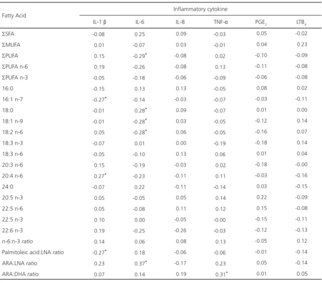
Table 4 shows the correlation coefficients between fatty acid concentrations and inflammatory cytokines. IL-1β was inversely
correlated with palmitoleic acid (C16:1n-7), AA (C20:4n-6) and the palmitoleic acid:AL ratio. IL-6 was inversely correlated with the sum of PUFA, stearic acid (C18:0), oleic acid (C18:1n-9) and LA (C18:2n-6) and directly correlated with the AA:AL ratio. TNF-α was the only inflammatory marker with a direct correlation with the AA:DHA ratio.
D I S C U S S I O N
The LA concentration observed in both children and adolescents in this study was low
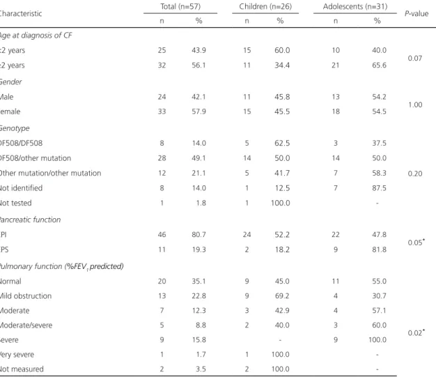
Table 1. Clinical characteristics of children and adolescents with Cystic Fibrosis treated at a Cystic Fibrosis reference center in Rio de Janeiro (RJ), Brazil, 2009-2010.
Characteristic Total (n=57) Children (n=26) Adolescents (n=31) P-value
n % n % n %
Age at diagnosis of CF
<2 years 25 43.9 15 60.0 10 40.0
0.07
≥2 years 32 56.1 11 34.4 21 65.6
Gender
Male 24 42.1 11 45.8 13 54.2
1.00
Female 33 57.9 15 45.5 18 54.5
Genotype
DF508/DF508 8 14.0 5 62.5 3 37.5
0.20
DF508/other mutation 28 49.1 14 50.0 14 50.0
Other mutation/other mutation 12 21.1 5 41.7 7 58.3
Not identified 8 14.0 1 12.5 7 87.5
Not tested 1 1.8 1 100.0
-Pancreatic function
EPI 46 80.7 24 52.2 22 47.8
0.05*
EPS 11 19.3 2 18.2 9 81.8
Pulmonary function (%FEV1 predicted)
Normal 20 35.1 9 45.0 11 55.0
Mild obstruction 13 22.8 9 69.2 4 30.7
0.02*
Moderate 7 12.3 3 42.9 4 57.1
Moderate/severe 5 8.8 2 40.0 3 60.0
Severe 9 15.8 - 9 100.0
Very severe 1 1.7 1 100.0
-Not measured 2 3.5 2 100.0
-Note: *P<0.05.
(<21%), lower than in pediatric EFA-deficient CF patients reported in other studies (11.23±1.15)
[4,11-13]. This result is noteworthy because the concentration of this fatty acid in EPI has become a clinically accepted biomarker for EFA deficiency and it is even more important than the triene:tetraene ratio (C20:3n-9:C20:4n-6) [10].
Normal plasma LA status is defined as plasma LA≥21mol% of total fatty acids [10]. Considering that approximately half the children and adolescents had EPI, it is reasonable to suggest that LA reduction may result from
Table 2. Erythrocyte-fatty acid composition in children and adolescents with Cystic Fibrosis treated at a cystic fibrosis reference center in Rio de Janeiro (RJ), Brazil, 2009-2010.
Fatty acids (%) Children (n=26) Adolescents (n=31) P-value
SSFA 53.9 ± 10.0 55.5 ± 11.6 0.59
SMUFA 16.8 ± 5.4 15.2 ± 5.0 0.25
SPUFA 29.3 ± 8.4 29.3 ± 8.4 0.99
SPUFA n-6 22.3 ± 6.8 21.8 ± 7.4 0.80
SPUFA n-3 7.0 ± 3.0 7.5 ± 3.0 0.52
16:0 27.1 ± 5.5 26.7 ± 5.5 0.78
16:1 n-7 0.2 (0.0 – 1.4) 0.3 (0.0 – 1.7) 0.27
18:0 20.4 ± 4.5 21.3 ± 6.3 0.55
18:1 n-9 8.1 ± 4.3 6.7 ± 3.6 0.19
18:2 n-6 7.0 ± 3.6 6.1 ± 3.5 0.34
18:3 n-3 0.2 ± 0.1 0.3 ± 0.7 0.40
18:3 n-6 1.0 (0.2 – 5.9) 0.9 (0.3 – 8.0) 0.74
20:3 n-6 1.4 ± 1.0 1.5 ± 1.1 0.82
20:4 n-6 6.3 ± 3.4 6.3 ± 3.1 0.96
24:0 3.5 ± 1.7 3.9 ± 1.5 0.31
20:5 n-3 1.0 ± 0.8 1.1 ± 0.6 0.68
22:5 n-6 1.0 ± 1.0 1.0 ± 0.7 0.78
22:5 n-3 1.4 ± 0.8 1.5 ± 0.9 0.46
22:6 n-3 1.2 ± 0.8 1.2 ± 0.9 0.86
n-6:n-3 ratio 3.6 ± 1.2 3.3 ± 1.4 0.42
Palmitoleic acid:LA ratio 0.0 (0.0 – 2.1) 0.0 (0.0 – 2.6) 0.23
AA:LA ratio 1.0 ± 0.4 1.9 ± 2.9 0.11
AA:DHA ratio 5.5 ± 2.4 6.9 ± 6.7 0.32
Note: Values expressed as Mean±Standard Deviation or Median (min. – max.).
S: Sum; SFA: Saturated Fatty Acids; MUFA: Monounsaturated Fatty Acids; PUFA: Polyunsaturated Fatty Acids; N-6 PUFAs: n-6 Polyunsaturated Fatty Acids; PUFA (n-3): n-3 Polyunsaturated Fatty Acids; 16:0: Palmitic Acid; 16:1n-7: Palmitoleic Acid; 18:0: Stearic Acid; 18:1n-9: Oleic Acid; 18:2n-6: Linoleic Acid (LA); 18:3n-3: Alpha-Linolenic Acid (ALA); 18:3n-6: Gamma-Linolenic Acid (GLA); 20:3n-6: Dihomo-Gamma-Linolenic Acid (DGLA); 20:4 n-6: Arachidonic Acid (AA); 24:0: Lignoceric Acid; 20:5n-3: Eicosapentaenoic Acid (EPA); 22:5n-6: Docosapentaenoic Acid (DPAn-6); 22:5n-3: Docosapentaenoic Acid (DPAn-3); 22:6n-3: Docosahexaenoic Acid (DHA).
Table 3. Serum inflammatory markers in children and adolescents with Cystic Fibrosis treated at a Cystic Fibrosis reference center in Rio de Janeiro (RJ), Brazil, 2009-2010.
Cytokine Children (n=26) Adolescents (n=31) P-value
IL-1 β (pg/mL) 12.3 (0.0 – 182.1) 14.1 (0.0 – 168.4) 0.81
IL-6 (pg/mL) 8.3 (1.5 – 42.4) 14.1 (1.1 – 51.4) 0.21
IL-8 (pg/mL) 1.9 (0.0 – 219.7) 3.0 (0.0 – 81.7) 0.59
TNF-α (pg/mL) 36.7 (0.0 – 284.3) 7.2 (0.0 – 337.8) 0.03*
PGE2 (pg/mL) 1006.0 (107.6 – 32659.3) 778.2 (73.9 – 5281.5) 0.34 LTB4 (pg/mL) 352.6 (94.2 – 2399.5) 332.0 (96.7 – 2296.9) 0.64 CRP (mg/dL)
<0.5 18 (47.4) 20 (52.6)
0.78
≥0.5 8 (42.1) 11 (57.9)
Note: *P<0.05.
Values expressed as Mean±Standard Deviation and Median (min. – max.) n(%).
IL-1β: Interleukin-1β; IL-6: Interleukin-6; IL-8: Interleukin-8; TNF-Α: Tumor Necrosis Factor-Α; PGE2: Prostaglandin E2; LTB4: Leukotriene B4; CRP: C-Reactive Protein.
Erythrocyte AA levels in this study were similar to plasma AA levels in CF children (5.99±0.83%) in another study [15]. Nonetheless, the AA levels in CF patients reported in the literature are contradictory since both low [4,11] and elevated values [16,17] have been found. In our study, the high turn-over of n-6 fatty acids and abnormally high AA release in CF, which result in reduced LA, was confirmed [5].
Incorporation of EFA into phospholipids is influenced by chloride channels, suggesting that CFTR may regulate aspects of EFA metabolism. Disturbances in lipid metabolism in CF, such as an increased release of AA from cell membrane phospholipids and low LA and DHA status, have been previously described [4,5]. Different hypotheses have been discussed; however, the link between CFTR, AA release and Docosahexaenoic Acid (DHA) abnormality have yet to be elucidated [2,6].
The concentration of erythrocyte fatty acids better indicates fatty acid status than plasma concentrations when monitoring long-term intake of fatty acids [15,18,19]. DHA, EPA, LA, Alpha-Linoleic Acid (ALA), oleic acid and palmitoleic acid concentrations in erythrocytes were low, which is in agreement with previous
studies. The concentration of AA was not as high as that reported in the literature [4,15-17] and an association between erythrocyte fatty acids and IL-1β and IL-6 was observed. These findings indicate that there is an association between PUFA profile and serum pro-inflammatory cytokines in the pediatric CF patients studied.
Analysis of erythrocyte fatty acid composition in children and adolescents in our study showed a higher percentage of SFA (53.9% and 55.5%, respectively) than in an Italian study with CF children aged 6-12 years (35% of total plasma fatty acids) [20].
Table 4. Correlation coefficients between concentrations of fatty acids and inflammatory cytokines in children and adolescents with Cystic Fibrosis treated at a Cystic Fibrosis reference center in Rio de Janeiro (RJ), Brazil, 2009-2010.
Fatty Acid
Inflammatory cytokine
IL-1 β IL-6 IL-8 TNF-α PGE2 LTB4
SSFA -0.08 0.25 0.09 -0.03 0.05 -0.02
SMUFA 0.01 -0.07 0.03 -0.01 0.04 0.23
SPUFA 0.15 -0.29* -0.08 0.02 -0.10 -0.09
SPUFA n-6 0.19 -0.26 -0.08 0.13 -0.11 -0.08
SPUFA n-3 -0.05 -0.18 -0.06 -0.09 -0.06 -0.08
16:0 -0.15 0.13 0.13 -0.05 0.08 0.02
16:1 n-7 -0.27* -0.14 -0.03 -0.07 -0.03 -0.11
18:0 -0.01 0.28* 0.09 -0.07 0.01 0.00
18:1 n-9 -0.01 -0.28* 0.03 -0.05 -0.12 0.14
18:2 n-6 0.05 -0.28* 0.06 -0.05 -0.16 0.07
18:3 n-3 -0.07 0.01 0.00 -0.19 -0.18 0.14
18:3 n-6 -0.05 -0.10 0.13 0.06 0.01 0.04
20:3 n-6 0.15 -0.19 -0.03 0.02 -0.18 -0.00
20:4 n-6 0.27* -0.23 -0.11 0.11 -0.03 -0.16
24:0 -0.07 0.22 -0.11 -0.14 0.03 -0.15
20:5 n-3 0.05 -0.05 0.05 0.14 0.22 -0.09
22:5 n-6 0.05 -0.08 0.11 0.12 0.15 -0.08
22:5 n-3 0.10 0.00 -0.05 -0.00 -0.15 -0.11
22:6 n-3 0.19 -0.25 -0.26 -0.03 -0.12 -0.13
n-6:n-3 ratio 0.14 0.06 0.08 0.13 -0.05 0.12
Palmitoleic acid:LNA ratio -0.27* 0.18 -0.06 -0.06 -0.01 -0.14
ARA:LNA ratio 0.23 0.37* -0.17 0.23 0.05 -0.14
ARA:DHA ratio 0.07 0.14 0.19 0.31* 0.01 0.05
Note: *P<0.05
IL-1Β: Interleukin-1β; IL-6: Interleukin-6; IL-8: Interleukin-8; TNF-Α: Tumor Necrosis Factor Α; PGE2: Prostaglandin E2; LTB4: Leukotriene B4; CRP: C-Reactive Protein; S: Sum; SFA: Saturated Fatty Acids; MUFA: Monounsaturated Fatty Acids; PUFA (n-6): n-6 Polyunsaturated Fatty Acids; PUFA (n-3): n-3 Polyunsaturated Fatty Acids; C16:0: Palmitic Acid; C16:1 n-7: Palmitoleic Acid; C18:0: Stearic Acid; C18:1n-9: Oleic Acid; C18:2n-6: Linoleic Acid (LNA); C18:3n-3: Alpha-Linolenic Acid (ALA); C18:3n-6: Gamma-Linoleic Acid (GLA); C20:3n-6: Dihomo-Gamma-Linolenic Acid (DGLA); C20:4n-6: Arachidonic Acid (AA); C24:0: Lignoceric Acid; C20:5n-3: Eicosapentaenoic Acid (EPA); C22:5n-6: Docosapentaenoic Acid (Dpan-6); C22:5n-3: Docosapentaenoic Acid (Dpan-3); C22:6n-3: Docosahexaenoic Acid (DHA); LNA: Linoleic Acid; ALA: Alpha-Linolenic Acid; ARA: Arachidonic Acid; EPA: Icosapentaenoic Acid; DHA: Docosahexaenoic Acid.
The levels of DHA and EPA in erythrocytes in our study were similar to those observed in plasma and tissues of EFA-deficient CF children in a previous study (1.23±0.18 and 0.94±0.09, respectively) [15]. Other authors have also observed low plasma and tissue DHA concentrations [4,11-13,16,25,26] as well as low erythrocyte EPA levels in CF patients [19,27]. DHA and EPA are converted to resolvins
importance of therapeutic supplementation with LC n-3 PUFA, such as EPA and DHA, for improving dietary therapy in CF patients.
Although DHA and EPA have the most potent anti-inflammatory effects, the therapeutic dose of n-3 PUFA in CF, a multifactorial disease, will also depend on the degree of severity of the disease, which, in turn, depends on the patient’s genetic predisposition and individual factors that affect the bioavailability of n-3 PUFA [2,28]. Several studies have shown that the optimal n-6:n-3 ratio may vary among diseases. Furthermore, there is no consensus on the ideal LA:ALA ratio intake in CF because of the imbalance and deficiency of LA and DHA in these patients [2,4]. Evidences are insufficient to understand the relationship between diet and inflammation and the ideal n-6:n-3 ratio for CF [6].
Our results confirmed that there was a low concentration of ALA, as it was observed in other studies with CF patients that found the following levels of erythrocytes and plasma fatty acids, 0.43±0.71% and 0.2%, respectively [20,25].
We found a higher AA:LA ratio than those observed in pediatric CF patients in Spain (0.27 and 0.28) [26], possibly indicating that the pathway from LA to AA was enhanced in children and adolescents in our study. In CF, the conversion of AL to AA is unregulated due to increased activity of the enzyme Phospholipase A2 (PLA2), which promotes an increase in AA, reducing the concentration of AL and DHA [2,5]. Our results support this hypothesis.
The AA:DHA ratio in our study was higher than the one found in individuals without CF, corroborating several studies [17,20,26]. When this ratio is high in CF patients, it is believed to be an important indicator of the inflammatory state [12], reflecting the ability of AA to generate eicosanoids that promote the production of pro-inflammatory cytokines and may be responsible for the early development of pulmonary disease [2,3]. This may explain the
direct correlations found between TNF-α and the AA:DHA ratio, IL-6 and the AA:LA ratio, and IL-1 and AA observed in this study.
The concentrations of palmitoleic and oleic acids (MUFA profile) in erythrocytes were lower in both groups than those found in another study, 0.9±0.4% [30] and 17.9±4.4 [25], respectively. The lower concentrations of oleic acid may be a result of conversion of this acid into eicosatrienoic acid, which was not measured in the present study. Although they are not eicosanoid precursors, MUFA may influence synthesis of these derivatives and the lipid metabolism of PUFA through enzymatic inhibition and saturation [18].
In EFA deficiency, the level of desaturases increases, despite the lower affinity of oleic acid for delta-6-desaturase, and this acid is converted into large quantities of eicosatrienoic acid [15]. The erythrocyte palmitoleic acid:LA
ratio was lower in adolescents than in children. This ratio is also considered an EFA deficiency marker in plasma, erythrocyte membranes and platelets in CF and it is a better marker than the triene:tetraene ratio [15]. This could explain the indirect correlation between IL1-β and the palmitoleic acid:LA ratio observed in our patients.
considered to reflect the level of inflammation to which CF patients are exposed during their disease. Moreover, most of our patients showed no evidence of acute inflammation, as revealed by their CRP levels (<0.5mg/dL). These are elevated when CF is exacerbated, and they can reveal early inflammation when other clinical parameters are equivocal [33].
TNF-α can be considered an early marker of inflammation in pediatric patients. Surprisingly, levels of this marker were extremely high in the children in our study and it was the only pro-inflammatory marker whose levels were statistically different in the two age groups. This finding is important because elevated plasma levels of TNF-α are also associated with reduced lean body mass, increased muscle proteolysis, increased respiratory exacerbations and poorer lung function, even in clinically stable patients [34].
As there are few studies comparing the profile of fatty acids and inflammatory markers in pediatric patients with CF, the scope for interpretation and comparison of our findings with the literature was limited. This was the first study conducted in Brazil to evaluate the composition of erythrocyte fatty acids and correlate them with inflammatory markers in pediatric CF patients.
C O N C L U S I O N
The pediatric CF patients presented EFA deficiency. An association between fatty acid profile in erythrocytes and serum pro-inflammatory cytokines was observed in the present study and imbalance in lipid metabolism may be associated with CFTR mutations.
These findings highlight the importance of assessing erythrocyte fatty acids and serum pro-inflammatory cytokines that may open new possibilities for studying the pathophysiology and treating CF patients, such dietary supplementation
with n-3 fatty acids (EPA and DHA). However, further longitudinal studies are needed for better clarification of the imbalance in lipid metabolism and inflammation in patients with CF.
C O N T R I B U T O R S
ALP Cunha conceptualized and designed the study, coordinated and supervised data collection, drafted the initial manuscript and made final revisions based on the critical reviews of the other authors. AC COSTA coordinated and carried out the statistical analyses, reviewed the manuscript and approved the final version of the manuscript. Z VASCONCELOS coordinated and carried out the cytokine measurements, reviewed the manuscript and approved the final version of the manuscript. MG TAVARES DO CARMO supervised the study, coordinated the fatty acid measurements, critically reviewed the manuscript and approved the final version of the manuscript. CR CHAVES conceptualized and designed the study, supervised the study, drafted the initial manuscript, critically reviewed the manuscript and approved the final version of the manuscript.
R E F E R E N C E S
1. Courtney JM, Ennis M, Elbom JS. Cytokines and inflammatory mediators in cystic fibrosis. J Cyst Fibros. 2004;3(4):223-31. http://dx.doi.org/10.10 16/j.jcf.2004.06.006
2. Strandvik B. Fatty acid metabolism in cystic fibrosis. Prostaglandins Leukot Essent Fatty Acids. 2010;83(3):121-9. http://dx.doi.org/10.1016/j.plefa. 2010.07.002
3. Calder PC, Innes JK. Omega-6 fatty acids and inflammation. Prostaglandins Leukot Essent Fatty Acids. 2018;132:41-8. http://dx.doi.org/10.1016/j. plefa.2018.03.004
4. Roulet M, Frascarolo P, Rappaz I, Pilet M. Essential fatty acid deficiency in well-nourished young cystic fibrosis patients. Eur J Pediatr. 1997;156:952-6.
6. Seegmiller AC. Abnormal unsaturated fatty acid metabolism in cystic fibrosis: Biochemical mechanisms and clinical implications. Int J Mol Sci. 2014;15(9):16083-99. http://dx.doi.org/10.3390/ ijms150916083
7. Borowitz D, Robinson KA, Rosenfeld M, Davis SD, Sabadosa KA, Spear SL, et al. Cystic Fibrosis foundation evidence-based guidelines for management of infants with Cystic Fibrosis. J Pediatr. 2009;155(6):S73-93. http://dx.doi.org/10. 1016/j.jpeds.2009.09.001
8. Pellegrino R, Viegi G, Brusasco V, Crapo RO, Burgos F, Casaburi R, et al. Interpretative strategies for lung function tests. Eur Respir J. 2005;26(5):948-68. http://dx.doi.org/10.1183/09031936.05.000352 05
9. Zock PL, Mensink RP, Harryvan J, Vries JHM, Katan MB. Fatty acids in serum cholesteryl esters as quantitative biomarkers of dietary intake in humans. Am J Epidemiol. 1997;145(12):1114-22.
10. Maqbool A, Schall JI, Garcia-Espana F, Zemel BS, Strandvik B, Stallings VA. Serum linoleic acid status as a clinical indicator of essential fatty acid status in children with cystic fibrosis. J Pediatr Gastroenterol Nutr. 2008;47(5):635-44. http://dx. doi.org/10.1097/MPG.0b013e31817fb76b
11. Strandvik B, Gronowitz E, Enlund F, Martinsson T, Wahlstrom J. Essential fatty acid deficiency in relation to genotype in patients with cystic fibrosis. J Pediatr. 2001;139:650-5. http://dx.doi. org/10.1067/mpd.2001.118890
12. Van Biervliet S, Vanbillemont G, Van Biervliet JP, Declercq D, Robberecht E, Christophe A. Relation between fatty acid composition and clinical status or genotype in cystic fibrosis patients. Ann Nutr Metab. 2007;51:541-9. http://dx.doi. org/10.1159/000114208
13. Van Biervliet S, Devos M, Van Biervliet JP, Robberecht E, Christophe A. Oral DHA supplementation in DF508 homozygous cystic fibrosis patients. Prostaglandins Leukot Essent Fatty Acids. 2008;78(2):109-15. http://dx.doi.org/10.1016/j. plefa.2007.12.005
14. Turck D, Braegger CP, Colombo C, Declercq D, Morton A, Pancheva R, et al. ESPEN-ESPGHAN-ECFS guidelines on nutrition care for infants, children, and adults with cystic fibrosis. Clin Nutr. 2016;35(3):557. http://dx.doi.org/10.1016/j.clnu. 2016.03.004
15. Lepage G, Levy E, Ronco N, Smith L, Galgano N, Roy CC. Direct transesterification of plasma fatty acids for the diagnosis of essential fatty acid deficiency in cystic fibrosis. J Lipid Res. 1989;30(10):1483-90.
16. Freedman SD, Blanco PG, Zaman MM, Shea JC, Ollero M, Hopper IK, et al. Association of cystic fibrosis with abnormalities in fatty acid metabolism. N Engl J Med. 2004;350(6):560-9. http://dx.doi.org/10.1056/NEJMoa021218
17. Olveira G, Olveira C, Acosta E, Espildora F, Garrido-Sanchez L, Garcia-Escobar E, et al. Fatty acid supplementation improves respiratory, inflammatory and nutritional parameters in adults with cystic fibrosis. Arch Bronconeumol. 2010;46(2):70-7. http://dx.doi.org/10.1016/j.arbres. 2009.11.001
18. Arab L, Akbar J. Biomarkers and the measurement of fatty acids. Public Health Nutr. 2002;5(6A):865-71. http://dx.doi.org/10.1079/PHN2002391
19. Cao J, Schwichtenberg KA, Hanson NQ, Tsai MY. Incorporation and clearance of omega-3 fatty acids in erythrocyte membranes and plasma phospholipids. Clin Chem. 2006;52(12):2265-72. http://dx.doi.org/10.1373/clinchem.2006.072322
20. Alicandro G, Faelli N, Gagliardini R, Santini B, Magazzu G, Biffi A, et al. A randomized placebo-controlled study on high-dose oral algal docosahexaenoic acid supplementation in children with cystic fibrosis. Prostaglandins Leukot Essent Fatty Acids. 2013;88(2):163-9. http://dx.doi. org/10.1016/j.plefa.2012.10.002
21. Katan MB, Deslypere JP, van Birgelen AP, Penders M, Zegwaard M. Kinetics of the incorporation of dietary fatty acids into serum cholesteryl esters, erythrocyte membranes, and adipose tissue: An 18-month controlled study. J Lipid Res. 1997;38(10):2012-22.
22. Zong G, Li Y, Wanders AJ, Alssema M, Zock P, Willet WC, et al. Intake of individual saturated fatty acids and risk of coronary heart disease in US men and women: Two prospective longitudinal cohort studies. BMJ. 2016;355:i5796. http://dx.doi. org/10.1136/bmj.i5796
23. Wu MY, Li CJ, Hou MF, Chu PY. New insights into the role of inflammation in the pathogenesis of atherosclerosis. Int J Mol Sci. 2017;18(10):2034. http://dx.doi.org/10.3390/ijms18102034
24. Smith C, Winn A, Seddon P, Ranganathan S. A fat lot of good: Balance and trends in fat intake in children with cystic fibrosis. J Cyst Fibros. 2012;11(2):154-7. http://dx.doi.org/10.1016/j.jcf.20 11.10.007
26. Aldamiz-Echevarria L, Prieto JA, Andrade F, Elorz J, Sojo A, Lage S, et al. Persistence of essential fatty acid deficiency in cystic fibrosis despite nutritional therapy. Pediatr Res. 2009;66(2009):585-9. http:// dx.doi.org/10.1203/PDR.0b013e3181b4e8d3
27. Hanssens L, Thiébaut I, Lefèvre N, Malfroot A, Knoop C, Duchateau J, et al. The clinical benefits of long-term supplementation with omega-3 fatty acids in cystic fibrosis patients: A pilot study. Prostaglandins Leukot Essent Fatty Acids. 2016;108:45-50. http://dx.doi.org/10.1016/j.plefa. 2016.03.014
28. Schuchardt JP, Hahn A. Bioavailability of long-chain ômega-3 fatty acids. Prostaglandins Leukot Essent Fatty Acids. 2013;89(1):1-8. http://dx.doi. org/10.1016/j.plefa.2013.03.010
29. Goyens PLL, Spilker ME, Zock PL, Katan MB, Mensink RP. Conversion of α-linolenic acid in humans is influenced by the absolute amounts of
α-linolenic acid and linoleic acid in the diet and not their ratio. Am J Clin Nutr. 2006;84(1):44-53. http://dx.doi.org/10.1093/ajcn/84.1.44
30. Keen C, Olin AC, Edentoft A, Gronowitz E, Strandvik B. Airway nitric oxide in patients with cystic fibrosis is associated with pancreatic function,
pseudomonas infection, and polyunsaturated fatty acids. Chest. 2007;131(6):1857-64. http://dx. doi.org/10.1378/chest.06-2635
31. Nixon LS, Yung B, Bell SC, Elborn JS, Shale DJ. Circulating immunoreactive interleukin-6 in cystic fibrosis. Am J Respir Crit Care Med. 1998;157(6);1764-9.
32. Dufresne V, Knoop C, Van Muylem A, Malfroot A, Lamotte M, Opdekamp C, et al. Effect of systemic inflammation on inspiratory and limb muscle strength and bulk in cystic fibrosis. Am J Respir Crit Care Med. 2009;180(2):153-8. http://dx.doi. org/10.1164/rccm.200802-232OC
33. Pepys MB. C-reactive protein fifty years on. Lancet. 1981;1(8221):653-7.
34. Ionescu AA, Nixon LS, Evans WD, Stone MD, Lewis-Jenkins V, Chatham K, et al. Bone density, body composition, and inflammatory status in cystic fibrosis. Am J Respir Crit Care Med. 2000;162(3):789-94. http://dx.doi.org/10.1164/ajrc cm.162.3.9910118