Cosmesis in patients with breast neoplasia
submitted to the hypofractionated radiotherapy
with of intensity-modulated beam
Fabiana Accioli Miranda1 Marina Tamm Lannes Vieira2
Fabio Ynoe de Moraes3
Gustavo Nader Marta4
Heloísa de Andrade Carvalho5
Samir Abdallah Hanna6
1. Radioteraphy of Hospital Sírio-Libanês – SP, São Paulo (SP), Brasil. 2. Radiation Oncologist of COI (Clínicas Oncológicas Integradas – RJ), Rio de Janeiro (RJ), Brasil. 3. Radiation Medicine Program, Princess Margaret Hospital University of Toronto, Toronto, Ontario, Canada. 4. Radiation Oncologist of Hospital Sírio-Libanês – SP and Instituto do Câncer do Estado de São Paulo, São Paulo (SP), Brasil. 5. Radiation Oncologist of Hospital Sírio-Libanês – SP and Instituto de Radiologia (InRad) – SP, São Paulo (SP), Brasil. 6. Radiation Oncologist of Hospital Sírio-Libanês – SP, São Paulo (SP), Brasil.
http://dx.doi.org/10.1590/1806-9282.64.11.1023
SUMMARY
OBJECTIVE: To assess the cosmetic satisfaction of patients diagnosed with breast cancer submitted to the hypofractionated radiothera-py with IMRT (hIMRT) technique and its correlation with dosimetric data of the radiotheraradiothera-py planning.
MATERIALS AND METHODS: The retrospective cohort study that assessed women with a diagnosis of malignant breast neoplasia sub-mitted to the conservative treatment or radical mastectomy and treated with hIMRT. In the period between August 2007 to December 2014, in a philanthropic / private institution, 170 records were selected. The cosmetic assessment was carried out by means of the Harvard/RTOG/NSABP scale with one-year minimum range after treatment. The collected dosimetric data were: breast / chest wall volume, volume that received 95% (V95%) and 107% (V107%) of the prescribed dose.
RESULTS: The volume of the treated breasts ranged from 169 to 2.103 ml (median = 702; IQR: 535 to 914 ml). Median V95% was 86.7% (54.6-96.6%; IQR: 80.0% to 90.6%); eight (5.7%) patients had V95% higher than 95%. Median V107% was 0% (0%-16.3%; IQR: 0.0% to 0.3% and 13); 9.3% patients had V107% higher than 2%. One hundred and thirty-three (78.2%) patients responded to the cosmetic assessment: 99 (74.4%) considered the cosmetic results excellent. Significant associations between cosmetic assessment and breast volume (p=0.875), V95% (p=0.294) e V107% (p=0.301) were not found.
CONCLUSION: The cosmetic results showed favorable when using hIMRT, and the lack of correlation with usual the dosimetric data illustrates the capacity of hIMRT to minimize the heterogeneity of the dose in this endpoint, even in voluminous breasts.
KEYWORDS: Hypofractionation. Breast neoplasia. Radiotherapy.
DATE OF SUBMISSION: 28-May-2018 DATE OF ACCEPTANCE: 05-Aug-2018 Corresponding Author: Fabiana A. Miranda Radioteraphy of Hospital Sírio-Libanês
Rua Adma Jafet, 91, São Paulo, SP, Brasil – CEP 01308-050 E-mail: fabimiranda10@hotmail.com, fabiana.amiranda@hsl.org.br
INTRODUCTION
The breast cancer is a malignant neoplasia more commonly in women. It is known that radiotherapy (RT) is an integral part of the adjuvant treatment for the most patients, regardless of the type of surgery
that is carried out, producing benefits in local control and survival1.
equip-ment, and planning systems, allowing better accu-racy in defining the target volume of treatment and more conformal plannings2. The latest techniques of
treatment, including conformal three-dimensional RT (RT3DC) and intensity modulated RT (IMRT), al-low a greater preservation of adjuvant bodies3,4.
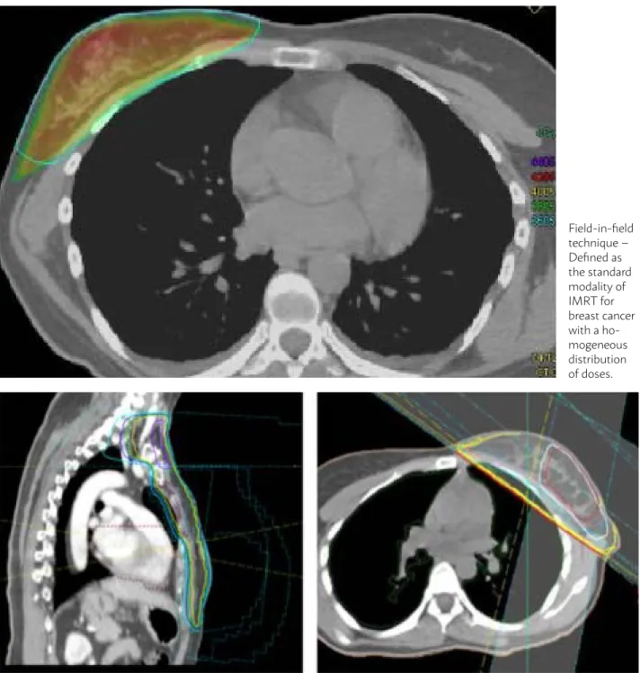
Field-in-field technique may be carried out with opposed tangential fields and it is considered a sim-ple way of IMRT (forward-planned IMRT), without the need for a reverse planning system or other more complex technologies. Smaller fields are added to the
main field in order to achieve a homogenization of the dose by handling the collimator blades. This tech-nique may be easily implanted and the treatment pe-riod is similar to the conventional techniques5,6
(Fig-ure 1).
The conventional treatments use profiles from five to six weeks of duration, total dose of 50 Gy. Therefore, for patients selected, the hypofractionat-ed regimes (shorter period of treatment with a high-er dose phigh-er fraction), such as 42.5 Gy in 16 fractions or 40 Gy in 15 fractions are equally efficient, besides
FIGURE 1: FIELD-IN-FIELD TECHNIQUE X 3D
Field-in-field technique – Defined as the standard modality of IMRT for breast cancer with a ho-mogeneous distribution of doses.
producing cosmetic results similar or better than the conventional profile7,8.
This study aims to assess the cosmetic satisfac-tion of patients diagnosed with breast cancer sub-mitted to hypofractionated RT with IMRT technique (hIMRT), and its correlation with dosimetric data of the radiotherapy planning.
PATIENTS AND METHODS
This is a retrospective uni-institutional co-hort study that assessed women with diagnosis of non-metastatic breast malignant neoplasia submit-ted to conservative treatment or radical mastectomy, whether or not followed by immediate reconstruc-tion, provided that no prosthesis, and submitted to the adjuvant irradiation of breast or chest wall (which corresponds to the surgical bed after mastec-tomy), with or without inclusion of classical lymph node sites with the hIMRT technique. The patients were assessed between August 2007 and December 2014. All included patients shall be a minimum fol-low-up of one year to assess the cosmesis.
The adopted exclusion criteria were: patients who did not complete the proposed RT, the presence of other neoplasia, except for non-melanoma skin or
in situ carcinoma of cervix uteri, as well as informa-tion on insufficient follow-up for analysis.
The cosmetic assessment was made by the Har-vard/RTOG/NSABP9 scale by simple questions and
comparison with the non-treated side (Table 1), from
Characteristic % Patients
Stage
0 20.2
IA 63.2
IB 0.6
IIA 12.3
IIB 2.5
III 1.2
Histological Grade
Invasive ductal carcinoma 72.1
Others 27.9
Histological grade
I 20.6
II 35.3
III 17.6
NA 26.5
TABLE 1 - ASSESSMENT OF COSMESIS
one year after treatment. The data were collected by researches by review of records, phone interview, and satisfaction questionnaire.
Demographic data and aspects involved in the tu-mor were collected, such as histological type, stag-ing, surgical extension, presence and type of breast reconstruction and additional treatments. In addi-tion, dosimetric data was also collected: prescribed dose, volume of breast / chest wall (Clinic Target Vol-ume, CTV), volumes that received 95% (V95%) and 107% (V107%) of the prescribed dose corresponding respectively to the coverage of target volume and vol-ume of “hot spots”, that is, it is assessed the homo-geneity for distribution of dose. The planning system used was Oncentra Masterplan (Nucletron)®.
The descriptive analysis was carried out by calcu-lating the frequencies, mean and standard deviation (dp) or median and interquartile range (IQR). The as-sessment of association between cosmetic satisfac-tion and dosimetric data was carried out when using Kruskal-Wallis test. It was admitted the level of sta-tistical significance p<0.05. The stasta-tistical analysis was carried out using the Stata™ program (version 11.2).
RESULTS
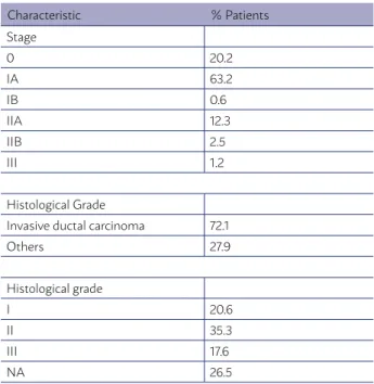
170 patients with a median age of 65.8 years (31 to 95 years, dp=3 years) were included. Among them, the majority (63.2%) presented neoplasias in IA stage, 33 (20.2%) in 0 stage and the other IB to III. The more frequent histological type was the invasive carcinoma with a special type (72.1%). Thirty-four (20%) patients carried out adjuvant chemotherapy and 135 (79.4%) were submitted to anti-hormone therapy. One hun-dred and nine (64%) patients were submitted to the conservative surgery and 61 (36%) to modified radical mastectomy (Table 2).
The adopted hypofractionated profile was 40.05 Gy in 15 fractions to 43.5% of patients and 42.4 Gy in 16 fractions in 56.5% of cases assessed. Twelve patients (7.0%) received irradiation of lymph node drainages and 28 (16.3%) received a booster dose in the operative bed with a dose of 10 Gy in five frac-tions.
Excellent: Little or no difference in size, symmetry or shape Good: Slight asymmetry in size or shape
Regular: Obvious differences in size and/or shape
Bad: Marked change in appearance, involving more than 14 of the breast
CHART 1 – BREAST VOLUME TABLE 2 - PATIENTS CHARACTERISTICS
Nuclear Grade
I 14.1
II 45.3
III 24.7
NA 15.9
Adjuvant Chemotherapy
Yes 20
No 80
Anti-Hormonal Treatment
Yes 79.4
No 20.6
Surgery
Conservative 64
Radical mastectomy 36
Reconstruction (without implants) 15.9
hIMRT scheme
15 fractions of 2.67Gy (40.05Gy) 43.5
16 fractions of 2.65Gy (42.40Gy) 56.5
RT of LM drainage
Yes 7
No 93
Boost use during surgery
Yes 16.3
No 83.7
Cosmetic Assessment
Excellent 74.4
Good 24.1
Regular 1.5
Bad 0
higher than 95%. Median V107% was 0% (0%-16.3%; IQR: 0.0% to 0.3% and 13); 9.3% patients had V107% higher than 2%. 133 patients (78.2%) responded to the questionnaire of cosmetic assessment. Among them, 99 (74.4%) considered cosmetic results excel-lent, 32 (24.1%) considered good, and two (1,5%) con-sidered reasonable (Table 2). Significant associations between cosmetic assessment and the breast volume were not found (p=0.875), V95% (p=0.294) and V107% (p=0.301).
DISCUSSION
Two decades ago, the first studies were designed, suggesting a hypofractionated treatment profile. It was sought an optimization of the dose-time relation to keep a maximum tumor response with rates of ac-ceptable toxicity. Moreover, a shorter treatment pro-file would offer an advantage of a more efficient and productive use of the funds from RT department10.
The safety and efficacy, besides better cosmet-ic results compared to the conventional treatment, were shown in four randomized trials, involving 5.685 patients treated with hypofractionated pro-file11.
The Canadian study randomized 1.234 women among the doses of 42.5 Gy in 16 fractions or 50 Gy in 25 fractions. After a median follow-up of 69 months, there was no difference in relation to the free survival of local relapse and global survivals and
free disease, with 77% of excellent or good cosmetic results in both arms12,13.
The Britain clinical trials Standardization of Breast Radiotherapy Trial, divided into (Start) A and B, sought to define the ideal fractionation, finding results similar to the prior studies14. With a mean
follow-up of five years, Start A included 2.236 oper-ated patients with breast neoplasia (T1-T3, N0-N1), without immediate reconstruction. The patients were treated with 50 Gy in 25 fractions, 41.6 Gy in 13 fractions or 39 Gy in 13 fractions. In the results, the local failure was similar to the groups of 50 Gy and 41.6 Gy, showing an equivalence among the profiles. Start B assessed 2.215 patients treated with 50 Gy in 25 fractions and 40 Gy in 15 fractions. After a mean follow-up of six years, the local failure was similar in both groups. Moreover, the dose of 40 Gy offered results of the local and esthetic control as good as the profile of 50 Gy15.
When the first works of hypofractionation started, the concern about increasing the breast fibrosis and the worsening of cosmetic results increased, especial-ly because of patients with voluminous breasts, there was a greater trend to a worst cosmetic result already showed in the results of works with conventional frac-tionation. In this way, many of these studies excluded women with voluminous breasts – in the Canadian studies, the patients were simply excluded if they had the distance between mean line and mean axillary line, measured in the breast center higher than 25 cm – and the trials that included these patients did not provide clear information about the impact on breast volume related to the toxicity and cosmesis, especially by the fact of using conventional radiotherapy16.
In the Canadian trials and Start, the toxicities were not worse when compared with patients who received the standard fractionation up to now (50 Gy in 25 daily fractions), in view of the study used as a radiobiological substrate for the equivalence of different treatments, the linear-quadratic model17.
Even though, the selection criteria of patients in these studies involved patients who did not receive prior chemotherapy, and in which there was no in-dication of irradiation of lymph nodes drainage nor immediate plastic reconstruction or voluminous breasts18. The latest works published tried to assess
the relation between cosmesis and dosimetry of the planning of these patients, who were submitted to breast hypofractionated RT because, besides radiobi-ological implications, it is important to consider the
practical advantages of hypofractionation, such as its convenience in terms of costs both for patients and health service, and also patient’s compliance to the treatment19.
It is known the importance of the correlation be-tween toxicity and cosmetic results with patients’ characteristics (age, comorbidities, body mass index) and medical treatments (neoadjuvant / adjuvant che-motherapy, hormonal deprivation, other concom-itant drugs). Therefore, besides these factors being assessed, the impact of the CTV volume must be analyzed (representing the breast volume) and do-simetric data, especially focused on maximum dose and homogeneity of the planning (absolute volumes of breast tissues exposed to ≥107% of the dose pre-scribed). In this way, patients with postoperative complications or voluminous breasts for which a maximum dose <107% is not reachable, or patients with implants for the increase or breast reconstruc-tion would have an increased risk of late fibrosis or cosmetic deterioration after RT20.
Recently, a Chinese clinic study21, published only
in a summary format, studied over 800 patients with post-mastectomy breast cancer and showed benefits of hypofractionated RT in the advanced disease with 43.5 Gy delivered over three weeks. After a five-year folup, the rates of tumor recurrence were not low-er than standard RT with conventional fractionation. The rates of locoregional recurrence were 8.3% for hypofractionated RT and 8.1% in the standard treat-ment (HR=1.10, IC 95%:0.67-1.83), with difference of 0.2% (IC 95% = –4.1 to 4.5). The rates of free survival of disease were from 74.6% to hypofractionation arm and 70.7% for the standard treatment arm (HR=0.88, IC 95%:0.67-1.16). The rates of global survival (GS) in five years were 83.2% after hypofractionation and 85.6% with standard treatment (HR=1.13, IC 95%:0.78-1.62). In addition, fewer side effects were evidenced in pa-tients of hypofractionation, indicating that hypofrac-tionated RT after mastectomy is a safe and efficient option for the locally advanced disease21.
The guidelines of American Society of Therapeu-tic Radiology and Oncology (Astro), initially located in 2010, recommended the breast hypofractionation for patients in initial stages, age above 50 years, treated with conservation surgery, without chemotherapy or indication of lymph nodes irradiation, and with dosi-metric parameters minimally acceptable, according to the techniques of conventional treatment22. These
expert panel, and not in formal contraindication. In this year of 201823, the same society joined again and
the panel updated the recommendations to a wider group of situations, such as the inclusion of systemic treatments, patients with advanced stages and with-out definitive age.
The breast volume as a relevant fact related to the skin toxicity is a contradictory topic in the per-tinent literature. Many authors reported a close correlation between the breast size and intensity of acute effects24. It is up to us a questioning in relation
to the criteria used for the definition of voluminous breast. Vicini et al. verified that patients with breast volume >1.600 cm3 presented higher acute toxicity in
the skin25. On the other hand, Harsolia et al. did not
show level 3 acute toxicity with breast volume <975 cm326. An interesting note of Moody et al. was the
evidence of the relation between the breast size and cosmesis associated with distributions of dose in its planning, finding a significant correlation between the breast size and the non-homogeneity of dose27.
This could explain the cosmetic changes in the ap-pearance of the voluminous breasts, once great vol-umes are frequently associated with the non- homo-geneity of the dose and maximum doses higher than 107% of the prescribed dose20. In addition to the acute
toxicity, a higher breast volume may be correlated to an increased risk of late effects28. In this sense,
IMRT could ensure higher homogeneity in the plan-ning with great breast volumes. Many works that used hIMRT and allowed any breast sizes found lack of acute toxicity in the skin and presented dose of ho-mogeneity lower than 7%29, data also showed in our
study, with lack of significant correlation between the cosmesis and the breast volume, most likely in terms of homogenous terms with median V107% of 0% (0%-16.3%; IQR: 0.0% to 0.3% and 13).
This study showed that breast volume did not have a correlation with prejudice to the cosmesis in the patients assessed, probably due to the benefit obtained with intensity-modulated planning. These findings allowed us to think about using hIMRT could expand the indications of hypofractionated RT for patients with voluminous breasts.
The more common changes in the appearance of the breast after RT are retraction, edema, and telangi-ectasia. These effects, in long-term, damage the cos-mesis and are the results of the breast atrophy, and fibrosis. There are specific responses of fibrosis to the irradiation that may increase the proliferation of these
cells and change and reabsorption of collagen30.
The late adverse effects usually show up after a mean follow-up ranging from five to ten years. The great critics to be carried out is if the cosmetic chang-es, in relation to the fractionation presented when was made the notes, are representing those that will develop over the patients’ lives31.
Curran et al. showed in their studies that the cosmesis after breast hypofractionated RT was the worst in the patients followed by five years more32. In
contrast, an English study did not show a significant difference in toxicity after RT between five and ten years of assessment33. Based on these considerations
and uncertainties, currently, we cannot consider the follow-up as a factor that restricts the interpretation of the hypofractionated studies34.
It is important to highlight, as a counterpoint to the study, the times of different follow-up, once it is about a retrospective cohort. Many patients did not complete the estimating period from five to ten years of follow-up for the appearance of adverse effects, despite the initially established objective has been defined with at least one-year follow-up.
In addition, another point of difficult interpreta-tion would be to know if the dissatisfacinterpreta-tion in relainterpreta-tion to the cosmesis happened after surgery or after ra-diotherapy since the patients were not assessed soon after the surgical procedure and the criteria were the comparison with contralateral breast. Moreover, it is currently hard to think about in patients with breast cancer treated with conservative surgery without ad-juvant irradiation.
Despite these variables, 98.5% of patients con-sidered the cosmetic results from good to excellent, with the lack of significant correlation between cos-mesis and breast volume. This result highlights the importance and the potential impact of the lack of homogeneity, areas of dose >107%, of a planning with short fractionation.
RESUMO
OBJETIVO: Avaliar a satisfação cosmética de pacientes diagnosticadas com câncer de mama submetidas à radioterapia hipofracionada com técnica IMRT (hIMRT) e sua correlação com dados dosimétricos do planejamento radioterápico.
MATERIAIS E MÉTODOS: Estudo de coorte retrospectivo que avaliou mulheres com diagnóstico de neoplasia maligna de mama submeti-das a tratamento conservador ou mastectomia radical e tratasubmeti-das com hIMRT. No período de agosto de 2007 a dezembro de 2014, em uma instituição filantrópica/particular, foram selecionados 170 prontuários. A avaliação cosmética foi feita por meio da escala de Harvard/RTOG/NSABP com um intervalo mínimo de um ano após o tratamento. Dados dosimétricos coletados foram: volume da mama/plastrão, volume que recebeu 95% (V95%) e 107% (V107%) da dose prescrita.
RESULTADOS: O volume das mamas tratadas variou de 169 a 2.103 ml (mediana = 702; IQR: 535 a 914 ml). O V95% mediano foi 86,7% (54,6-96,6%; IQR: 80,0% a 90,6%); oito (5,7%) pacientes tiveram o V95% superior a 95%. O V107% mediano foi 0% (0%-16,3%; IQR: 0,0% a 0,3% e 13); 9,3% pacientes tiveram o V107% superior a 2%. Cento e trinta e três (78,2%) pacientes responderam à avaliação cosmética: 99 (74,4%) consideraram o resultado cosmético excelente. Não foram encontradas associações significativas entre a aval-iação cosmética e o volume da mama (p=0,875), V95% (p=0,294) e V107% (p=0,301).
CONCLUSÕES: Os resultados cosméticos mostraram-se favoráveis com o uso de hIMRT, e a ausência de correlação com os dados dosimétricos usuais ilustra a capacidade do hIMRT em minimizar a heterogeneidade da dose neste desfecho, mesmo em mamas volumosas.
PALAVRAS-CHAVE: Hipofracionamento. Neoplasia de mama. Radioterapia.
REFERENCES
1. M. Clarke, R. Collins, S. DarbyThe Early Breast Cancer Trialists Collabo-rative Group. Effects of radiotherapy and of differences in the extent of surgery for early breast cancer on local recurrence and 15-year survival: An overview of the randomised trials. Lancet, 366 (2005), pp. 2087-2106.
2. Gersem WD, Claus F, Wagter CD, et al. Leaf position optimization for step- and-shoot IMRT. Int J Radiat Oncol Biol Phys 2001; 51:1371-88.
3. Taylor C, et al. Estimating the Risks of Breast Cancer Radiotherapy: Evi-dence From Modern Radiation Doses to the Lungs and Heart and From Previous Randomized Trials. Journal of Clinical Oncology. March, 2017.
4. Mukesh B.et al. Randomized Controlled Trial of Intensity-Modulated Ra-diotherapy for Early Breast Cancer: 5-Year Results Confirm Superior Over-all Cosmesis. JCO. Vol 31, N 36, Dec 2013.
5. Coles CE, Moody AM, Wilson CB, Burnet NG. Reduction of radiotherapy- induced late complications in early breast. Clin Oncol. 2005 17(2):98-110.
6. E.M. Donovan, U. Johnson, G. Shentall, P.M. Evans, A.J. Neal, J.R. Yarnold Evaluation of compensation in breast radiotherapy: a planning study using multiple static fields. Int J Radiat Oncol Biol Phys, 46 (2000), pp. 671-679.
7. Donovan E, Bleakley N, Denholm E, et al. Randomised trial of standard 2D radiotherapy (RT) versus intensity modulated radiotherapy (IMRT) in patients prescribed breast radiotherapy. Radiother Oncol 2007;82:254–64.
8. Holloway C, Panet-Raymond V, Olivotto IA. Hypofractionation should be the new ‘standard’ for radiation therapy after breast conserving surgery.
Breast. 2010;19:163–167.
9. National Surgical Adjuvant Breast and Bowel Project: NSABP Protocol B-39 Form COS.
10. Awwad HK. Dose-Time-Volume relationships in nor- mal tissue to radia-tion. In Radiation Oncology: Ra- diobiological and Physiological Prespec-tive. Kluwer Academic, Dordecht/ Boston/ London. 1990, 129- 187.
11. Owen JR, Ashton A, Bliss JM, et al. Effect of radiotherapy fraction size on tumour control in patients with early-stage breast cancer after local tumour excision: long-term results of a randomised trial. Lancet Oncol. 2006;7:467– 471.
12. Whelan T, MacKenzie R, Julian J, Levine M, Shelley W, Grimard L, Lada B, Lukka H, Perera F, Fyles A, Laukkanen E, Gulavita S, Benk V, Szechtman B.Randomized trial of breast irradiation schedules after lumpectomy for women with lymph node-negative breast cancer. J Natl Cancer Inst. 2002 Aug 7;94(15):1143-50
13. Whelan TJ, Pignol JP, Levine MN, Julian JA, MacKenzie R, Parpia S, Shelley W, Grimard L, Bowen J, Lukka H, Perera F, Fyles A, Schneider K, Gulavita S, Freeman C. Long-term results of hypofractionated radiation therapy for breast cancer. N Engl J Med. 2010 Feb 11;362(6):513-20.
14. The START Trialists’ Group.The UK Standardisation of Breast Radiothera-py (START) Trial A of radiotheraRadiothera-py hypofractionation for treatment of ear-ly breast cancer: a randomised trial. Lancet Oncol 2008; published online March 19. DOI:10.1016/S1470-2045(08)70077-9.
15. The START Trialists’ Group. The UK Standardisation of Breast Radio-therapy (START) Trial B of radioRadio-therapy hypofractionation for treatment of early breast cancer: a randomised trial. Lancet 2008; published online March 19. DOI:10.1016/S0140-6736(08)60348-7.
16. Taher AN, El-Baradie MM, Essa H, Zaki O, Ezzat S: Hypofractionation ver-sus conventional fractionation radiotherapy after conservative treatment of breast cancer: early skin reactions and cosmetic results. J Egypt Natl Canc Inst 2004, 16:178–187.
17. Bentzen SM. Steepness of the radiation response curve for dose-per-fraction escalation keeping the number of fractions fixed. Acta Oncol. 2005;44:825-828.
18. Haviland JS, Owen JR, Dewar JA, START Trialists’ Group, et al. The UK Standardisation of breast radiotherapy (START) trials of radiotherapy hy-pofractionation for treatment of early breast cancer: 10- year follow-up results of two randomised controlled trials. Lancet Oncol. 2013;14:1086– 1094.
19. Cox JD, Stetz J, Pajak TF: Toxicity criteria of the Radiation Therapy On-cology Group (RTOG) and the European Organization for Research and Treatment of Cancer (EORTC). Int J Radiat Oncol Biol Phys 1995, 31:1341– 1346.
CONCLUSION
The cosmetic results of the hIMRT for patients with breast cancer showed to be consistent with what is noted in the current literature, which is fa-vorable to the use of hIMRT. The lack of correlation of the results with dosimetric data suggests that
hIMRT may minimize the heterogeneity of the dose and ensure this benefit even in voluminous breasts.
20. Rubin P, Constine LS: (RTOG Late Effects Working Group). Overview: late effects of normal tissues (LENT) scoring system. Int J Radiat Oncol Biol Phys 1995, 31:1041–1042.
21. Sun, G.Y. et al. Hypofractionated Radiation Therapy After Mastectomy for the Treatment of High-Risk Breast Cancer: 5-Year Follow-Up Result of a Randomized Trial. International Journal of Radiation Oncology • Biology • Physics , Volume 99, Issue 2 , S3 - S4.
22. BD Smith, SM Bentzen, CR Correa, et al.Fractionation for whole breast irradiation: An American Society for Radiation Oncology (ASTRO) ev-idence- based guideline Int J Radiat Oncol Biol Phys, 81 (2011), pp. 59-68.
23. Smith BD, Bellon JR, Blitzblau R, et al. Radiation therapy for the whole breast: Executive summary of an American Society for Radiation Oncolo-gy (ASTRO) evidence-based guideline. Pract Radiat Oncol 2018.
24. Deantonio L, Gambaro G, Beldì D, Masini L, Tunesi S, Magnani C, Krengli M: Hypofractionated radiotherapy after conservative surgery for breast cancer: analysis of acute and late toxicity. Radiat Oncol 2010, 5:112.
25. Vicini FA, Sharpe M, Kestin L, Martinez A, Mitchell CK, Wallace MF, Mat-ter R, Wong J: Optimizing breast cancer treatment efficacy with intensity- modulated radiotherapy. Int J Radiat Oncol Biol Phys 2002, 54(5):1336– 1344.
26. Harsolia, H. et al. Intensity-Modulated Radiotherapy results in significant decrease in clinical toxicities compared with conventional wedge-based breast radiotherapy. J. Radiation Oncology. Volume 68, Number 5, 2007.
27. Moody AM, Mayles WP, Bliss JM, Owen JR, Regan J, Broad B, et al. The
influence of breast size on late radiation effects and association with radio-therapy dose inhomogeneity. Radiother Oncol. 1994, 33: 106-112.
28. Plataniotis GA, Dale RG: FIPEM., FRCR. Biologically effective dose- re-sponse relationship for breast cancer treated by conservative surgery and postoperative radiotherapy. Int J Radiat Oncol Biol Phys 2009, 75:512–517.
29. Freedman GM, Anderson PR, Goldstein LJ, Ma CM, Li J, Swaby RF, Lit-win S, Watkins-Bruner D, Sigurdson ER, Morrow M: Four-week course of radiation for breast cancer using hypofractionated intensity modulated radiation therapy with an incorporated boost. Int J Radiat Oncol Biol Phys 2007, 68(2):347–353.
30. Archambeau JO, Pezner R, Wasserman T. Pathophysiology of irradiated skin and breast. Int J Radiat Oncol Biol Phys. 1995, 31: 1171-1185.
31. Yarnold J, Bentzen SM, Coles C, Haviland J: Hypofractionated whole- breast radiotherapy for women with early breast cancer: myths and reali-ties. Int J Radiat Oncol Biol Phys 2011, 79:1–9.
32. Curran D, van Dongen JP, Aaronson NK, Kiebert G, Fentiman IS, Mignolet F, Bartelink H: Quality of life of early-stage breast cancer patients treated with radical mastectomy or breast-conserving procedures: results of EO-RTC Trial 10801. Eur J Cancer 1998, 34:307–314.
33. Yarnold J, Ashton A, Bliss J, Homewood J, Harper C, Hanson J, Haviland J, Bentzen S, Owen R: Fractionation sensitivity and dose response of late adverse effects in the breast after radiotherapy for early breast cancer: long-term results of a randomised trial. Radiother Oncol 2005, 75:9–17.