Acetylcholine Release and Choline Uptake by
Cuttlefish (Sepia officinalis) Optic Lobe Synaptosomes
M. ALEXANDRA NUNES1, SOFIA SANTOS1, J. MIGUEL CORDEIRO1,2, PAULO NEVES1, VIRGI´LIA S. SILVA1, ANTO´ NIO SYKES3, FERNANDO MORGADO1, YVES DUNANT2, AND
PAULA P. GONC¸ ALVES1,*
1
CESAM, Departamento de Biologia, Universidade de Aveiro, 3810-193 Aveiro, Portugal;2De´partment de Neurosciences, CMU, 1211 Geneva 4, Switzerland; and3Centro de Cieˆncias do Mar do Algarve,
Faculdade de Cieˆncias do Mar e do Ambiente, Universidade do Algarve, Campus de Gambelas, 8005-139 Faro, Portugal
Acetylcholine (ACh), which is synthesized from choline (Ch), is believed to hold a central place in signaling mech-anisms within the central nervous system (CNS) of cuttlefish (Sepia officinalis) and other coleoid cephalopods. Although the main elements required for cholinergic function have been identified in cephalopods, the transmembrane translo-cation events promoting the release of ACh and the uptake of Ch remain largely unsolved. The ACh release and Ch uptake were quantitatively studied through the use of in
vitro chemiluminescence and isotopic methods on a
subcel-lular fraction enriched in synaptic nerve endings (synapto-somes) isolated from cuttlefish optic lobe. The ACh release evoked by K⫹ depolarization was found to be very high (0.04 pmol ACh䡠s-1䡠mg-1protein). In response to stimulation by veratridine, a secretagogue (a substance that induces secretion) that targets voltage-gated Na⫹ channels, the release rate and the total amount of ACh released were significantly lower, by 10-fold, than the response induced by KCl. The high-affinity uptake of choline was also very high (31 pmol Ch䡠min-1䡠mg-1protein). The observed ACh release
and Ch uptake patterns are in good agreement with pub-lished data on preparations characterized by high levels of ACh metabolism, adding further evidence that ACh acts as a neurotransmitter in cuttlefish optic lobe.
Cuttlefish, like other coleoid cephalopods, has been the subject of many neurobiological studies. Although the
func-tional anatomy of the cuttlefish nervous system has been well illustrated, including investigation by electrical stimu-lation (1, 2), much less information is available on signaling mechanisms within the neural networks of cephalopods (3). The CNS of cuttlefish consists of distinct but intercon-nected lobes, of which the paired optic lobes correspond to more than half (4). The cuttlefish optic lobe comprises an outer granule cell layer, a neurophil layer, an inner granule cell layer (the outer cortex), and numerous clusters of cell bodies separated by areas of nerve fibers and synaptic areas (the central medulla) (5). The optic lobes contain a reper-toire of neuronal circuits, which integrate the central visual processing, visuomotor, learning and memory systems (6). ACh has been suggested to mediate neuronal signaling at the CNS of cuttlefish, given that the Koelle-Friedenwald stain revealed a neuropilar localization of the reaction of cholinesterases (7). Moreover, it has been shown that ACh catabolism increases in the optic lobes as a response to memory formation with a long-term retention delay (8). Additionally, the optic lobes, in which nicotinic-like recep-tors are widely present (9), are among the cuttlefish CNS regions with the highest levels of enzyme activity respon-sible for the synthesis (choline acetyltransferase) and deg-radation (acetylcholinesterase) of ACh (10). Furthermore, recent work on cuttlefish optic lobe slices (11) has shown that mecamylamine hydrochloride, a nicotinic receptor an-tagonist, blocks excitatory postsynaptic currents, which are due to spontaneously released ACh. As far as we know, our recent paper (12) was the first to provide a brief insight into the release of ACh and the uptake of its precursor (Ch) in cuttlefish optic lobe. The present study further characterizes
Received 24 August 2007; accepted 5 November 2007.
* To whom correspondence should be addressed: E-mail: pgoncalves@ua.pt
Abbreviations: ACh, acetylcholine; Ch, choline; CNS, central nervous
system; HC-3, hemicholinium-3. © 2008 Marine Biological Laboratory
these major processes of cholinergic function, by analyzing the time-course of ACh release and Ch uptake and by comparing the effect of two secretagogues, KCl and vera-tridine, in pinched-off nerve terminals (synaptosomes) from cuttlefish optic lobes.
The synaptosomal fraction was isolated from the optical lobes of cuttlefish (Sepia officinalis L. 1758) captured from the Atlantic coast of Portugal (Algarve and Costa Nova). Isolation was by the method described by Crispino et al. (13), which involves tissue homogenization in ice-cold 0.7 mol l-1sucrose buffered with 20 mmol l-1Tris-Cl (pH 7.3)
and sedimentation of nuclei and cell debris at low-speed centrifugation, before the final centrifugation step at 13,170 ⫻ g for 30 min. The resulting floating particulate layer (synaptosomal fraction) was collected by decantation, washed with the homogenizing medium, and gently resus-pended in the same medium. After the protein content of the preparations was determined (14), freshly prepared synap-tosomes were immediately used in either assays of acetyl-choline release (15) or assays of acetyl-choline uptake (16).
The procedure for the measurement of ACh released from
synaptosomes was as follows: Aliquots of synaptosomal suspension were transferred to continuously stirred lumi-nometer tubes containing isosmotic Na⫹-enriched medium and various chemiluminescence agents: 460 mmol l-1NaCl,
10 mmol l-1 KCl, 55 mmol l-1 MgCl
2, 11 mmol l-1 CaCl2,
0.6 mmol l-1KHCO
3, 10 mmol l-1Tris (pH 7.5);
acetylcho-linesterase (10 U䡠ml-1); choline oxidase (5 U䡠ml-1);
peroxi-dase (6 U䡠ml-1); and 33mol l-1luminol. After incubation
for several minutes at room temperature, the depolarization stimulus was applied by addition of KCl (final concentration 40 mmol l-1) or veratridine (final concentration 40mol l-1).
Finally, the amount of ACh occluded in synaptosomes was estimated after detergent permeabilization with Triton X-100 (final concentration 0.01%). All responses were calibrated in-dividually by injecting a known amount of standard acetylcho-line perchlorate.
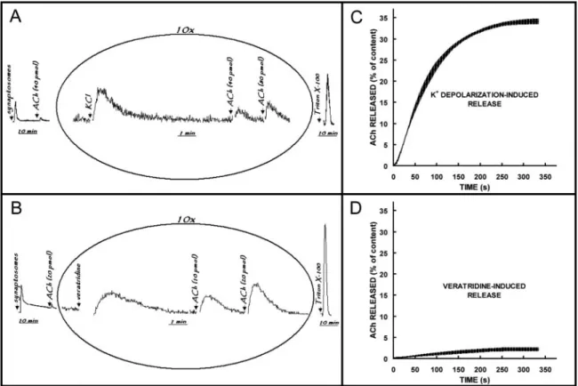
An initial strong light emission fell to a constant resting emission (Fig. 1A, B). Then, when KCl was added to the assay tube in the presence of external Ca2⫹, the light emis-sion once again showed a strong increase (Fig. 1A). In contrast, addition of veratridine produced, after a short
Figure 1. Chemiluminescent detection of endogenous ACh released from synaptosomes isolated from cuttlefish optic lobe. The synaptosomal fractions were prepared from optic lobes (8 to 12 per preparative procedure) rapidly dissected from decapitated heads. Aliquots (30l) of the synaptosomal suspension were transferred to continuously stirred luminometer tubes containing 300l of reaction mixture consisting of isosmotic Na⫹-enriched medium and the chemiluminescence agents. Representative records of chemilumines-cent response to 40 mmol l-1 KCl (A) and 40mol l-1veratridine (B). Arrows denote time point at which
synaptosomal suspension, ACh, KCl/veratridine, and Triton X-100 were added. Profiles for cumulative ACh release triggered by K⫹depolarization (C) and veratridine (D). Values are the mean⫾ SE of three experiments.
latency, a moderate and slow enhancement of light emission (Fig. 1B). Accordingly, the final ACh content of synapto-somes, detected as light emission after permeabilization with Triton X-100, was higher after treatment with veratri-dine than after KCl.
To further evaluate the effects of high potassium concen-tration and of veratridine on the neurotransmitter release, we analyzed the cumulative release profiles. The cumulative release was calculated as the area under the release curve and above baseline at a given time, divided by the area of the peaks corresponding to the total amount of released ACh during secretion and liberation after the addition of Triton X-100, which was considered to correspond to the total ACh content of synaptosomes (100%). The cumulative profile of the release induced by K⫹ depolarization (Fig. 1C) rose rapidly between 15 and 45 s (0.04 pmol ACh䡠s-1䡠mg-1
pro-tein), then slowly decreased, approaching zero in about 300 s. Hence the light emission returned to baseline in about 300 s (Fig.1A). Benech et al. (17) found that changing the external K⫹ concentration from 10 to 30 mmol l-1 KCl
evoked a transient increase in cytoplasmic Ca2⫹in synap-tosomes isolated from squid optic lobe. The Ca2⫹transient in that study was calculated to be around 340 nmol l-1and
reached its maximal level in less than 10 s, which seems to be consisent with the typical behavior of voltage-gated Ca2⫹ channels that inactivate rapidly. The increase we recorded in the chemiluminescent response to KCl addition thus seems to represent the Ca2⫹-dependent release of ACh by cuttlefish synaptosomes. We estimated the total amount of ACh released to be 0.9 ⫾ 0.3 (mean ⫾ SE) nmol䡠mg-1
protein, which corresponds to 34% ⫾ 1% of the total content of the synaptosomal fraction. When veratridine, a hydrophilic toxin that targets the type 2 receptor site of voltage-gated Na⫹ channels (18), was applied to synapto-somes, the release rate and the total amount of ACh released were significantly lower, by 10-fold, than the response induced by K⫹depolarization (Fig. 1D). Voltage-gated Na⫹ channels are responsible for the generation of action poten-tials in nerve cells. Veratridine causes Na⫹channels to stay open, producing a sustained membrane depolarization that leads to many secondary effects, including exocytosis. Con-sequently, veratridine is frequently used as a secretagogue (19).
As shown in Figure 1, the veratridine-induced release was much lower than the KCl-induced release of ACh by syn-aptosomes isolated from cuttlefish optic lobe. Putative volt-age-gated Na⫹channels were found to be expressed in the squid optic lobe, mostly at the second-order visual giant neurons (20, 21), suggesting an unequal distribution of Na⫹ channels among different cell types in the cuttlefish optic lobe. Although the chemiluminescent method has often been applied to follow ACh release from tissue slices and synaptosomal fractions, one can argue that it detects Ch in addition to ACh. In the present study, we evaluated this
possibility by measuring light emission upon stimulation with KCl or veratridine of synaptosomes pretreated with 47 mol l-1
of echothiophate iodide, an acetylcholinesterase inhibitor. When the acetylcholinesterase inhibitor was present, we observed no significant enhancement of light emission after the addition of the secretagogues (data not shown); therefore we had neglected Ch release under our experimental conditions. Synaptosomal fractions contain acetylcholinesterase that hydrolyses ACh to Ch; and Ch, which is actively transported back to the intrasynaptosomal space, is used as a substrate for ACh synthesis by choline acetyltransferase. Both enzymatic activities have been eval-uated in cuttlefish optic lobe by Bellanger et al. (22). Ac-cording to the authors, up to 16 pmol of ACh can be formed and more than 20 nmol of ACh can be degraded per milli-gram of protein within the time-scale resolution (1 s) of ACh release experiments. In the present work we explored the ability of synaptosomes to take up Ch. High-affinity Ch uptake is considered the primary mechanism by which cholinergic nerve endings accumulate Ch for ACh synthesis (23, 24).
Figure 2 depicts the time course of [3H]Ch uptake by synaptosomes isolated from cuttlefish optic lobe. The sus-pension of synaptosomes was first allowed to recover for 15 min in isosmotic Na⫹-enriched medium at room tempera-ture, in the absence or in the presence of hemicholinium-3 (HC-3), a potent and selective inhibitor of the high-affinity uptake system for Ch at presynaptic nerve terminals (25). Then, choline⫹[3H]choline ([3H]Ch) was added to obtain a
final Ch concentration of 5mol l-1and a specific radioac-tivity of 50 Ci䡠mol-1. At designated reaction times, the reactions were stopped by rapid filtration of aliquots, and the values for [3H]Ch uptake were calculated after subtrac-tion of blank values obtained by filtering aliquots of reacsubtrac-tion media containing [3H]Ch (0.25Ci䡠ml-1).
The synaptosomal fraction isolated from cuttlefish optic lobe was capable of taking up Ch when incubated in a Na⫹-enriched medium containing 460 mmol l-1 NaCl, 10
mmol l-1KCl, 55 mmol l-1MgCl2, 11 mmol l -1
CaCl2, 0.6
mmol l-1 KHCO3, 10 mmol l -1
Tris at pH 7.5 (Fig. 2A). Exposure to micromolar concentrations of HC-3 signifi-cantly decreased the amount of [3H]Ch retained by synap-tosomes throughout the assay (60 min). The results suggest that cuttlefish optic lobes contain a high-affinity Ch uptake system localized at nerve terminals, given that the degree of inhibition of [3H]Ch uptake remained constant when the HC-3 concentration was increased from 10 to 100mol l-1
(Fig. 2A, B). The time course of the high-affinity Ch uptake by cuttlefish optic lobe synaptosomes displayed a hyper-bolic trend (Fig. 2C), approaching steady-state in less than 10 min. It is evident from our results that [3H]Ch uptake by the high-affinity transporter can proceed at rates higher than 20 pmol Ch䡠min-1䡠mg-1protein, promoting the accumulation
of [3H]Ch up to 176⫾ 42 (mean ⫾ SE) pmol䡠mg-1protein
when the synaptosomes were exposed to 5mol l-1[3H]Ch.
We know of no published articles describing ACh release and Ch uptake in the CNS of cuttlefish. Dowdall and Simon (26) and Matsumura (27) showed that Ch is transported by synaptosomes isolated from squid optic lobes. The uptake of Ch by synaptosomes isolated from cuttlefish optic lobe exhibited similar characteristics, including a high initial rate of accumulation and sensitivity to HC-3.
In this study, we characterized the depolarization-trig-gered release of ACh and the high-affinity uptake of Ch (ACh precursor) in cuttlefish optic lobe synaptosomes. Once again, we wish to draw attention to the evidence, provided in our recent paper (12), that synaptosomal frac-tions isolated from cuttlefish optic lobe are suitable in vitro models to study neurotransmission. In particular, we have demonstrated that the uptake of Ch is inhibited by the selective inhibitor of the (Na⫹/K⫹)ATPase, ouabain, sug-gesting that the high-affinity Ch transporter of cuttlefish optic lobe also requires a transmembrane Na⫹ gradient to mediate active transport of Ch. In fact, dependence on Na⫹ and high sensitivity to HC-3 represent the main functional characteristics of the high-affinity choline transporter 1 (CHT1), which is specifically expressed in cholinergic neu-rons (23).
In conclusion, the results described here, together with preliminary data from earlier studies (2, 5, 7–12), provide further strong evidence that ACh performs an important physiological role in mediating neuronal signaling at syn-apses of the cuttlefish optic lobe.
Acknowledgments
We are grateful to Wyeth-Ayerst Research (USA) for a generous gift of echothiophate iodide. This work was sup-ported by Portuguese Foundation for Science and Technol-ogy (POCTI/BSE/46721/2002, SFRH/BPD/14677/2003, SFRH/BD/1079/2000, SFRH/BD/6403/2001 and SFRH/ BD/18101/2004) and a European Commission grant (to Y.D.) “LIPIDIET” QLK1-CT-2002-00172.
Literature Cited
1. Boycott, B. B. 1961. The functional organization of the brain of the cuttlefish Sepia officinalis. Proc. R. Soc.Lond. B153: 503–534. 2. Chichery, R., and J. Chanelet. 1976. Motor and behavioural
re-sponses obtained by stimulation with chronic electrodes of the optic lobe of Sepia officinalis. Brain Res. 105: 525–532.
3. Messenger, J. B. 1996. Neurotransmitters of cephalopods.
Inver-tebr. Neurosci. 2: 95–114.
4. Wells, M. J. 1962. Brain and Behavior in Cephalopods. Stanford
University Press, Stanford, CA.
5. Chrachri, A., and R. Williamson. 2003. Modulation of spontane-ous and evoked EPSCs and IPSCs in optic lobe neurons of cuttlefish
Sepia officinalis by the neuropeptide FMRF-amide. Eur. J. Neurosci.
17: 526 –536.
6. Williamson, R., and A. Chrachri. 2004. Cephalopod neural net-works. Neurosignals 13: 87–98.
7. Chichery, M. P., and R. Chichery. 1974. Histochemical study of the localization of cholinesterases in the central nervous system of
Sepia officinalis. Cell Tissue Res. 148: 551–560.
8. Bellanger, C., F. Dauphin, M. P. Chichery, and R. Chichery. 2003. Changes in cholinergic enzyme activities in the cuttlefish brain during memory formation. Physiol. Behav. 79: 749 –756.
9. Bellanger, C., M. P. Halm, F. Dauphin, and R. Chichery. 2005. In
Figure 2. Uptake of exogenous Ch by synaptosomes isolated from cuttlefish optic lobe. The synaptosomal fractions were prepared from optic lobes (12 to 30 per preparative procedure) rapidly dissected from decapitated heads. Synaptosomal suspensions were incubated in isosmotic Na⫹-enriched medium, in the absence and in the presence of 10mol l-1or 100mol l-1HC-3. After 15 min, the reactions were started by the addition of [3H]Ch
(5mol l-1 and 0.25Ci/ml), and were stopped at designated times by rapid filtration of aliquots through
glass-fiber filters (Whatman GF/B) prewashed with ice-cold isosmotical media without Ch. The filters were then washed with the same ice-cold media and plunged into scintillation cocktail (UniversolTM ES) for further radioactivity measurement by liquid scintillation. The points are the mean⫾ SE of two to five experiments for each time. (A) The time-course of [3H]Ch uptake in the absence and in the presence of HC-3. Data from panel
A are replotted in panels B and C to depict the inhibitory effect of HC-3 at 60 min (B) and the time-course of the HC-3 sensitive uptake of [3H]Ch (C).
vitro evidence and age-related changes for nicotinic but not muscarinic
acetylcholine receptors in the central nervous system of Sepia
offici-nalis. Neurosci. Lett. 387: 162–167.
10. Bellanger, C., F. Dauphin, L.P. Belzunces, C. Cancian, and R.
Chichery. 1997. Central acetylcholine synthesis and catabolism ac-tivities in the cuttlefish during aging. Brain Res. 762: 219 –222. 11. Chrachri, A., and R. Williamson. 2004. Cholinergic and
glutama-tergic spontaneous and evoked excitatory postsynaptic currents in optic lobe neurons of cuttlefish, Sepia officinalis. Brain Res. 1020: 178 –187.
12. Silva, V. S., M. A. Nunes, J. M. Cordeiro, A. I. Calejo, S. Santos,
P. Neves, A. Sykes, F. Morgado, Y. Dunant, and P. P. Gonc¸alves. 2007. Comparative effects of aluminum and ouabain on synaptoso-mal choline uptake, acetylcholine release and (Na⫹/K⫹)ATPase.
Tox-icology 236: 158 –177.
13. Crispino, M., E. Castigli, C. P. Capano, R. Martin, E. Menichini,
B. B. Kaplan, and A. Giuditta. 1993. Protein synthesis in a syn-aptosomal fraction from squid brain. Mol. Cell. Neurosci. 4: 366 –374. 14. Gornall, A. G., C. J. Bardawill, and M. M. David. 1949. Deter-mination of serum proteins by means of the biuret reaction. J. Biol.
Chem. 177: 751–766.
15. Israe¨l, M., and B. Lesbats. 1981. Continuous determination by a chemiluminescent method of acetylcholine release and compartmen-tation in Torpedo electric organ synaptosomes. J. Neurochem. 37: 1475–1483.
16. Kristofikova´, Z., H. Tejkalova´, and J. Klaschka. 2001. Amyloid beta peptide 1– 40 and the function of rat hippocampal hemicho-linium-3 sensitive choline carriers: effects of a proteolytic degradation
in vitro. Neurochem. Res. 26: 203–212.
17. Benech, J. C., P. A. Lima, J. R. Sotelo, and E. R. Brown. 2000. Ca2⫹dynamics in synaptosomes isolated from the squid optic lobe.
J. Neurosci. Res. 62: 840 – 846.
18. Wang, S.-Y., and G. K. Wang. 2003. Voltage-gated sodium chan-nels as primary targets of diverse lipid-soluble neurotoxins. Cell.
Signal. 15: 151–159.
19. Meyer, E. M., and D. H. Otero. 1989. Differential effects of 4-ami-nopyridine on acetylcholine release triggered by K⫹depolarization, veratridine, or A23187 in rat cerebral cortical synaptosomes.
Neuro-chem. Res. 14: 157–160.
20. Liu, T. I., and W. F. Gilly. 1995. Tissue distribution and subcellular localization of Na⫹channel mRNA in the nervous system of the squid,
Loligo opalescens. Receptor. Channel. 3: 243–254.
21. Sato, C., and G. Matsumoto. 1992. Primary structure of squid sodium channel deduced from the complementary DNA sequence.
Biochem. Biophys. Res. Commun. 186: 61– 68.
22. Bellanger, C., F. Dauphin, L. P. Belzunces, and R. Chichery. 1998. Parallel regional quantification of choline acetyltransferase and cho-linesterase activity in the central nervous system of an invertebrate (Sepia officinalis). Brain Res. Brain Res. Protoc. 3: 68 –75. 23. Okuda, T., and T. Haga. 2003. High-affinity choline transporter.
Neurochem. Res. 28: 483– 488.
24. Ribeiro, F. M., S. A. Black, V. F. Prado, R. J. Rylett, S. S.
Ferguson, and M. A. Prado. 2006. The “ins” and “outs” of the high-affinity choline transporter CHT1. J. Neurochem. 97: 1–12. 25. Happe, H. K., and L. C. Murrin. 1993. High-affinity choline
transport sites: use of [3H]hemicholinium-3 as a quantitative marker.
J. Neurochem. 60: 1191–1201.
26. Dowdall, M. J., and E. J. Simon. 1973. Comparative studies on synaptosomes: uptake of (N-Me-3H)choline by synaptosomes from
squid optic lobes. J. Neurochem. 21: 969 –982.
27. Matsumura, F. 1988. Deltamethrin induced changes in choline transport and phosphorylation activities in synaptosomes from the optic lobe of squid, Loligo pealei. Comp. Biochem. Physiol. C 89: 179 –183.