ANA INÊS LOURENÇO DE ALMEIDA
AN APPROACH TO MOLECULAR GENETICS OF THYROID
CANCER: FROM NOVEL MUTATIONS TO A ZEBRAFISH MODEL
Tese de candidatura ao grau de Doutor em Patologia e Genética Molecular submetida ao Instituto de Ciências Biomédicas Abel Salazar da Universidade do Porto
Orientador – Doutora Ana Paula Soares Dias Ferreira
Categoria – Professora Auxiliar, Faculdade de Medicina da Universidade do Porto e Coordenadora do Grupo Cancer Signaling &
Metabolism, IPATIMUP/Instituto de
Investigação e Inovação em Saúde
Afiliação – Faculdade de Medicina da Universidade do Porto e IPATIMUP/Instituto de Investigação e Inovação em Saúde
Coorientador – Miguel Godinho Ferreira Categoria – Coordenador do Grupo Telomere and Genome Stability, Instituto Gulbenkian de Ciência
Financial Support
The candidate was supported by a PhD fellowship (SFRH/BD/79135/2011) from Fundação para a Ciência e Tecnologia (FCT).
There are many people in my life And then there’s you,
Acknowledgments
Thank you to every one of you that came along on my five-year journey.Believe me, I haven’t forgotten each one of you. Yes, you! Thank you Paula.
Thank you Miguel.
Thank you Professor Sobrinho Simões. A BIG THANK YOU to my Dad and my Mum.
Prefácio
Eu acredito que cada pessoa tem a sua própria filosofia. Na minha filosofia há uma grande disposição para absorver a experiência da vida e com ela apreciar o mundo. Grande parte da minha satisfação pessoal provém das minhas opções profissionais. Há cinco anos atrás lancei um desafio. Candidatei-me a um programa doutoral. E tracei o meu caminho. Ao longo dele encontrei fraquezas, encontrei esperanças. E fui guardando tudo o que vi, tudo o que fiz. A minha tese reflecte o meu percurso destes cinco anos.Comecei o meu projecto de doutoramento no grupo Cancer Biology no IPATIMUP, o qual reportou uma elevada prevalência de mutações BRAF em carcinomas papilares da tiróide esporádicos e linhas celulares derivadas destes carcinomas. A procura de factores adicionais que explicassem a tumorigénese do cancro da tiróide potencialmente relacionadas com as mutações do BRAF e a descoberta de mutações no promotor da telomerase permitiram ao grupo explorar estas últimas mutações nas várias séries existentes no banco de tumores. Entretanto, enquanto outros elementos do grupo procuravam perceber mecanisticamente o efeito das mutações no promotor da telomerase, eu foquei-me no desenvolvimento de um modelo animal que permitisse inicialmente compreender o efeito de alterações frequentemente encontradas no cancro da tiróide (mutações nos genes BRAF e p53) e futuramente estudar factores adicionais que agora se sabem terem um papel relevante na tumorigénese tais como as mutações no promotor da telomerase. Por esta altura foi estabelecida uma colaboração com o grupo Telomeres and Genome Stability no IGC passando de uma ciência translacional para uma ciência básica. O desenvolvimento de um modelo para o estudo do efeito das mutações nos genes BRAF e p53 em peixe-zebra foi o fruto dessa colaboração. Deixarei um legado de ferramentas que muitos poderão usufruir.
Publications
Ao abrigo do disposto do nº 2, alínea a) do artigo 31º do Decreto-Lei n.º 115/2013 de 7 de Agosto fazem parte integrante desta tese de doutoramento os seguintes trabalhos já publicados ou submetidos para publicação:Artigo I - Vinagre J, Almeida A, Pópulo H, Batista R, Lyra J, Pinto V, Coelho R, Celestino R, Prazeres H, Lima L, Melo M, da Rocha AG, Preto A, Castro P, Castro L, Pardal F, Lopes JM, Santos LL, Reis RM, Cameselle-Teijeiro J, Sobrinho-Simões M, Lima J, Máximo V, Soares P. 2013. Frequency of TERT promoter mutations in human cancers. Nat Commun. 4:2185.
Artigo II – Almeida A, Sobrinho-Simões M, Ferreira MG, Soares P. (Submetido).
O seguinte capítulo de livro não faz parte do corpo principal de resultados desta tese, mas é parte integrante da mesma, tendo sido utilizado na sua Introdução e Discussão.
Appendix I - Almeida AL, Boaventura P, Soares P, Clinical Management of Thyroid Cancer: Etiopathogenic factors of thyroid cancer, Pages 46-62, Future Medicine. 2013
Table of contents
Abbreviations iv
Abstract ix
Resumo xi
Chapter I – Introduction 1
I.1 Thyroid gland 2
I.1.1 Thyroid physiology 2
I.1.2 Thyroid disorders 3
I.1.2.1 Goiter 4
I.1.2.2 Neoplasias 5
Papillary thyroid cancer 7
Molecular genetics of papillary thyroid cancer 9 Signaling pathways altered in papillary thyroid carcinomas 19
I.2 Zebrafish as a model system 23
I.2.1 Zebrafish in Cancer Research 23
I.2.2 Studies on zebrafish thyroid physiology and function 25
I.3 Aims 28
Chapter II – Material & Methods 30
II.1 Plasmid cloning 31
II.1.1 p5E-tg promoter plasmid 31
II.1.2 pME-mCherry and p3E-polyA plasmids 31
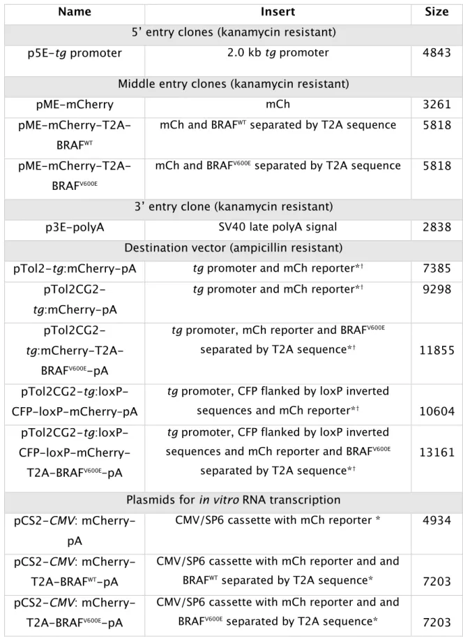
II.1.3 pME-mCherry-T2A-BRAFWT and
pME-mCherry-T2A-BRAFV600E plasmids
32
II.1.4 pTol2A2-tg:mCherry–pA and pTol2CG2-tg:mCherry–pA plasmids
32
II.1.5 pTol2CG2-tg:mCherry-T2A- BRAFV600E–pA plasmids 33
II.1.6 tg:loxP-CFP-loxP-mCherry–pA and pTol2CG2-tg:loxP-CFP-loxP-mCherry-T2A-BRAFV600E–pA plasmids
34
II.1.8 pCS2-CMV:mCherry-T2A-BRAFWT-pA and
pCS2-CMV:mCherry-T2A-BRAFV600E-pA plasmids
36
II.1.9 Summary of the plasmids generated 36
II.2 Cloning-auxiliary techniques 38
II.2.1 PCR 38
II.2.2 DNA sequencing 38
II.2.3 DNA quantification 38
II.2.4 Restriction digestion and ligation 39
II.2.5 Isolation of DNA by agarose gel electrophoresis 39 II.2.6 Plasmid transformation in competent E. Coli 39 II.2.7 Plasmid growth in solid and liquid cultures 40
II.2.8 Plasmid purification 40
II.3 Capped mRNA synthesis 40
II.3.1 Capped transposase mRNA synthesis 40
II.3.2 Capped mCherry mRNA, mCherry-T2A-BRAFWT mRNA and
mCherry-T2A-BRAFV600E mRNA synthesis
41
II.4 Microinjections 41
II.4.1 DNA plasmid microinjections 41
II.4.2 Capped mRNA microinjections 42
II.5 Transgenesis 42
II.5.1 WT lines 43
II.5.2 tp53M214K lines 44
II.6 Fish strains and husbandry 45
II.7 Screening and Imaging 46
II.8 Fin clip and gDNA extraction 46
II.9 Genotyping 46
II.10 Histopathology 47
II.11 Measurement of standard length in larvae 47
II.12 Measurement of body mass index (BMI) 48
II.13 Measurement of thyroid volume in adult fish 48
II.14 Preparation of embryo lysates 49
II.15 Dissection of thyroid tissue in adult zebrafish 49
II.16 Preparation of tissue lysates 49
II.17 Immunoblotting 50
II.18 Heatshock and drug treatment 51
Chapter III – Results 52 III.1 Frequency of TERT promoter mutations in human cancers 53 III.2 Targeted expression of BRAFV600E in thyroid cells of transgenic
zebrafish induces hyperplasia reverted by loss of WT p53
83
Chapter IV – Discussion 139
IV.1 Frequency of TERT promoter mutations in human cancers 140 IV.2 Targeted expression of BRAFV600E in thyroid cells of transgenic
zebrafish induces hyperplasia reverted by loss of WT p53
143
Chapter V – Conclusion 151
References 154
Abbreviations
4-OHT 4-Hydroxyl-TamoxifenAKAP9 A Kinase (PRKA) Anchor Protein 9
AKT V-Akt Murine Thymoma Viral Oncogene Homolog ALK Anaplastic Lymphoma Receptor Tyrosine Kinase AMP Adenosine Monophosphate
ARAF A-Raf Proto-Oncogene, Serine/Threonine Kinase ATC Anaplastic Thyroid Cancer
ATP Adenosine Triphosphate attL Left End of Prophage attR Right End of Prophage
BAC Bacterial Artificial Chromosome BCL2 B-Cell Lymphoma 2
BF Bright Field BMI Body Mass Index
bp Base Pairs
BRAF B-type Raf kinase
cAMP Cyclic Adenosine Monophosphate CCDC6 Coiled-Coil Domain Containing 6 CDE Cell Cycle-Dependent Element CDK Cyclin-Dependent Kinases
CDKN2A Cyclin-Dependent Kinase Inhibitor 2A CFP Cyan Fluorescent Protein
CHR Cell Cycle Genes Homology cmcl2 Cardiac Myosin Light Chain CMV Cytomegalovirus
CNS Central Nervous System CPT Carcinoma Papilar da Tiróide
CRAF C-Raf Proto-Oncogene, Serine/Threonine Kinase Cre Cre Recombinase
CreER Cre Recombinase-Estrogen Receptor ctsb Cathepsin B
DBD DNA-Binding Domain DEPC Diethylpyrocarbonate
dpf Days Post Fertilization DNA Deoxyribonucleic Acid dpi Days Post Injection
EDTA (Ethylenedinitrilo)tetraacetic Acid EGFP Enhanced Green Fluorescent Protein EGTA Ethylene Glycol Tetraacetic Acid Erg ETS-Related gene
ERK Extracellular Regulated Kinase
ETS Erythroblast Transformation-Specific FCT Fundação para a Ciência e Tecnologia
Fos FBJ murine Osteosarcoma Viral Oncogene Homolog FTC Follicular Thyroid Carcinoma
GABP GA-Binding Protein GBM Glioblastomas
GDP Guanosine-5'-Diphosphate GFRα GDNF family receptor-α
GIST Gastrointestinal Stromal Tumor GTP Guanosine-5'-Triphosphate HE Hematoxylin-Eosin
hhex Hematopoietically Expressed Homeobox hpf Hours Post Fertilization
hpi Hours Post Injection
HPT Hypothalamus-Pituitary-Thyroid HRP Horseradish Peroxidase
hspl70 Heat shock cognate 70-kd protein, like IGC Instituto Gulbenkian de Ciência
IGFBP7 Insulin-like Growth Factor-Binding Protein 7 IgG Immunoglobulin G
IPATIMUP Institute of Molecular Pathology and Immunology at the University of Porto
JUN Jun proto-oncogene
LB Lysogeny Broth
LKB1 Tumor Suppressor Liver Kinase B1 LOF Loss Of Function
MAPK Mitogen-Activated Protein Kinases mCh Monomeric mCherry
MCS Multicloning Site
MDM2 MDM2 proto-oncogene, E3 ubiquitin protein ligase ME Middle Entry Clone
MEK MAP Kinase 1
mpf Months Post Fertilization mRNA Messenger Ribonucleic Acid MS222 Tricaine Methanesulfonate
mTOR Mechanistic Target Of Rapamycin
mTORC1 Mammalian Target Of Rapamycin Complex 1 mTORC2 Mammalian Target Of Rapamycin Complex 2
Myc V-Myc Avian Myelocytomatosis Viral Oncogene Homolog NCOA4 Nuclear Receptor Coactivator 4
NICD Notch Intracellular Domain NIS Sodium-Iodine Symporter nk2-1a NK2 Homeobox 1a
NRAS Neuroblastoma RAS Viral (v-ras) Oncogene Homolog NTRK1 Neurotrophic Tyrosine Kinase, Receptor, Type 1
pA PolyA
PAX2.1 Paired Box Gene 2.1 PAX8 Paired Box 8
PCCL3 Rat Thyroid Follicular Cell Line PCNA Proliferating Cell Nuclear Antigen PCR Polymerase Chain Reaction
PDK1 Pyruvate Dehydrogenase Kinase, Isozyme 1 PDTC Poorly Differentiated Thyroid Cancer
phf20l1 PHD Finger Protein 20-Like 1
PI3K Phosphatidylinositol-4,5-Bisphosphate 3-Kinase
PIK3CA Phosphatidylinositol-4,5-Bisphosphate 3-Kinase, Catalytic Subunit Alpha
PKA Protein Kinase A
PMSF PhenylMethylSulfonyl Fluoride
PPAR Peroxisome Proliferator-Activated Receptor
PPARG Peroxisome Proliferator-Activated Receptor Gamma
PRKAR1A Protein Kinase, cAMP-Dependent, Regulatory, Type I, alpha PSI Pounds per Square Inch
PtdIns Phosphatidylinositol
PTEN Phosphatase and Tensin Homolog PVDF Polyvinylidene Fluoride
Rag2 Recombination Activating Gene 2 RAS Rat Sarcoma Viral Oncogene Homolog RET Rearranged during Transfection
RIPA Radioimmunoprecipitation Assay Buffer RPM Revolutions Per Minute
SAP Shrimp Alkaline Phosphatase SDS Sodium Dodecyl Sulfate
SDS PAGE Sodium Dodecyl Sulfate Polyacrylamide Gel Electrophoresis
Ser Serine
SETDB1 SET Domain, Bifurcated 1
siRNA Small Interfering Ribonucleic Acid
SL Standard Length
slc5a5 Solute Carrier Family 5 (Sodium/Iodide Cotransporter), Member 5
SV40 Simian Vacuolating Virus 40 T2A Thoseaasigna virus 2A T3 Tri-Iodothyronine
T4 Thyroxine
TAE Tris base, acetic acid and EDTA solution T-ALL T-cell Acute Lymphoblastic Leukemia
TAM Tamoxifen
TBE Tris base, boric acid and EDTA solution TBS Tris-Buffered Saline
TERT Telomerase Reverse Transcriptase
tg Thyroglobulin
TGB Thyroxine-Binding Globulin TGFβ Transforming Growth Factor Beta
TH Thyroid Hormone
Thr Threonine
TK Tyrosine Kinase TPO Thyroid Peroxidase
TRAP Telomeric Repeat Amplification Protocol TRH Thyrotropin-Releasing Hormone
Tris-Cl Tris(hydroxymethyl)aminomethane Chloride TSH Thyroid Stimulating Hormone
TshR Thyroid Stimulating Hormone Receptor WDTC Well-Differentiated Thyroid Cancer WHO World Health Organization
Abstract
Thyroid diseases are extremely frequent and are most often of benign nature. Thyroid cancer is the most common endocrine malignancy in humans and the majority of tumors harbor genetic alterations such as the BRAF mutation. Most of this mutation is an activating mutation in the kinase domain of the BRAF. In sporadic papillary thyroid carcinomas (PTCs), BRAF gene mutations are found in 29%-83% of all cases and almost never co-exist with RAS mutations or RET (RET/PTC) and NTRK1 rearrangements. As a result of BRAF mutation, the MAPK pathway is activated and cellular processes such as proliferation, survival, motility and invasion are promoted. Recently, mutations in the telomerase reverse transcriptase (TERT) promoter have been described in thyroid cancer and considered one of the possible mechanisms that underlies TERT reexpression in several types of human tumors including those of the thyroid.
In the first part of the thesis, I describe the study regarding TERT promoter mutations in which I was deeply involved. Our study highlighted the presence of recurrent somatic mutations in the TERT promoter in cancers of the central nervous system (43%), bladder (59%), thyroid (follicular cell-derived, 10%) and skin (melanoma, 29%). Concerning thyroid cancer, the presence of TERT promoter mutations was found to be significantly associated with higher TERT mRNA expression and with older age of the patients. We concluded that TERT promoter mutations are relatively frequent in several specific types of human cancer and that such mutations may enhance expression of telomerase.
Over the years, thyroid cancer has been studied using mice models. These models have provided evidence showing that thyroid-specific expression of BRAFV600E
induced goiter as well as invasive PTCs which progress to poorly differentiated carcinoma closely recapitulating some human thyroid tumor phenotypes. Very successful, mice models of thyroid cancer are being used to explore molecular mechanisms involved in thyroid tumorigenesis. Mice models can also be used to monitor tumors and to perform drug screening in the setting of thyroid cancer but such tasks remain time-consuming.
In the second part of the thesis, I developed a thyroid-targeted BRAFV600E
during all stages of development up to 12 months of age. I observed that thyroid-specific expression of BRAFV600E induced abnormal thyroid morphogenesis early in
life that developed later on into hyperplasia by ~2-3 months of age and colloid goiter by 12 months of age. BRAFV600E–expressing cells disclosed upregulation of
proliferation, concomitant with MAPK pathway activation, and there was promotion of apoptosis. High levels of p53 suggested that this protein may be restraining progression to malignancy. Loss of WT p53 using a tp53M214K zebrafish prevented
impairment of thyroid morphogenesis induced by BRAFV600E and surprisingly no
evidence of thyroid hyperplasia, goiter and/or neoplasia was detected in those animals up to 12 months of age. tp53M214K BRAFV600E–expressing cells were low
proliferative, consistent with downregulation of the MAPK pathway; suppression of apoptosis was also observed.
In conclusion, my work showed that TERT promoter mutations are relatively frequent in specific types of human cancer, including those of the thyroid, and that may enhance telomerase expression. Also, thyroid-specific expression of BRAFV600E
induces hyperplasia and colloid goiter in zebrafish and together with the absence of WT p53, BRAFV600E was not able to develop thyroid cancer. This data provides
evidence that BRAF activation is sufficient for thyroid cell transformation and that BRAF and p53 pathways must interact genetically in zebrafish thyroid.
Resumo
As doenças na tiróide são extremamente frequentes e são geralmente de natureza benigna. O cancro da tiróide é o tumor endócrino maligno mais comum em humanos e a maioria destes tumores possui alterações genéticas tais como a mutação no gene BRAF. A maioria das mutações do BRAF activam o domínio de cinase da proteína BRAF. Em carcinomas papilares da tiróide (CPT) esporádicos, as mutações no gene BRAF são encontradas em 29% a 83% do total de casos e quase nunca co-existem com as mutações do RAS e rearranjos do RET (RET/PTC) e NTRK1. Como resultado das mutações do BRAF, a via de sinalização das MAP cinases é activada e processos celulares, tais como proliferação, sobrevivência, motilidade e invasão, são promovidos. Recentemente, foram descritas mutações no promotor da telomerase transcriptase reversa (TERT) e estas são consideradas um dos possível mecanismos de reexpressão da telomerase em vários tipos de cancro humano incluindo os da tiróide.
Na primeira parte da tese eu descrevo o estudo relativo às mutações no promotor da TERT no qual eu estive envolvida. O nosso estudo realçou a presença de mutações somáticas recorrentes no promotor da TERT em tumores como os do sistema nervoso central (43%), da bexiga (59%), da tiróide com origem nas células foliculares (10%) e da pele (melanoma) (29%). Relativamente ao cancro da tiróide, foi encontrada uma associação significativa entre a presença de mutações no promotor da TERT e níveis elevados de expressão de mRNA e também uma associação com pacientes mais velhos. Concluímos que as mutações no promotor da TERT são relativamente frequentes em determinados tipos de cancro humanos e que estas mutações podem aumentar a expressão da TERT.
Ao longo dos anos, o cancro da tiróide tem sido estudado usando modelos de ratinho. Estes modelos deram evidências que demonstram que a expressão específica de BRAFV600E induziu bócio bem como CPT invasivos que progrediram
para carcinomas pouco diferenciados recapitulando alguns dos fenótipos dos tumores de tiróide humanos. Os modelos de ratinho ainda são usados para explorar mecanismos moleculares envolvidos na tumorigénese da tiróide. Estes modelos podem também ser usados para monitorizar tumores e para realizar ensaios de drogas no contexto do cancro da tiróide mas estas tarefas são morosas.
Na segunda parte do tese eu desenvolvi uma linha transgénica com expressão específica de BRAFV600E na tiróide de peixe-zebra e avaliei os fenótipos na tiróide
durante todos os estádios de desenvolvimento e até aos doze meses de idade. Observei que a expressão específica de BRAFV600E na tiróide induziu uma
morfogénese anormal deste tecido em estádios iniciais que se desenvolvem em hiperplasia aos 2-3 meses de idade e bócio colóide ao fim de 12 meses. Células da tiróide que expressavam BRAFV600E tinham uma sobreregulação da proliferação,
concomitante com a activação da via de sinalização das MAP cinases, e foi observada indução da apoptose. Nível elevados de p53 sugerem que esta proteína pode ter contido a progressão para malignidade. Perda da proteína selvagem de p53, usando uma linha homozigota para a mutação M214K do tp53, preveniu morfogénese anormal da tiróide induzida pelo BRAFV600E e surpreendentemente não
foram encontradas evidências de hiperplasia, bócio e/ou carcinomas em peixes até doze meses de idade. Células da tiróide que expressavam BRAFV600E eram pouco
proliferativas, consistente com desregulação da via de sinalização das MAP cinases, e foi observada supressão da apoptose.
Em conclusão, o meu trabalho demonstrou que as mutações no promotor da TERT são relativamente frequentes em determinados tipos de cancro humanos, incluindo os da tiróide, e podem aumentar a expressão da TERT. A expressão específica de
BRAFV600E induziu hiperplasia e bócio colóide em peixe-zebra e que em conjunto
com a ausência da proteína p53, o BRAFV600E não foi capaz de induzir cancro na
tiróide. Estas observações demonstram evidências de que a activação do BRAF é suficiente para a transformação de células da tiróide e que as vias de sinalização da qual fazem parte o BRAF e o p53 devem interagir geneticamente na tiróide do peixe-zebra.
Chapter I
THE HUMAN ENDOCRINE SYSTEM comprises part of the body’s communication system connecting the brain to the organs which in turn control the metabolism, growth and reproduction. Tight control of the system is possible through complex feedback mechanisms that maintains homeostasis. Any disruption to an endocrine gland or to the feedback mechanisms may result in endocrine disturbance. Ultimately, cancer may either contribute or be the outcome of such disturbance.
I.1 Thyroid gland
The human thyroid gland is a butterfly-shaped gland located on the trachea and comprises two lobes connected by an isthmus (VanPutte et al., 2010). The gland is highly vascularized and is one of the largest endocrine glands in the human body (VanPutte et al., 2010). The thyroid gland comprises numerous and varying sized follicles consisting of a thin-layer of cuboidal epithelial cells and a central lumen. The lumen is filled with a homogeneous protein-rich colloid named thyroglobulin which is essential to thyroid hormone (TH) synthesis (Manson et al., 1973; VanPutte et al., 2010). Two hormones are produced in the thyroid gland by the follicular cells in response to thyroid-stimulating hormone (TSH) released from the pituitary: tri-iodothyronine (T3) and thyroxine (T4). These hormones have an effect on all body systems at all stages of life regulating the basal metabolic rate and tissue growth and maturation (Manson et al., 1973; Kumar et al., 2005; VanPutte et al., 2010). Parafollicular cells secreting calcitonin are also found in clusters surrounding the follicles and in the connective tissue (VanPutte et al., 2010).
I.1.1 Thyroid physiology
Thyroid follicles are the factory and the storage of THs. The presence of TSH is indispensable for the synthesis and secretion of THs as well as an adequate iodide nutrition.
The first step in the synthesis of THs is the uptake of iodide by sodium-iodine symporter (NIS) which is converted to iodine and then condensed onto tyrosine residues from the backbone of thyroglobulin, a protein produced inside the thyroid cells. The newly formed iodothyroglobulin can be either mono-iodinated or di-iodinated. When coupled, two di-iodotyrosine molecules result in the formation of
T4 whereas a di-iodotyrosine coupled with a mono-iodotyrosine results in T3. Although the T3 is more biologically active than the T4, the production of T3 occurs preferentially outside the thyroid gland by peripherical conversion from T4. THs are stored inside the thyroid follicles composing the majority of the colloid material. T3 and T4 are released by proteolysis from the thyroid to the bloodstream where they bind to TH binding proteins namely the thyroxin binding globulin (TBG) (Figure 1) (Kumar et al., 2005; Brix et al., 2011).
Figure 1. TH synthesis. TSH signaling via the TSH receptor controls TH synthesis. NIS at the basolateral membrane takes up iodide from the blood. Iodide is organified in the tyrosyl residues of tg in a reaction catalyzed by thyroid peroxidase (TPO). T3, and T4 are stored in colloid until they are released into the blood.
I.1.2 Thyroid disorders
Thyroid disorders can range from an enlarged thyroid gland that does not need treatment to thyroid cancer. The most common thyroid problems include goiter and benign thyroid nodules. Also relatively frequent, and clinically more evident, is the abnormal production of TH that can be classified into two groups: hyperthyroidism and hypothyroidism (Manson et al., 1973; VanPutte et al., 2010).
In hyperthyroidism, the thyroid gland is overactive producing high levels of thyroid hormones and speeding up the metabolism. Graves’ disease also known as toxic diffuse goiter is the most common cause of hyperthyroidism (Manson et al., 1973; VanPutte et al., 2010). In hypothyroidism, the thyroid gland is underactive producing inadequate levels of thyroid hormones and slowing down the metabolism. Hashimoto’s thyroiditis, congenital hypothyroidism and irradiation are some of the causes of hypothyroidism (Manson et al., 1973; VanPutte et al., 2010).
The proper treatment of hyperthyroidism and hypothyroidism depends on the symptoms of the disease and the aetiology. In hyperthyroidism, treatments include thiouracils or thioamides, radioiodine therapy, thyroidectomy, radioactive iodine and/or beta blockers. In hypothyroidism, levothyroxine is a hormone replacement used for treatment (VanPutte et al., 2010).
I.1.2.1 Goiter
A goiter is an enlarged thyroid gland, it can be either diffuse or nodular and it may extend into the retrosternal space with or without substantial anterior enlargement (Kumar et al., 2005; Lam et al., 2014).
A deficiency in iodine intake or in TH synthesis leads to an increased TSH production which in turn sustains increased cellularity and hyperplasia of the thyroid gland as an attempt to normalize the levels of the TH. TH deficiency can be due to defects on hormone synthesis, iodine deficiency and goitrogens (Figure 2). Also, a goiter may appear as a result of the stimulation of the thyroid gland by thyroid stimulating hormone receptor (TshR) agonists such as TSH receptor antibodies, pituitary resistance to thyroid hormone, adenomas of the hypothalamus or pituitary gland and human chorionic gonadotropin-producing tumours (Kumar et al., 2005; Lam et al., 2014).
Figure 2. Relation between hypothalamus-pituitary-thyroid (HPT) axis and human goiter development. Iodine deficiency and/or goitrogens disrupt the TH synthesis in many ways. Upon insufficient TH levels, the negative feedback inhibition is lost resulting in increased secretion of tropic hormones (TRH and TSH). High TSH levels stimulate thyroid cells promoting goiter. Also, TSH receptor agonists can ilicitly mimick the biological activity of TSH.
Small benign euthyroid goiters do not require any treatment, however their size may be reduced with levothyroxine suppressive therapy. Large and complicated goiters usually require surgical and/or radiation treatment followed by TH replacement (Kumar et al., 2005; Lam et al., 2014).
I.1.2.2 Neoplasias
Thyroid cancer is the 16th most common cancer worldwide and accounts for
approximately 2% of total of human malignancies with around 298.000 new cases diagnosed in 2012 worldwide (latest data reported) (Nikiforov, 2012; Ferlay et al., 2013). Incidence rates of thyroid cancer are highest in Northern America and other developed countries and lowest in western Africa but this is partly due to data quality and reflects different prevalence of risk factors and screening and
diagnostic methods used (Nikiforov, 2012; Ferlay et al., 2013). Although incidence rates have increased in most countries, mortality rates have decreased and remained low. There were approximately 37.800 deaths worldwide from thyroid cancer in 2012 corresponding to 0.5% of total cancer deaths (Nikiforov, 2012; Ferlay et al., 2013).
Thyroid cancer represents the most common type of endocrine malignancy although it is relatively rare when compared to benign thyroid tumors. Thyroid cancer occurs primarily in youngsters and middle aged adults whilst in children is rare. The incidence of thyroid cancer is four times higher in females than in males and this sex difference it less pronounced in children and older adults (DeLellis et al., 2004; Nikiforov, 2012).
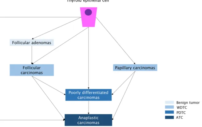
According to the current World Health Organization (WHO) classification, thyroid tumors can be subdivided into primary and secondary or metastatic tumors (DeLellis et al., 2004). While most of the primary tumors are epithelial and originated from the thyroid follicular cells, a lower number of cancers derive from neural crest-derived parafollicular C cells. Follicular thyroid cell-derived tumors can be benign (follicular adenomas) or malignant (well differentiated follicular and papillary carcinomas, poorly differentiated and undifferentiated/anaplastic carcinomas) and tumors with an uncertain malignant potential (Soares et al., 2011; Nikiforov, 2012) (Figure 3). Also found are the medullary carcinomas which have a C-cell origin (DeLellis et al., 2004). Among malignant tumors, papillary thyroid carcinomas (PTC) constitute about 80% of all thyroid cancer cases and the increased incidence observed in thyroid cancer is mostly due to PTCs. The follicular thyroid carcinomas (FTC) represent 15% and medullary and anaplastic carcinomas about 3% and 2%, respectively, of all thyroid cancer cases. For these tumors, the incidence has not changed significantly (DeLellis et al., 2004; Nikiforov, 2012).
Figure 3. Follicular thyroid cell-derived tumors and its putative progression. Scheme of step-wise dedifferentiation of follicular cell-derived thyroid cancer from benign to malignant tumors.
Conventional surgical thyroidectomy and adjuvant ablation by radioiodine treatment are the standard treatments for follicular thyroid cell-derived tumors. These treatments are not curative in all cases and a big effort is still being made towards understanding the molecular pathogenesis of thyroid cancer, namely of those that show clinical progression (DeLellis et al., 2004; Nikiforov, 2008; Nikiforov, 2012).
Papillary thyroid cancer
The PTC is a malignant epithelial tumor that shows evidence of follicular cell differentiation and is characterized histologically by distinctive nuclear features (DeLellis et al., 2004; Kumar et al., 2005; Nikiforov, 2012).
PTCs are rare in childhood but still are the most common pediatric thyroid tumor. Incidence of PTCs begins to rise in the second decade of life up to the fifth and
sixth decades. Female to male ratio is as high as 4:1 but in older adults the prevalence for females is less pronounced (DeLellis et al., 2004; Kumar et al., 2005; Nikiforov, 2012). Increased accuracy in the diagnosis and changes in the histological WHO criteria have contributed to a very high prevalence of the papillary type among other thyroid carcinomas (DeLellis et al., 2004). Nevertheless, one cannot exclude a real increase in the incidence of PTCs due to iodine supplementation or exposure to radiation and endocrine disruptors especially during childhood (Farahati et al., 2004; Tronko et al., 2006; Frasca et al., 2008; Almeida et al., 2013). Indeed, development of PTC is linked to environmental (mainly radiation exposure), genetic, hormonal factors and interactions between them. Radiation either external or internal from radioactive iodine is a major risk factor and children are the most susceptible as thyroid gland grows during childhood (DeLellis et al., 2004; Kumar et al., 2005; Nikiforov, 2012; Almeida et al., 2013).
PTC is presented as a cold mass on radioactive scan or as a cervical lymphadenopathy. On gross examination, PTCs present a variety of patterns but most masses are grey-white and firm with irregular borders that may infiltrate the surrounding thyroid tissue. Tumor size ranges from less than 1mm to several centimeters and multicentricity and cystic change are commonly observed (DeLellis et al., 2004; Kumar et al., 2005; Nikiforov, 2012).
The diagnosis of a PTC relies mainly in the cytological features observed in the nuclei and on the architecture. This includes enlargement, oval shape, elongation and overlapping of the nuclei, chromatin clearing, irregular nuclear contours, nuclear pseudoinclusions and nuclear grooves. Papillary growth pattern with branching is frequently seen but not required for diagnosis. Also found are intratumoral sclerosis, peritumoral lymphocytic infiltration and psamomma bodies (DeLellis et al., 2004; Kumar et al., 2005; Nikiforov, 2012).
Some PTCs present particular features which represent specific histological variants. The two most frequent variants are the papillary microcarcinomas (30-40%) usually found incidentally and with a tumor size less than 1cm and the follicular variant (15-20%) which has a follicular pattern instead of a papillary pattern, very often absent. Other variants, exhibiting variable clinical behaviors, are less prevalent (solid, tall-cell, sclerosing, etc.) (DeLellis et al., 2004; Kumar et al., 2005; Lloyd et al., 2011; Nikiforov, 2012).
Prognosis is overall excellent with a 20-year survival of 98% and a mortality rate of less that 1% (DeLellis et al., 2004). However, rate of recurrence is highly variable ranging from 5% to 40%. Commonly, PTCs metastasize via lymphatic vessels often in the cervical lymph nodes and rarely to the lungs and bones as bilateral, small and diffuse lesions (DeLellis et al., 2004; Kumar et al., 2005; Nikiforov, 2012). Treatment of PTCs remains surgical. Total thyroidectomy is advised for tumors greater that 1cm and since well-differentiated PTCs are radiosensitive, iodine-131 ablation may be employed as adjuvant therapy (DeLellis et al., 2004; Kumar et al., 2005; Nikiforov, 2012).
Molecular genetics of papillary thyroid cancer
The rapidly understanding of cancer molecular genetics has led not only to a deep insight of cell biology but also to seek prevention and new treatments for cancer. In thyroid cancer as in other human cancers, the success of molecular genetics to further contribute with novel therapeutic approaches, such as targeted molecular therapy, is yet to be fully achieved.
PTCs are characterized by nonoverlapping genetic alterations in more than 70% of the cases. These alterations include rearrangements in the tyrosine kinase (TK) receptors Rearranged-during-transfection (RET), Neurotrophic Tyrosine Kinase, Receptor, Type 1 (NTRK1), Anaplastic Lymphoma Receptor Tyrosine Kinase (ALK) and A Kinase (PRKA) Anchor Protein 9/B-type Raf kinase (AKAP9/BRAF) and also point mutations in Rat Sarcoma Viral Oncogene Homolog (RAS) and BRAF genes. Ultimately, most cases will lead to an aberrant activation of the RAS-RAF-MEK-ERK kinase pathway (DeLellis et al., 2004; Kumar et al., 2005; Nikiforov, 2012).
Despite the high frequency of chromosomal rearrangements in PTCs, the predisposition of follicular thyroid cells to undergo those rearrangements is not fully understood. Chromosomal rearrangements are likely to reflect the sensitivity of the follicular thyroid cells to ionizing radiation and/or the intrinsic capacity of cells to repair radiation-induced deoxyribonucleic acid (DNA) double-strand breaks and proneness to aberrant DNA repair instead of apoptosis (Galleani et al., 2014).
BRAF mutationsBRAF serine-threonine kinase belongs to the RAF family together with A-Raf Oncogene, Serine/Threonine Kinase (ARAF) and C-Raf Proto-Oncogene, Serine/Threonine Kinase (CRAF). BRAF is an intracellular effector mediating the response to growth factor signaling through the Mitogen-Activated Protein Kinases (MAPK) pathway which controls cell proliferation and differentiation (McKay et al., 2007). From all the RAF proteins, BRAF has the highest basal kinase activity and it is the most potent activator of the MAPK signaling pathway (Marais et al., 1997).
Most mutations in BRAF are activating mutations in the kinase domain. In sporadic PTCs, BRAF gene mutations are found in 29%-83% of the cases and are almost exclusive to RAS mutations, RET (RET/PTC) and NTRK1 rearrangements (Soares et al., 2003; Xing, 2005; Frasca et al., 2008).
Up to 90% of the BRAF gene mutations are a thymine-to-adenosine transversion at position 1799 in exon 15 of the BRAF gene leading to the substitution of a valine (V) by glutamic acid (E) at the residue 600 (V600E) of the protein (Garnett et al., 2004; Frasca et al., 2008). When BRAF is in its inactive and dephosphorylated conformation, the adenosine triphosphate (ATP)-binding site and the phosphoregulatory activation loop are bound through hydrophobic interactions and the kinase domain of BRAF is closed. In the mutant form of BRAF, glutamic acid introduces a negative charge adjacent to the Thr599 residue mimicking phosphorylations at Thr599 and Ser602 residues. This disrupts the hydrophobic interactions and allows new interactions that maintain the protein in a catalytically competent conformation resulting in a continuous phosphorylation of MAP Kinase 1 (MEK), a downstream effector (Dhillon et al., 2004; Wan et al., 2004).
BRAFV600E mutation are highly frequent in classical PTCs and in the tall-cell variant
but rare in the follicular variant of PTCs. Additionally, BRAFV600E mutation is found
in poorly differentiated and undifferentiated carcinomas arising from preexisting PTCs which may still contain well-differentiated areas (Nikiforova et al., 2003; Begum et al., 2004; Soares et al., 2004; Frasca et al., 2008).
BRAFV600E mutation is also found in many other human cancers with a high
prevalence in cutaneous melanoma and in a smaller subset of serous ovarian carcinoma and colorectal carcinoma (Davies et al., 2002).
Besides the BRAFV600E mutation, other rare BRAF mutations are found comprising
nucleotides around the codon 600 which also constitutively activate the BRAF kinase (Soares et al., 2003; Xu et al., 2003). One example is the BRAFK601E mutation
which lead to the substitution of a lysine (K) by glutamic acid (E) at the residue 601 (K601E). This mutation has a higher prevalence in the follicular variant of PTCs (Trovisco et al., 2005).
BRAFV600E and thyroid tumorigenesis The initial evidence that BRAFV600E is required
for the cell proliferation, transformation and tumorigenicity of follicular thyroid cells was demonstrated in a xenograft mice model (Liu et al., 2007) although many studies had already supported the role of that BRAF mutation in tumor initiation. By stably and specifically knocking down BRAF using small interfering ribonucleic acid (siRNA) expression vectors in BRAF mutation-harboring papillary thyroid cells, suppression of cell proliferation, colony formation in monolayer culture and anchorage-independent cell growth in soft agar was achieved (Liu et al., 2007). Furthermore, it was observed inhibition of in vivo tumorigenicity and tumor growth of BRAF mutation-harboring papillary thyroid cells after stable knockdown of BRAF and xenografted in nude mice. These data suggest that BRAFV600E is a maintainer of
PTCs phenotype (Liu et al., 2007).
Targeted expression of the BRAFV600E in follicular thyroid cells of transgenic FVB/N
mice with a bovine thyroglobulin promoter was also studied. BRAF induced thyroid dysfunction which was compensated by increased levels of TSH and goiter development. In this model, multifocal tumors involving both lobes of the thyroid gland with mixed papillary and follicular growth pattern were observed in 12 and 22-week-old mice. PTCs presented the classical architecture, tall-cell features and a high potential for invasiveness. Tumors from one of the transgenic lines progressed into poorly differentiated carcinomas (Knauf et al., 2005).
PTCs were also observed in a thyrocyte-specific knock-in of BRAFV600E in mice but
with a very short latency and complete penetrance by 3 weeks. When this model was crossed with a TshR knockout mice to genetically ablate TSH signaling, thyroid growth was reduced and low-grade PTCs were only observed by 9 weeks of age (Franco et al., 2011).
Along with these observations, a study has explored the consequences of induced BRAFV600E expression in the thyroid of adult mice. Shortly after inducing BRAFV600E
expression, one-month-old mice developed hypothyroidism and a dramatically enlarged, goiterous and hypercellular thyroid gland that was 10 times larger than controls and up to 300 times larger by 12 months. Only after 6-to-9 months, all
mice developed PTC that closely recapitulated the phenotype in humans. Strikingly, treatment of these mice with a MEK inhibitor reduced thyroid size, restored the production of THs and inhibited tumorigenesis (Charles et al., 2011).
More evidence of BRAFV600E involvement in mice thyroid tumorigenesis was observed
when BRAFV600E expression was induced in follicular thyroid cells in a
doxycycline-inducible manner. As early as one week after doxycycline treatment, development of highly penetrant and high-grade PTCs with poorly differentiated features and a reversible activation of the MAPK pathway were observed. Upon doxycycline withdrawal, follicular architecture was reestablished but a second induction not only resulted in hypothyroidism but also reduced thyroid-specific genes expression (Chakravarty et al., 2011).
It is reasonable to assume that BRAFV600E is an early event in thyroid tumorigenesis
due to a high prevalence of BRAFV600E in papillary microcarcinomas and development
of tumors with histological features of human PTCs induced by expression of
BRAFV600E in transgenic mice in the absence of any other genetic alterations (Park et
al., 2010).
Intriguingly, it has been showed that some human PTCs have intratumor heterogeneity of the BRAF genotype as there are two distinct cell populations either with the wild-type (WT) or the BRAFV600E. This may suggest that the clonal occurrence
of BRAF mutation is a rare event, occuring only in a subpopulation of cells, and BRAF mutations are rather a late subclonal event in PTCs (Guerra et al., 2012). Also, genome-wide allelotyping and BRAF mutation analysis of foci in multifocal human PTCs showed that BRAFV600E mutation is an early event during clonal
evolution in most but not all cases. In fact, BRAFV600E is not always present in all
tumor foci which suggests that other genetic factors in the primary tumor clone may have triggered neoplastic transformation (Jovanovic et al., 2008).
Nonetheless, in most mice studies BRAFV600E is expressed in all thyroid cells very
early in life (fetal or during the first month) and BRAF-induced suppression of thyroid function led to TSH elevation which in turn promoted thyroid tumorigenesis. One particular study generated a model in which BRAFV600E
expression was temporally and spatially restricted so that it can recapitulates the human sporadic PTC that usually arises postnatally from follicular cells under physiological serum TSH concentrations. With this approach, thyroid carcinomas under normal TSH levels were not found (Shimamura et al., 2013). This shows that
the timing of BRAF activation may be the key to determine cell transformation as the induction of an oncogene in poorly dividing cells, such as the follicular thyroid cells, during adulthood may not trigger tumorigenesis (Shimamura et al., 2013). These data questions whether BRAFV600E initiates thyroid tumorigenesis or BRAFV600E
is a consequence of tumor development and not a driver mutation. An alternative scenario is that BRAFV600E does initiate the formation of a PTC however as secondary
genetic alterations and/or epigenetic changes take over to maintain tumor sustainability, BRAFV600E is no longer selected and/or important for tumor
maintenance (Xing, 2012).
RET/PTC and NTRK1 rearrangementsThe RET gene is a member of the cadherin superfamily and encodes one of the first TK receptors that were found to have a role in human cancer (Phay et al., 2010). RET ligands belong to the glial cell-derived neurotrophic factor family and, when bound to RET co-receptors (GFRα-1), brings together two RET molecules leading to the autophosphorylation of the intracellular tyrosine portion (Manie et al., 2001; Airaksinen et al., 2002; Trovisco et al., 2007). In turn, there is the recruitment and binding of adaptor proteins and subsequent activation of signaling pathways such as the MAPK pathway which are able to control cell proliferation, differentiation, motility and survival (Manie et al., 2001; Airaksinen et al., 2002; Trovisco et al., 2007).
NTRK1 gene encodes a member of the neurotrophic TK receptor family. Upon neurotrophin binding, this membrane-bound receptor auto-phosphorylates and activates other members of the MAPK signaling pathway leading to cell differentiation (Teng et al., 2004; Trovisco et al., 2007).
Rearrangements of RET and NTRK1 usually involve the fusion with heterologous genes resulting in RET/PTC and NTRK1 chimeric transcripts. The chimeric proteins have an aberrant and persistent activation of their TK domains (Trovisco et al., 2007).
Somatic rearrangements of RET gene are found in 3% and up to 60% of sporadic PTCs and lead to a de novo expression of the TK on RET domain in the cytoplasm of follicular thyroid cells (Nikiforov, 2002; Santoro et al., 2002; Frasca et al., 2008). The most common RET rearrangements are the RET/PTC1 and RET/PTC3. RET/PTC1 is by far the most prevalent type comprising 60-70% of all the
rearrangements however RET/PTC3 is the most frequent rearrangement found early after radiation exposure. RET/PTC1 and RET/PTC3 are paracentric rearrangements with CCDC6 (coiled-coil domain containing 6) and NCOA4 (nuclear receptor coactivator 4) genes, respectively (Trovisco et al., 2007). Another rearrangement, RET/PTC2, involves reciprocal translocations with the protein kinase, cAMP-dependent, regulatory, type I, alpha (PRKAR1A) gene. RET gene can still be involved in other rearrangements but it is mainly associated with radiation (Trovisco et al., 2007). RET/PTC fusions leave intact the TK domain of the RET receptor enabling the protein to induce activation of signaling cascades including MAPK and phosphatidylinositol-4,5-bisphosphate 3-kinase (PI3K)-V-Akt murine thymoma viral oncogene homolog (AKT) pathways (Kuroda et al., 2003; Knauf et al., 2009).
Rearrangements of NTRK1 gene are rare and are found in less than 10% of sporadic PTCs (Trovisco et al., 2007).
RAS mutationsThe RAS gene encodes for a family of related proteins that stay at the center of a cascade of molecular interactions. Most proteins are activated by RAS upon phosphorylation as Ras switches between its “on” and “off” state. Usually, RAS binds to guanosine-5'-diphosphate (GDP) but upon a receptor activation, GDP is expelled allowing guanosine-5'-triphosphate (GTP) to bind. In turn, GTP causes a subtle rearrangement of the RAS protein ultimately leading to the activation of MAPK signaling pathway. As GTP is hydrolyzed to GDP, RAS turns itself “off”, self-limiting its activity (Lodish et al., 2000).
RAS mutations lead to a loss of the GTPase activity in RAS protein in such a way that it locks RAS in a constitutively active GTP-bound state which potentiate uncontrolled proliferative signals (Lodish et al., 2000).
In PTCs, RAS gene mutations are more frequently found in the follicular variant of PTC (Zhu et al., 2003, Giordano et al., 2005, Frasca et al., 2008). The prevalence of RAS mutations in PTCs ranges from 0% to 16% and neuroblastoma RAS viral (v-ras) oncogene homolog (NRAS) gene is the most predominantly mutated namely on codon 61 (Zhu et al., 2003; Trovisco et al., 2007).
PAX8-PPAR gamma rearrangements The paired box 8 (PAX8) gene encodes for a member of the paired box family of transcription factors involved in follicular
thyroid cell development and expression of thyroid-specific genes (Kimura, 2011). Peroxisome proliferator-activated receptor gamma (PPARG) gene encodes for a peroxisome proliferator-activated receptor that regulates the expression of target genes involved in cell proliferation, differentiation and immune and inflammatory responses (Kroll et al., 2000).
PAX8-PPAR gamma rearrangements are tipically found in follicular thyroid adenomas, FTCs and on the follicular variant of PTCs, in the latter with a prevalence up to 38% (Castro et al., 2006).
PTEN and PIK3CA mutations The phosphatase and tensin homolog (PTEN) gene encodes for a phosphatidylinositol-3,4,5-triphosphate 3-phosphatase which regulates dephosphorylation of phosphoinositide substrates thereby negatively regulating the PI3K-AKT signaling pathway (Sun et al., 1999; Hou et al., 2007). PTEN mutations are found in 1-2% of PTCs and mutations in this tumor suppressor gene activate the PI3K-AKT pathway (Hou et al., 2007).
Phosphatidylinositol-4,5-bisphosphate 3-kinase, catalytic subunit alpha (PIK3CA) encodes for a catalytic subunit that uses ATP to phosphorylate phosphatidylinositol-4-monophosphate and phosphatidylinositol-4,5-diphosphate (Samuels et al., 2004). PIK3CA mutations have also a very low prevalence in PTCs (1-3%) and most are found in the helical and kinase domains of the p110α protein producing variants that are independent of the regulatory subunits and are capable of inducing cell proliferation, invasiveness and resistance to apoptosis (Gymnopoulos et al., 2007; Santarpia et al., 2010). PIK3CA copy gain prevalence goes up to 14% (Wang et al., 2007; Hou et al., 2007). Either mutations in the PIK3CA gene or increase in copy number result in the gain of function reflected by high or constitutive activation of the PI3K activity and promotion of tumorigenesis (Santarpia et al., 2010).
p53 mutations The TP53 gene encodes for a protein that maintains genome integrity by binding specifically to a DNA consensus sequence to induce growth inhibitory genes or nonspecifically to damaged sites leading to DNA repair or apoptosis (Liu et al., 2001). The levels of p53 in normal cells are very low but upon p53 activation in response to environmental challenges such as cellular stress, p53 protein is accumulated and stabilized (Liu et al., 2001). p53 is capable of arresting the cell cycle at G1, G2 or in S-phase by inducing p21 which in turn blocks the
cycling-dependent kinases (CDKs) responsible for checkpoint regulation and progression of the cell cycle (Bai et al., 2006). This allows time to repair damaged DNA or induce cell death (Bai et al., 2006). p53 is also an activator of the MDM2 proto-oncogene, E3 ubiquitin protein ligase (MDM2) gene which negatively auto-regulates p53 maintaining low levels of the p53 protein in normal cells (Bai et al., 2006).
At variance with other human tumors, TP53 mutations are not frequent in thyroid cancer (only 10%) and most have been documented in anaplastic carcinomas. Indeed, well-differentiated thyroid cancers do not harbor mutations in TP53 suggesting a role on thyroid cancer progression to poorly differentiated and aggressive phenotypes (Morita et al., 2008). Of note, studies on thyroid tumor samples revealed an accumulation of p53 in poorly differentiated and anaplastic forms but also well-differentiated tumors in the absence of any p53 mutation suggesting that p53 inactivation may result from loss of interaction with MDM2 (Soares et al., 1994). In thyroid cancer, TP53 mutations are commonly found at codons 213 and 238 and in anaplastic carcinomas are also found at codons 248 and 273 (Bai et al., 2006).
Modeling thyroid cancer in mice have shown that acquired mutations drive tumor progression and in general BRAFV600E is sufficient to initiate PTCs. Already
demonstrated is evidence that p53 constrains progression from papillary to anaplastic thyroid carcinoma (Preto et al., 2004). By generating a thyroid-specific Cre recombinase-estrogen receptor (CreER) transgenic mouse and using a Cre-regulated BRAFV600E and a conditional Trp53, it was found that p53 loss does enable
progression to aggressive anaplastic thyroid cancer but additional events may be required for full anaplastic conversion (McFadden et al., 2014).
Telomerase promoter mutationsTelomerase is a ribonucleoprotein complex that adds telomeres repeats sequences to the ends of telomeric DNA. The protein component has reverse transcriptase (TERT) activity while the RNA component serves as a template for the telomere repeat (Capezzone et al., 2009; Hanahan et al., 2011).
Telomerase is active in the vast majority of human cancer cells (80-90%) enabling their replicative immortality (Figure 4) (Hanahan et al., 2011) and sporadic thyroid carcinomas are no exception. In PTCs, telomerase activity measured by TRAP assay
ranged from 20% to 87.5% (Capezzone et al., 2009) but the high levels reported in some studies may be due to the presence of lymphocytic infiltration coexisting with the neoplasia (Brousset et al., 1997; Umbricht et al., 1997; Saji et al., 1999). Of note, telomerase activity in normal thyroid tissue is almost absent (Capezzone et al., 2009).
Figure 4. Regulation of telomere length by telomerase. (A) In normal somatic cells, telomerase is absent and the telomere repeat sequence (light blue boxes) is lost everytime the cell divides. After many cell divisions, telomeres reach a critically short length triggering senescence and cessation of proliferation. (B) In cancer cells, reexpression of the telomerase expression bypasses senescence and telomere length is compatible with proliferation. Telomere elongation sustains cancer.
Telomerase activity is considered the result of clonal selection as telomeres become critically shortened (Skvortzov et al., 2009). It was shown that genetic mechanisms that promote telomerase reactivation in human tumors include TERT alternative splicing, TERT gene amplification and TERT promoter mutations (Figure 5) (Skvortzov et al., 2009). As recently reported two recurrent, non-overlapping somatic mutations on chromosome 5, -124C>T (C228T) and -146C>T (C250T) (where -1 is the base just upstream of the ATG translation start site of the TERT
gene), in the TERT promoter are very frequent in sporadic melanoma (Huang et al., 2013, Horn et al., 2013). These mutations were subsequently found in several tumors including thyroid cancer lines and either well-differentiated or poorly and anaplastic thyroid carcinomas (Vinagre et al., 2014).
TERT promoter mutations create an eleven-base nucleotide stretch 5’-CCCCTTCCGGG3’ which contains a new consensus binding site GGAA, generating de novo consensus binding motifs for E-twenty-six (erythroblast transformation-specific –ETS- transcription factors). It was shown that it increases the transcriptional activity of the telomerase promoter by two-to-six-fold in human cancer cell lines (Huang et al., 2013; Horn et al., 2013; Liu et al., 2013). This is consistent with a mildly enhanced TERT expression with respect to normal tissues (Muzza et al., 2015). One of the transcription factors found to be recruited specifically to the mutant promoter is the multimeric GA-binding protein (GABP) (Bell et al., 2015).
TERT promoter mutations are found in 8% to 22% of PTCs, being the -124C>T mutation more prevalent (Liu et al., 2013; Landa et al., 2013; Vinagre et al., 2013; Melo et al., 2014; Liu et al., 2014; Wang et al., 2014; Muzza et al., 2015). Noticeably, 33% of PTCs with distant metastasis harbored TERT promoter mutations (Melo et al., 2014; Xing et al, 2014; Gandolfi et al., 2015).
It was proposed that TERT promoter mutations may be more common in cancers derived from terminally differentiated cells which have a low self-renewing capacity. Indeed, follicular thyroid cells have a very low mitotic rate postnatally (proliferative rate lower than 0.1 in adults). Also, well-differentiated PTCs are usually indolent lesions with low rate of growth. A significant rate of TERT promoter mutations in those carcinomas suggests that thyroid cancer cells may benefit from this mechanism to maintain telomerase lengthening (Saad et al., 2006; Killela et al., 2013).
Moreover, it was found a significant overrepresentation of TERT promoter mutations in thyroid tumors harboring alterations in BRAF or RAS genes. TERT promoter mutations seemed to be more frequent in BRAFV600E that in BRAFWT-PTCs
(Liu et al., 2013; Melo et al., 2014; Xing et al., 2014).
In differentiated thyroid carcinomas, TERT promoter mutations are associated with older age at diagnosis, tumor size and male gender and are correlated with a
reduced progression free survival and overall survival (Vinagre et al., 2013; Xing et al., 2014; Melo et al., 2014).
This comes as no surprise that TERT promoter mutations are associated with older age at diagnosis which is consistent with a progressive shortening of telomeres in follicular cells during lifetime. When these cells acquire genetic alterations, such as the BRAFV600E mutation, and begin to reply to the oncogenic stimuli by inducing
proliferation, very short telomeres also trigger telomere dysfunction which may be compensated by telomerase reactivation via TERT promoter mutations (Londoño-Vallejo, 2008; Muzza et al., 2015). Furthermore, BRAF mutations leading to MAPK activation can be conceived as inducers of expression of members of the ETS transcription factor family. Having de novo consensus binding sites for ETS factors promoted by TERT promoter mutations, the lifespan of BRAF-driven clones is extended. Further accumulation of additional genetic defects is promoted which in turn allows progression to advanced tumor stages (Pratilas et al., 2009; Huang et al., 2013; Horn et al., 2013; Liu et al., 2013).
These hypothesis could explain why well differentiated PTCs harboring BRAF mutations are more likely to harbor TERT promoter mutations than those PTCs without BRAF mutations. It is also consistent with an enrichment of TERT promoter mutations in poorly differentiated thyroid carcinomas and anaplastic thyroid carcinomas which have partially and completely lost differentiation, respectively, and are the most aggressive thyroid carcinomas (Landa et al., 2013; Liu et al., 2013). Of note, TERT promoter mutations must be only one of the several mechanisms that illegitimately activate telomerase in human cancer.
Signaling pathways altered in papillary thyroid carcinomas
MAPK signaling pathway The MAPK pathway comprises evolutionarily conserved kinase modules that join extracellular signals to the machinery responsible for cell growth, proliferation, differentiation, migration and apoptosis. One of the groups of MAPK characterized in mammals is the extracellular signal-regulated kinase (ERK)1/2 (Dhillon et al., 2007).
Constitutive activation of the MAPK signaling pathway is a frequent event in human cancers particularly in PTCs and melanomas (Xing, 2013).
For most cancers, constitutive activation of ERK signaling is established by sustained autocrine or paracrine production of activating ligands, overexpression and activating mutations of the TK receptor and activating mutations in RAS and BRAF (Dhillon et al., 2007; Knauf et al., 2009). In thyroid cancers, besides RAS and BRAF mutations, RET-PTC, NTRK1 and ALK mutations also mediate tumorigenesis via the MAPK pathway (Xing, 2013).
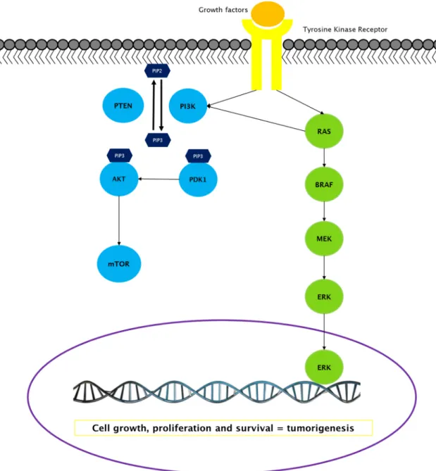
In this pathway, ligand-mediated activation of TK receptor promote RAS GTPase conversion which recruits and activates RAF kinases to the plasma membrane. ERK1 and ERK2 are activated upon phosphorylation by MEK1 and MEK2 which are themselves activated when phosphorylated by RAF proteins. Activated ERKs phosphorylate cytoplasmic and nuclear targets including kinases, phosphatases, transcription factors and cytoskeletal proteins (Figure 5) (Dhillon et al., 2007). The effect of ERK signaling activation is consistent to the cellular processes that itself regulates. Sustained ERK signaling promotes phosphorylation and stabilization of FBJ murine osteosarcoma viral oncogene homolog (Fos), Jun proto-oncogene (Jun), V-Myc avian myelocytomatosis viral proto-oncogene homolog (Myc) and ETS-Related (Erg-1) genes and also cyclin D1 thereby promoting cell-cycle entry and can repress genes responsible for inhibiting proliferation (Yamamoto et al., 2006). On the other hand, high levels of ERKs can induce cell-cycle arrest by expression of CDK-inhibitor proteins such as p21 and p27 that must be counteracted by elevated RhO signaling or activation of AKT so that cells continue to proliferate (Dhillon et al., 2007).
PI3K-AKT signaling pathway PI3Ks represent a family of kinases that phosphorylate the 3’-hydroxyl group of phosphatidylinositol inositides that are activated by many TK receptors. Class I of PI3Ks consists of heterodimers of regulatory (p85) and catalytic (p110) subunits. p110α and p110β subunits have an important role in tumorigenesis as RAS function is mediated by its interaction with the RAS-binding site present in those subunits. Also, activation of TK receptor by extracellular signals is itself sufficient to activate p110 subunits which in turn phosphorylates phosphatidylinositol (PtdIns)-3,4-P2 producing PtdIns-3,4,5-P3 leading to the recruitment of AKT to the cytosolic membrane. AKT is then phosphorylated and activated by PDK1 resulting in the phosphorylation of downstream effector such as the mammalian target of rapamycin (mTOR) (Figure
5). This result in a broad cascade of signaling responsible for cell growth and proliferation, glucose uptake, migration and apoptosis resistance (Saji et al., 2010). Constitutive activation of the PI3K-regulated signaling pathway is relevant in a wide variety of human tumors including thyroid cancer. This is particularly valid in Cowden’s syndrome, that present thyroid carcinomas, which is characterized by mutations in the PTEN gene that encodes a phosphatase that dephosphorylate PtdIns-3,4,5-P3 and thereby negatively regulates the PI3K-AKT pathway. Additionally, RAS mutations, RET/PTC rearrangements and PIK3CA and AKT1 mutations are further evidences that PI3K signaling pathway has a fundamental role in thyroid tumorigenesis (Saji et al., 2010, Xing, 2010).
mTOR signaling pathway mTOR is a serine/threonine kinase that belongs to the phosphoinositide 3-kinase (PI3K)-related kinase family and functions as a regulator of cell growth-related processes. mTOR can form two distinct complexes with other proteins, mTOR complex 1 (mTORC1) and complex 2 (mTORC2), that have different upstream inputs and downstream outputs. Regulation of mTOR by growth factors occurs through the PI3K/AKT pathway which is counteracted by PTEN. There is evidence of overactivation of AKT/mTOR pathway in PTCs when compared to other differentiated thyroid carcinomas and correlation with BRAFV600E
mutation which could be explained by BRAF-induced phosphorylation of tumor suppressor liver kinase B1 (LKB1) Ser428, a main upstream kinase of AMP-activated protein kinase (Faustino et al., 2012).
Figure 5. MAPK and PI3K-AKT-mTOR signaling pathways. In green are the proteins from the MAPK pathway; in blue those belonging to the PI3K-AKT-mTOR pathway. These two classical pathways are associated to TK receptors at the cell membrane that transduces extracellular growth signals into intracellular signals downstream of the pathways. RAS protein can couples the signaling from the receptor to both MAPK and PI3K-AKT-mTOR pathways. These two pathways, driven by genetic alterations in one or more of their components, have a fundamental role in thyroid tumorigenesis.
THROUGHOUT HISTORY, ethical and religious considerations have prevented experimental studies of human biology. Most of the knowledge of human biology, physiology, endocrinology and pharmacology has come first from studies in animal models. Often practical, economical and scientific reasons are also crucial in understanding why animals are the best models for studies of biological phenomena.
I.2 Zebrafish as a model system
Zebrafish is a freshwater fish that was originally found in slow streams and rice paddies and in the Ganges River in East India and Burma. In these water, zebrafish rarely grow larger than 4 centimeters long (Clark et al., 2013).
In comparison to other animal models, zebrafish has a far shorter relationship with research. Zebrafish started to be studied for developmental genetics by George Streisinger from the University of Oregon in the late 1960s. His idea was to apply mutational analysis to study zebrafish embryonic development as he thought this analysis was needed in vertebrates. Initially studies were set to study features of the organization and embryological development of the nervous system using mutant vertebrate strains (Grunwald et al., 2002; Clark et al., 2013).
Zebrafish was and it still is an attractive model due to its brood size, short life cycle, transparent embryos, ex utero development of embryos, ease of keeping many fish in a small space, frequent mating possibility, sexual maturity at 3 months post fertilization (mpf) and conservation of vertebrate tissues (White et al., 2013; Liu et al., 2011). Zebrafish was only later used in large forward genetic screens that produced genetic mutants for virtually any phenotype (Dooley et al., 2000; Liu et al., 2011) and nowadays this model reached its full potential to study vertebrate biology and physiology as well as human diseases (Dooley et al., 2000).
I.2.1 Zebrafish in Cancer Research
The cancer field is heading towards a post-genomic state and, as most human cancers are being extensively sequenced, new tools are required so that the extensive genomic alterations found can be studied in a biological context and be translated into a more effective therapy (Mione et al., 2010; Liu et al., 2011; White et al., 2013).
Although no single model can fully reflect the complexity of cancer biology in vivo, zebrafish has been considered a great addition to animal models of human disease (Liu et al., 2011; White et al., 2013).
In 1982, zebrafish was first proposed as a model for cancer with a study showing that exposure to carcinogens induced low-penetrance tumors (Pliss et al., 1982). Remarkably, despite over 300 million years of divergence between fish and humans, tumors in zebrafish strongly resemble human tumors at the histological, gene expression and genomic levels. Moreover, crucial genes and pathways involved in tumorigenesis are highly conserved between fish and humans (Amatruda et al., 2002).
Two decades later more studies were able to demonstrate various neoplasms in zebrafish after mutagen exposure but the breakthrough came when T-cell leukemia was induced by expressing oncogenic myc under the control of rag2 in transgenic animals (Langenau et al., 2003).
Since then, many zebrafish models of cancer expressing dominant-acting oncogenes have been established using various technologies. The two main approaches are either to select genetic alterations in human tumors and then test it in transgenic animals or to identify evolutionarily drivers conserved between human tumors and those tumors arising in transgenic animals (Liu et al., 2011; White et al., 2013).
Taking into account the first approach, probably the most extensively studied transgenic zebrafish model of cancer is the melanoma model. Human melanoma is a lethal form of skin cancer with an incidence and mortality rates that are rapidly increasing (Wangari-Talbot, 2012). Molecular genetic analysis of precursor lesions such as the nevi and melanoma tumors have pointed to alterations in key genes of the RAS-RAF-MEK-ERK and PI3K-AKT pathways regulating signal transduction and other genes involved in cell cycle regulation such as the cyclin-dependent kinase inhibitor 2A (CDKN2A) (Wangari-Talbot, 2012). The majority of human melanomas exhibit mutations in BRAF, the same found in PTCs, and this may have a critical role in melanoma initiation (Wangari-Talbot, 2012).
To study the role of activated BRAF, a transgenic zebrafish expressing BRAFV600E
under the control of a melanocyte promoter was generated. Expression of the oncogene in the melanocytes lead to nevi which rapidly develop into invasive melanomas only in the absence of WT p53 (Patton et al., 2005). Furthermore, to