Thyroid Hormone Stimulates the Proliferation of
Sertoli Cells and Single Type A Spermatogonia in
Adult Zebrafish (
Danio rerio
) Testis
R. D. V. S. Morais, R. H. Nóbrega, N. E. Gómez-González, R. Schmidt, J. Bogerd, L. R. França, and R. W. Schulz
Laboratory of Cellular Biology (R.D.V.S.M., L.R.F.), Department of Morphology, Institute of Biological Sciences, Federal University of Minas Gerais, Belo Horizonte, Brazil; Reproductive Biology Group (R.D.V.S.M., R.H.N., N.E.G.-G., R.S., J.B., R.W.S.), Division of Developmental Biology, Department of Biology, Faculty of Science, Utrecht University, Utrecht, The Netherlands; and Department of Morphology (R.H.N.), State University of São Paulo, Botucatu, Brazil
Thyroid hormones participate in regulating growth and homeostatic processes in vertebrates, including development and adult functioning of the reproductive system. Here we report a new stimulatory role of thyroid hormone on the proliferation of Sertoli cells (SCs) and single, type A undifferentiated spermatogonia (Aund) in adult zebrafish testes. A role for T3in zebrafish testis is
suggested by in situ hybridization studies, which localized thyroid receptor␣(thr␣) in SCs and the (thr) mRNA in Sertoli and Leydig cells. Using a primary zebrafish testis tissue culture system, the effect of T3on steroid release, spermatogenesis, and the expression of selected genes was
eval-uated. Basal steroid release and Leydig cell gene expression did not change in response to T3.
However, in the presence of FSH, T3potentiated gonadotropin-stimulated androgen release as
well as androgen receptor (ar) and 17␣-hydroxylase/17,20 lyase (cyp17a1) gene expression. More-over, T3alone stimulated the proliferation of both SCs and Aund, potentially resulting in newly
formed spermatogonial cysts. Additional tissue culture studies demonstrated that Igf3, a new, gonad-specific member of the IGF family, mediated the stimulatory effect of T3on the proliferation
of Aundand SCs. Finally, T3induced changes inconnexin 43mRNA levels in the testis, a known
T3-responsive gene. Taken together, our studies suggest that T3expands the population of SCs and
Aundinvolving Igf signaling and potentiates gonadotropin-stimulated testicular androgen
pro-duction as well as androgen sensitivity.(Endocrinology154: 4365– 4376, 2013)
T
he relationship between thyroid hormones (THs) and reproduction has been investigated in different verte-brate species (1). The effects of THs are mediated by spe-cific intracellular thyroid hormone receptors (THRs), members of the nuclear receptor superfamily, and similar to the situation in vertebrates in general, TH effects on reproduction in fish are complex (2).Although potentially not valid for all mammalian spe-cies investigated (1), in rodents (rats, mice, and hamsters) T3(the biologically most active TH) regulates testicular growth and pubertal maturation by stimulating the ter-minal differentiation of Sertoli cells (SCs), thereby
deter-mining the period during which they can proliferate (3–5). However, the differentiation state of adult mammalian SCs seems less terminal than has been thought for many years (6). With regard to sex steroid signaling, SC differ-entiation also involves T3-mediated down-regulation of aromatase gene transcription (7) and up-regulation of an-drogen receptor gene expression (8). In the early 1990s, Cooke and coworkers (9, 10) showed that testis size and sperm output were enhanced in hypothyroid rats. Further studies demonstrated that T3can directly suppress prolif-eration and induce differentiation of cultured neonatal rat SCs (11), involving up-regulation of cyclin-dependent
ki-ISSN Print 0013-7227 ISSN Online 1945-7170 Printed in U.S.A.
Copyright © 2013 by The Endocrine Society Received April 4, 2013. Accepted August 15, 2013. First Published Online September 3, 2013
Abbreviations: Aund, type A undifferentiated spermatogonia; BrdU, bromodeoxyuridine; CX, connexins; DIG, digoxigenin; IGF1R, IGF 1 receptor; ISH, in situ hybridization; 11-KT, 11-ketotestosterone; qPCR, quantitative PCR; SC, Sertoli cell; TH, thyroid hormone; THR, thyroid hormone receptor.
nase inhibitors (12), thereby eliminating mitogenic effects of FSH (13). Moreover, TH reduced the expression of the neural cell adhesion molecule in SC-gonocyte cocultures isolated from neonatal rat testis (14). Another marker for SC differentiation/maturation in mammals is the expres-sion of connexins (CXs) (15). Gilleron and coworkers (16) demonstrated that T3 increased the levels of CX43, an important gap junctional protein that participates in the control of cell proliferation. Male mice with a SC-specific loss ofCX43show an arrest of spermatogenesis at the level of spermatogonia or display a SC-only syndrome (17). In two fish species, rainbow trout (Oncorhynchus mykiss) and brook trout (Salvelinus fontinalis), four different Cxs/
cxswere identified in the testis (18, 19), and Cx43 may be involved in maturation of trout testis because its expres-sion levels increased during the onset of spermatogenesis. TH plays a critical role in the onset of Leydig cell dif-ferentiation and stimulation of steroidogenesis in postna-tal rat (20) by increasing the expression of steroidogenic acute regulatory protein. Stimulatory effects on gonadal steroidogenesis have been reported for different species, such as rat (21) and goldfish (22). TH effects on steroid-ogenesis can be direct or via modulation of gonadotropin-stimulated steroid release, at least in mammals (21).
In contrast to the situation in mammals, fish (and am-phibians) show the cystic type of spermatogenesis (23), in which cytoplasmic extensions of SCs form cysts by envel-oping a synchronously develenvel-oping germ cell clone derived from a single spermatogonial cell, the type A undifferen-tiated spermatogonia (Aund). The cyst-forming SCs retain their capacity to proliferate also in the adult fish testis (24), and because growth and development of spermatogenic cysts in the adult testis involve an increase in the number of SCs per cyst (24, 25), SC proliferation can be expected in the adult, spermatogenetically active fish testis. The cy-toskeleton of SCs and dynamic junctional complexes be-tween SC-SC and SC-germ cell are important for SC func-tions (26, 27). Both gap juncfunc-tions and tight juncfunc-tions form between SC as part of the SC barrier (previously known as blood-testis barrier), which in fish forms later than in mammals, namely at the beginning of the spermiogenic phase of cyst development (25, 28). It is known that gap junctions between SCs and SCs and germ cells are essential for spermatogenesis and for male fertility (27). Because TH modulate the expression of CXs (19, 20), connexin gene expression may be a useful parameter in studies on T3 effects in zebrafish testis tissue culture.
In all vertebrates, THRs are expressed by several dif-ferent cell types, and hence, THs have pleiotropic effects, including effects on the gonads. In goldfish, recent in vivo and in vitro studies suggested that THs affect gonadal steroid synthesis and steroid receptor expression (2). In
fact, most experimental studies to date have focused on TH effects on the developing testes and only limited data are available regarding to adult testis functions, whereas direct effects of TH on the fully mature testis have not been studied yet.
The present study addresses the following questions regarding potentially direct effects of TH on adult ze-brafish testis: 1) Which testicular cell types express THRs? 2) Does TH modulate testicular expression of selected tes-ticular target genes? 3) Does TH alone, or in association with recombinant zebrafish FSH (rzf FSH), modulate the steroidogenic process? 4) Does TH change the prolifera-tion and differentiaprolifera-tion status of SCs and germ cells?
Materials and Methods
Animals
Sexually mature male zebrafish (Danio rerio) between 6 and 12 months of age were used in the present study. The animals were kept and handled according to the Dutch national regula-tion and experiments were approved by the Utrecht University animal use and care committee.
Testicular explants
To study effects of T3(Sigma-Aldrich) on androgen release,
spermatogenesis, and gene expression, a previously described ex vivo organ culture system for zebrafish testis was used (29). Sev-eral experiments were conducted (Supplemental Figure 1, pub-lished on The Endocrine Society’s Journals Online web site at
http://endo.endojournals.org), including T3dose response (2,
10, and 50 ng/mL) experiments to study gene expression and androgen release. For morphological evaluation, a concentra-tion of 50 ng T3/mL was selected. The two testes from each fish
were incubated in parallel, one testicle (randomly chosen) serv-ing as control for the contralateral one; eight replicates were used for each of the different conditions.
Morphological analysis
After 4 days in culture, zebrafish testes (n⫽8) were fixed in 4% buffered glutaraldehyde at 4°C overnight, dehydrated, em-bedded in Technovit 7100 (Heraeus Kulzer), sectioned at 4m thickness, and stained with toluidine blue, according to conven-tional histological procedures.
Germ and somatic cell proliferation analysis
To evaluate whether T3affects the proliferative activity of
single spermatogonia Aundor SCs, 50g/mL
bromodeoxyuri-dine (BrdU; Sigma-Aldrich) was added to the medium during the last 6 hours of culture (Supplemental Figure 1A). To study if T3
action involves the IGF signaling pathway, zebrafish testes were exposed to T3 in the absence or presence of 10 M
Figure 1, A and B, where tissue was exposed to 50 ng/mL of T3,
were compared. Because no significant difference was found, the data were combined to form a single group (n⫽16). This group was compared to the basal condition (n⫽8) and to T3⫹NVP
(n⫽8). Zebrafish testes were fixed at 4°C overnight in freshly prepared methacarn (60% [v/v] absolute ethanol, 30% chloro-form, and 10% acetic acid), after which the tissue was dehy-drated, embedded in Technovit 7100, sectioned at 3m thick-ness, and submitted to BrdU immunodetection, as described previously by Leal and coworkers (29).
In vitro 11-ketotestosterone (11-KT) release by zebrafish testes
The androgen release capacity of zebrafish testicular tissue was measured after 1 day of ex vivo culture in two experiments. First, zebrafish testes were incubated in control medium or me-dium containing 50 ng/mL T3(Supplemental Figure 1A). In the
second experiment, testes were exposed to medium containing
25 ng/mL rzf FSH or to medium containing 25 ng/mL rzf FSH and 2, 10, or 50 ng/mL T3(Supplemental Figure 1C). The 11-KT
release into culture medium was quantified using a steroid re-lease bioassay previously adapted for zebrafish testis (32). The results were calculated as nanograms of 11-KT released per mil-ligram of testis tissue.
Gene expression analysis by real-time, quantitative PCR (qPCR)
The capacity of T3(2, 10, or 50 ng/mL) to modulate testicular
gene expression was investigated after incubation periods of 1 or 4 days. Total RNA was extracted from the samples using an RNAqueous-Micro Kit (Ambion), following the manufacturer’s instructions. To estimate relative mRNA levels of selected genes (Table 1), qPCR was performed as described by de Waal and coworkers (32). The levels of elongation factor 1␣(ef1␣) mRNA served as endogenous control RNA, which remained stably ex-pressed under the different experimental conditions
(Supplemen-Table 1. Primers Used for Gene Expression Studies and to Generate DNA Templates for DIG-labeled cRNA Probe Syntheses for In Situ Hybridization
Target
Gene Primers Sequence (5ⴕ33ⴕ)
Amh AD (Fw) CTCTGACCTTGATGAGCCTCATTT
AE (Rv) GGATGTCCCTTAAGAACTTTTGCA
AF (probe) FAM-ATTCCACAGGATGAGAGGCTCCCATCC-TAMRA
Ef1␣ AG (Fw) GCCGTCCCACCGACAAG
AH (Rv) CCACACGACCCACAGGTACAG
AI (probe) FAM-CTCCAATTTTGTACACATCCTGAAGTGGCA-TAMRA
igf3 2680 (Fw) TGTGCGGAGACAGAGGCTTT
2681 (Rv) CGCCGCACTTTCTTGGATT
igf1r␣ 2362 (Fw) TACATCGCTGGCAACAAGCA
2363 (Rv) TCATTGAAACTGGTCCTTATGCAAT
igf1r 2595 (Fw) GTGCTGGTCCTCTCCACACTCT
2596 (Rv) TTACCGATGTCGTTGCCAATATC
cx43 3856 (Fw) CTACAGGGCTCTCCACTCTTTACTTCT
3858 (Rv) CGCACTCCAGTCACCCATCT
cx43.4 3859 (Fw) CGTAGCTGAGGAAAAGAGTGGAAA
3860 (Rv) CGTAAGAAAACTCCAGCTCATGGT
ar 2412 (Fw) ACGTGCCTGGCGTGAAAA
2413 (Rv) CAAACCTGCCATCCGTGAAC
insl3 2466 (Fw) TCGCATCGTGTGGGAGTTT
2467 (Rv) TGCACAACGAGGTCTCTATCCA
star 2546 (Fw) CCTGGAATGCCTGAGCAGAA
2547 (Rv) ATCTGCACTTGGTCGCATGAC
cyp17a1 2773 (FW) GGGAGGCCACGGACTGTTA
2774 (Rv) CCATGTGGAACTGTAGTCAGCAA
dazl 3104 (Fw) AGTGCAGACTTTGCTAACCCTTATGTA
3105 (Rv) GTCCACTGCTCCAAGTTGCTCT
piwil1 2542 (Fw) GATACCGCTGCTGGAAAAAGG
2543 (Rv) TGGTTCTCCAAGTGTGTCTTGC
piwil2 2994 (Fw) TGATACCAGCAAGAAGAGCAGATCT
2995 (Rv) ATTTGGAAGGTCACCCTGGAGTA
thr␣ 3691a(Fw) T3Rpps-TCAAACAAATAACATACTAACACTTTCCTTCTAAGTGGA 3692b(Rv) T7Rpps-CCATTGCGTCTCATCTCCTTCTG
thr 3695a(Fw) T3Rpps-TCATTTCAGGCCACGTATGTCCGATC 3696b(Rv) T7Rpps-TAACTTGGTATGTACCCATTCTGCATGGCCTC
a
Primer 3691 and 3695 contain the T3 RNA polymerase promoter sequence (underlined) at its 5⬘-end (T3Rpps; 5⬘-GGGCGGGTGTTATTAACCCTCACTAAAGGG-3⬘).
bPrimer 3692 and 3696 contain the T7 RNA polymerase promoter sequence (underlined) at its 5
tal Figure 2). All qPCRs were performed in 20L reaction vol-ume and quantification cycle (Cq) values were determined in a 7900HT Real-Time PCR System (Applied Biosystems) using de-fault settings. Relative mRNA levels were calculated as reported previously (32, 33).
In situ hybridization
Zebrafishthr␣andthr-specific PCR products were gen-erated with primers 3691–3692 and 3695–3696 (Table 1), respectively, containing either T3 (primers 3691 and 3695) or T7 (primers 3692 and 3696) RNA polymerapromoter se-quences attached at their 5⬘-ends. The⬃450- and⬃455-bp PCR products obtained were gel purified and served as tem-plates for digoxigenin (DIG)-labeled cRNA probe syntheses, as described by Vischer and coworkers (34). Whole-mount in situ hybridization (ISH) was used to localize thyroid hormone receptor (thr␣andthr) mRNAs in testicular tissue fixed in 4% paraformaldehyde in PBS (pH 7.4), according to previ-ously described methods (35). The tissue was treated with proteinase K (20g/mL; Sigma-Aldrich) at 37°C for 20 min-utes, and acetic anhydride (0.25% in 0.1 M triethanolamine [pH 8.0]; Merck) was included to reduce background after postfixation and before prehybridization. Hybridization with T3 and T7 DIG-riboprobes was performed overnight at 72°C, and DIG immunostaining was performed on the following day using anti-conjugated alkaline phosphatase (1:2000; Roche). Staining was revealed with nitro blue
tetrazolium/5-bromo-4-chloro-3⬘-indolyphosphate (both Sigma-Aldrich), followed by three consecutive PBS washings. Then, tissue was fixed in 4% paraformaldehyde in PBS, dehydrated, and embedded in Technovit 8100 (Heraeus Kulzer) for plastic sectioning. Sec-tions of 7 m thickness were counterstained with 0.1% Nuclear Fast Red (in 5% aluminum sulphate solution) for 2 minutes and washed in running tap water for 5 minutes. After a rinse in deionized water, air-dried sections were mounted with Aqueous Mounting Medium (Dako North America Inc).
Statistical analysis
For the ex vivo experiments, differences between control and treatment for the measured parameters (ie, BrdU-positive SCs [Figure 6C], relative mRNA levels, androgen release) were tested for statistical significance using the Student’sttest for paired observations; significant differences are indicated by an asterisk. When comparisons were made over three or more conditions (ie,connexinmRNA levels, mitotic index of SCs, and type A spermatogonia), data were analyzed by one-way ANOVA, followed by a Tukey’s multiple comparison test; significant differences are indicated by different lowercase letters. In some cases, data were log transformed to achieve an equal variance. A significance level (P)⬍.05 was applied in all statistical analyses, for which we used the Prism4 software package (GraphPad). Data are presented as the mean⫾SEM.
Results
Thyroid hormone receptor mRNA localization in zebrafish testis
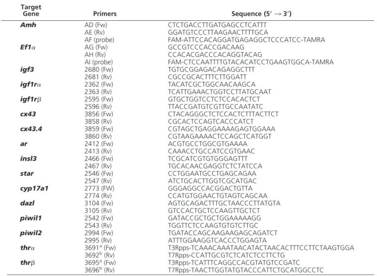
Identification of the cell types ex-pressing the mRNAs coding for the two THRs described for zebrafish (thr␣andthrmRNA) was accom-plished by ISH. Based on the shape and localization of the stained cells in the tubular or intertubular compart-ments, the cells specifically labeled were identified as Sertoli and Leydig cells (Figure 1). Thethr␣mRNA sig-nal was detected in SCs only (Figure 1A). Labeled SCs were in contact with germ cells at different stages of differentiation (Figure 1B). Thethr
mRNA signal was localized in both Sertoli and Leydig cells (Figure 1, C and D). For both receptors, labeling also appeared as thin stretches be-tween germ cells in the lumen of sper-matogenic cysts (eg, in cysts contain-ing spermatocytes [Figure 1B] or spermatids [Figure 1D]). No specific Figure 1. Localization of THR mRNAs in zebrafish testis using whole-mount ISH and post
signal was obtained when sections were incubated with the sensethr␣andthrprobes (Figure 1,insets).
Triiodothyronine effects on gene expression and androgen release
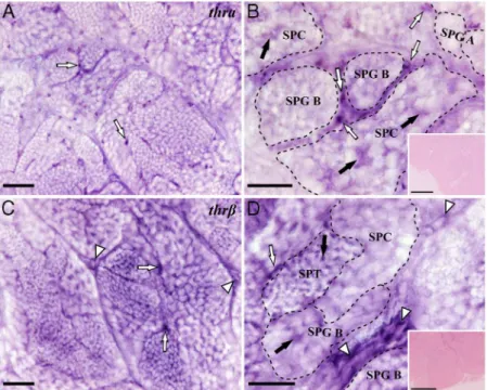
To investigate if T3changes gene expression in the ze-brafish testis, mRNA levels of selected Sertoli, Leydig, and germ cell genes (Table 1) were quantified. In a first exper-iment using 50 ng T3/mL, only the mRNA levels for IGF 3 (igf3), a gonad-specific IGF subtype recently discovered in teleost fish (36), changed significantly on day 1 of the culture (Figure 2A). We repeated the analysis using dif-ferent doses of T3, which showed thatigf3mRNA levels were up-regulated after 1 day in response to the interme-diate and highest dose of T3, 10 and 50 ng/mL, respectively (Figure 2C). No significant response to 50 ng/mL T3was observed after 4 days of incubation (Figure 2, A and B). Studying the effect of T3on the mRNA levels of two CXs, a transient down-regulation of cx43mRNA levels was observed after 1 day of culture (Figure 2D), which
recov-ered after 4 days. No change was observed in the mRNA levels ofcx43.4in response to T3.
The release of 11-KT in culture medium was measured after 1 day of culture (Supplemental Figure 1, A and C). Exposure to T3alone did not change 11-KT release (Figure 3A). However, T3-dose response studies in combination with rzf FSH demonstrated that at the highest dose (50 ng T3/mL) the androgen release was significantly enhanced when compared to rzf FSH only, which in turn clearly stimulated androgen release above the levels found under basal conditions or in the presence of T3alone. Moreover,
arandcyp17a1gene expression was not different from the one under basal conditions when testis tissue was incu-bated with T3alone, while T3enhanced FSH-stimulatedar andcyp17a1gene expression significantly in the presence of 50 ng/mL T3(Figure 3B). Previous results showed that rzf FSH alone did not changearmRNA levels but led to a fourfold increase in cyp17a1 mRNA levels (37). These results suggest that T3plays a permissive role for
stimu-Figure 2. Gene expression analysis after 1 or 4 days of zebrafish testis tissue culture. (A, B) Relative mRNA levels of Sertoli, Leydig, and germ cell-marker genes in testicular explants incubated with T3(50 ng/mL) for 1 or 4 days. Bars represent the relative mRNA levels (fold of basal; mean⫾
SEM; n⫽8). Solid lines (—) indicate genes repeatedly localized to the indicated cell types. Dotted lines (• • • •) indicate genes reported to be expressed in the indicated cell types in some but not all studies. (C) Relative mRNA levels ofigf3after T3 dose response after 1 day of incubation (fold of control; mean⫾SEM; n⫽6). (D) Testicular mRNA levels of two CXs (cx43andcx43.4) in tissue after 1 or 4 days of exposure to T3(fold of
latory effects of FSH on androgen production and andro-gen sensitivity in adult zebrafish testis tissue.
Triiodothyronine stimulates germ and SC proliferation
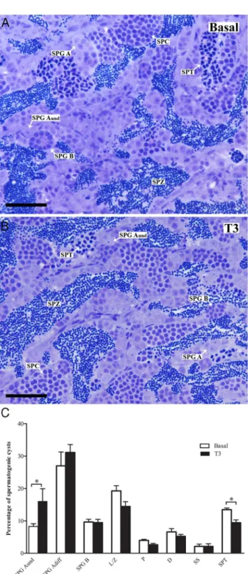
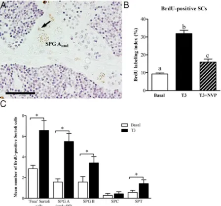
Morphological analysis showed that in testes incubated with basal medium or with T3, the testicular cyto-archi-tecture remained intact. The spermatogenic process ap-peared normal and we observed cysts with germ cells at all stages of differentiation, including free spermatozoa in the lumen of seminiferous tubules (Figure 4, A and B). Ana-lyzing the effects of T3on spermatogenesis after 4 days of tissue culture (Figure 4C) revealed a higher volume frac-tion (P⬍.05) of single type Aundspermatogonia cysts and fewer cysts with spermatids (P⬍.05) in comparison with the control testes. To determine if T3 also changed the proliferation activity of these spermatogonia, their mitotic index was obtained. Quantitative evaluation of the sec-tions after immunocytochemical detection of BrdU showed a significantly higher proportion of BrdU-positive type Aundspermatogonia in T3-treated tissue (Figure 5), suggesting that a stimulation of proliferation of this germ cell type is the basis for the increased volume fraction of these cells in morphometric analysis (Figure 4).
Proliferation of SCs was observed as well (Figure 6A). Quantitative evaluation showed that ⬃30% of the SCs were BrdU-positive after T3 treatment compared with only⬃9% in the control group (Figure 6B). BrdU-labeled SCs can occur in association with germ cells forming sper-matogenic cysts, or as “free” SCs (ie, not associated with germ cells). Intriguingly, closer analysis of the BrdU-la-beled SCs showed that the majority was either free or as-sociated with type A spermatogonia, whereas the minority was associated with type B spermatogonia, spermato-cytes, and spermatids (Figure 6C).
Members of the IGF family are known for their mito-genic activity and we observed that T3induced an increase of testicularigf3mRNA levels (Figure 2, A and C). There-fore, we studied whether T3-stimulated proliferation of Aundand SCs depended on the functioning of IGF recep-tors (Supplemental Figure 1B). Incubating testis tissue with T3 in the absence or presence of the IGF receptor inhibitor NVP-AEW541 showed that T3-induced BrdU incorporation into Aund(Figure 5) or SCs (Figure 6) was abolished or reduced, respectively, leading to a significant decrease (P⬍.05) of the BrdU-labeling index of these two cell types.
Discussion
In vertebrates, THs have several functions and THR expression in the testis suggests THs participate in reg-ulating male reproduction. In the present study, we investigated poten-tial roles of T3, alone or in combina-tion with rzf FSH, on adult zebrafish steroidogenesis and spermatogene-sis, using morphological, physiolog-ical, and molecular approaches.
It is known that THs work via their specific intracellular THRs, and studies on the thr␣ and thr
mRNA levels in testis tissue of the seasonally reproducing brook trout demonstrated seasonal variations with a constant expression through-out spermatogenesis while the higher expression pattern was observed af-ter spawning season (20). However, the testicular cell types expressing the THRs have not been clearly iden-tified in fish. In mammals, THRs have been localized to SCs by differ-ent laboratories, while the staining in Figure 3. Ex vivo androgen release and gene expression analysis in adult zebrafish testis. (A)
Androgen release after 1 day of incubation under basal conditions and with 50 ng/mL T3or after
exposure to different doses of T3 in the presence or absence of 25 ng/mL rzf FSH. Results are given as the amount of 11-KT produced in nanogram per milligram weight of testis tissue incubated. (B) Testiculararandcyp17␣1mRNAs levels (fold of basal; mean⫾SEM; n⫽8) after 1 day of exposure to different doses of T3, alone or in combination with rzf FSH. *, significant
germ cells and in interstitial cells is debated (38, 39). Also in zebrafish, information on the testicular cell types ex-pressing THRs is missing but is needed to direct further studies as regards physiological functions of THs in the testis. We found that SCs, contacting germ cells in different stages of spermatogenesis, expressed both THR forms while Leydig cells expressed thr only. We also found stained areas between germ cells in a number of (often larger) cysts. In cod testis intermediate cytoskeletal protein of SCs, vimentin, also was detected in between germ cells (40). Similarly, FSH receptor protein localization by im-munohistochemistry in eel testis revealed staining in be-tween germ cells (41). These data suggest that SCs forming a spermatogenic cyst do not form a simple, saclike space but may show a more complex structure toward the germ cell-contacting luminal side of the cyst, perhaps with cy-toplasmic extensions inserted between germ cells. Future morphological/ultrastructural work may provide evi-dence for this assumption. Collectively, our data indicate that THs modulate testicular functions via two important somatic cell types, the Sertoli and Leydig cells, whereas direct effects on germ cells seem unlikely in zebrafish. As in other vertebrates, the localization ofthr␣ also in ze-brafish SCs indicates that this cell type is an evolutionary conserved target for THs. As in some (but not all) other models (42, 43), we detectedthralso in Leydig cells. In zebrafish germ cells were THR negative, different from reports in rat where germ cells from intermediate sper-matogonia to pachytene spermatocytes expressedThr␣1
(38, 43). Our results suggest that TH effects on SC and germ cell proliferation are mediated by THRs in SC, whereas T3-modulation of steroidogenesis is mediated by Thrin Leydig cells. Modulatory effects of TH on andro-gen levels were described in rat in vivo (44), but our study provides original information on the fact that TH directly modulates zebrafish testicular androgen production (see next page).
After 1 day of tissue culture, T3significantly increased theigf3mRNA levels. Interestingly, this gonad-specificigf
type, recently discovered in fish but not present in tetrapod vertebrates (36), has been localized to SCs in adult ze-brafish testis and may play a role in the recovery following a cytotoxic insult in adult zebrafish testis (45, 46). In hy-pothyroid rats T3greatly stimulated SC IGF1 production in vivo, also incubation of cultured SCs with T3increased the production of IGF1 (47). It appears that a stimulatory role of TH on the release of IGF family members by SCs is an evolutionary conserved mechanism in vertebrates.
Incubation of testicular fragments collected from ma-turing brook trout during the rapid growth phase of the testis with T3 increasedcx43mRNA levels (19). In our study, exposure of the fully grown adult testis to T3did not Figure 4. Morphological analysis of zebrafish testis after tissue
culture. (A, B) Histological sections stained with toluidine blue showing normal cyto-architecture of zebrafish testis after 4 days of tissue culture with or without 50 ng/mL of T3. All germ cell types were
present in the testis. (C) Morphometric quantification (% of the total number of cysts at different stages of germ cell development; mean⫾
SEM; n⫽8) of the different germ cell cysts in zebrafish. D, diplotene primary spermatocytes; L/Z, leptotene/zygotene primary spermatocytes; P, pachytene primary spermatocytes; SPG Adiff, type A differentiated
spermatogonia; SPG Aund, type A undifferentiated spermatogonia; SPG
increase cx43 mRNA levels significantly above control levels. However, comparingcx43mRNA levels over time, the levels first decreased after 1 day, before increasing in T3-exposed tissue fragments toward day 4. In a murine SC line, THs increasedCx43mRNA levels and (16, 48) in-hibited SC proliferation, which was associated with the expression of cell-cycle regulatory proteins such as S-phase kinase-associated protein 2 and cyclin-dependent kinase inhibitor p27Kip1 (also called CDKN1B) (16, 48 – 50). Conversely, in the SC-specific connexin 43 knockout mice (SC-Cx43KO), SC number was increased 73% com-pared with wild-type mice, indicating that CX43 is in-volved in the pubertal cessation of SC proliferation (13). In adult zebrafish testis, on the other hand, THs stimulated SC proliferation. Although THs may play distinct roles in piscine and mammalian testes, the downstream mecha-nisms underlying SC proliferation in both models may involve the down-regulation of Cx43. However, no changes were recorded for another extracellular matrix gene transcript (cx43.4mRNA).
THRs are ligand-regulated transcription factors that bind TH while being bound to DNA sequences of target genes where they can then interact with corepressor and coactivator complexes (51). Analyzing the T3effects on the expression of selected target genes, we observed that the two Leydig cell genes starand insl3did not change
significantly, and neither did basal androgen release. Similarly, in gold-fish, in vitro experiments demon-strated that T3did not change andro-gen levels significantly (2). However, studying gonadotropin-stimulated estrogen release from maturing (early vitellogenic) ovarian follicles isolated from rainbow trout THs were found to amplify the steroido-genic gonadotropin effect (52). The present study showed that also for the adult zebrafish testis, T3 poten-tiated FSH effects on steroid release and gene expression. In the adult rat testis recovering from a cytotoxin-induced loss of Leydig cells, TH treatment stimulated Leydig cell covery and increased androgen re-lease (53), whereas in adult rat ren-dered transiently hypothyroid, the circulating testosterone levels were significantly decreased (44). In ze-brafish, T3 alone did not modulate the acute androgen release, but clearly enhanced FSH-stimulated
cyp17a1 and ar gene expression and androgen release. Piscine Leydig cells express FSHr/fshr(41, 54) and FSH is a potent steroidogenic hormone in fish (37, 55), while previous studies (42) have shown that androgens do not stimulatecyp17a1gene expression, so that we can attri-bute the increased expression of this gene to FSH. Hence, our data suggest that in the presence of rzf FSH, T3 po-tentiates the trophic effects of FSH on the steroidogenic system in the adult zebrafish testis. At the same time, T3 had a permissive effect because it was only in concert with T3that FSH increasedargene expression. Hence, T3 in-creased both androgen production and sensitivity of adult zebrafish testis tissue. In general, our findings support the concept of a direct, stimulatory crosstalk between THs and the androgen-producing system that was recently pro-posed to operate in vertebrates (56).
In testes treated with T3for 4 days, the volume fraction of type Aund spermatogonia increased. Analyzing the BrdU-labeling index showed that proliferation of this par-ticular germ cell type (but also of SCs) was stimulated. Interestingly, treatment of African catfish (Clarias gariepi-nus) with thiourea, a TH inhibitor, compromised sper-matogenesis, leading to a decrease in spermatid/sperma-tozoa counts (57). This observation is compatible with the assumption that TH-stimulated production of type A sper-matogonia and SCs (in conjunction with the enhanced Figure 5. Localization and quantification of BrdU incorporated during the last 6 hours of
incubation into zebrafish testis tissue explants after 4 days in culture. Transversal section showing BrdU immunodetection in tissue incubated in basal medium (A), in the presence of 50 ng/mL T3
(B), or in medium containing 50 ng/mL T3⫹10M NVP-AEW541 (C). (black arrowheads)
BrdU-positive single type A undifferentiated spermatogonia (Aund). (white arrowheads) unlabeled Aund.
(D) BrdU labeling index of type Aund(% of total number of BrdU-positive Aund). Bars, mean⫾
androgen signaling) are relevant in vivo. Remarkably, in zebrafish, an IGF receptor inhibitor abolished the T3 -stim-ulated increase of the mitotic index of type Aund spermato-gonia, demonstrating that T3-stimulated proliferation of Aundspermatogonia is mediated via a factor activating IGF receptors in the zebrafish testis.
In mammals, the effect of T3on SC proliferation has been comprehensively discussed. Several studies demon-strated that neonatal hypothyroidism and hyperthyroid-ism affect the number of SC by extending or shortening, respectively, the period of proliferation (3, 58, 59). Our observation of T3-stimulated proliferation of SCs con-trasts with the mammalian situation. On the other hand, SC proliferation is an expected event in the adult fish testis that accompanies growth and development of spermato-genic cysts (24) and probably involves activation of FSH signaling (60). Interestingly, in the present study, an im-portant fraction of proliferating SCs was either associated with type A spermatogonia or was not associated with germ cells at all (referred to as “free” SCs). This suggests
that this type of SC proliferation cre-ated additional germ cell support ca-pacity, perhaps mediated by the THR forms expressed by SCs. In ad-dition, analyzing SC proliferation in the presence of the IGF receptor in-hibitor, the BrdU-labeling index de-creased, suggesting that T3 -stimu-lated SC proliferation is in part mediated by IGF/Igfr signaling. Our observations in zebrafish seem con-gruent with conclusions based on in vitro studies on mouse SC, where in-activation of the IGF1 receptor gene decreased SC proliferation and also increased SC apoptosis (30). More-over, a recent study demonstrated that SC-specific loss of insulin/IGF signaling in mice strongly reduced SC proliferation and testis size (61). We propose that an evolutionary conserved mechanism to stimulate SC proliferation is mediated in ze-brafish by the gonad-specific Igf3.
Considering that germ cells are THR negative, the T3-induced in-crease in number and mitotic index of type Aundspermatogonia could be mediated by somatic cells, possibly SC. Elevated SC numbers may have allowed this spermatogonial popula-tion to expand, potentially sup-ported by an increased availability of SC-derived growth factors stimulating spermatogonial development. Be-cause our results have shown that the IGF signaling pathway mediated T3-stimulated proliferation of SC and type Aundspermatogonia proliferation, it is tempt-ing to speculate that T3triggered an autocrine Igf3 loop to stimulate SC proliferation, and a paracrine loop to stimulate spermatogonial proliferation. Interestingly, in rainbow trout testis IGF receptor expression has been found in both somatic and germ cells (62). Assuming that newly formed SCs associate with single type Aund spermatogonia and hence form new spermatogenic cysts, the spermatogenic capacity of the testis would increase. On the other hand, hypothyroidism induced in vivo in pubertal Nile tilapia (Oreochromis niloticus) increased SC and germ cell numbers per cyst (63). This effect is not exactly opposite to the one that we have observed in adult zebrafish, because it was the size, not the number, of cysts that changed in pubertal tilapia testis. Hence, as described previously for mammals Figure 6. Localization and quantification of BrdU incorporated during the last 6 hours of
incubation into zebrafish testis explants incubated with 50 ng/mL T3in the absence or presence
of 10M NVP-AEW541 ex vivo for 4 days. (A) Labeled SC nucleus (arrow) associated with a BrdU-positive single type Aundspermatogonium. (B) BrdU labeling index (% of total number of
cells) of SCs. Different letters indicate significant differences (P⬍.05) between incubation conditions. (C) Treatment with T3increased the labeling index of BrdU-labeled SCs associated, or
not (“free”), with different germ cell cysts. SPG A (und⫹diff), type A undifferentiated and differentiated spermatogonia; SPG B, type B spermatogonia; SPC, spermatocytes; SPT,
(14), T3may play different roles depending on the on-togenetic stage of development and the species investi-gated (14).
Conclusion
The direct effects of T3on the zebrafish testis are coordi-nated by receptors expressed by Sertoli and Leydig cells. When present alone, T3stimulates SC and type Aund sper-matogonia proliferation. When T3and FSH are present at the same time, we can expect an increase in the number of SC and single type Aundspermatogonia, but also a poten-tiation of the steroidogenic activity of FSH and of the androgen sensitivity of the testis. Because androgens are known to stimulate in particular the somewhat more ad-vanced stages of germ cell differentiation in fish (23, 64) and mammals (65), we speculate that the joint action of T3 and FSH would result in more newly formed spermato-genic cysts that would then be stimulated to differentiate in an androgen-driven manner.
Acknowledgments
We thank Wytske van Dijk, Mara Lívia Santos, and Henk Schriek for technical help.
Address all correspondence and requests for reprints to: Rü-diger W. Schulz, Department of Biology, Utrecht University, Padualaan 8, 3584 CH, Utrecht, The Netherlands. E-mail: [email protected].
Financial support from the Minas Gerais State Foundation (FAPEMIG, Brazil), the European Union LIFE CYCLE project no. FP7/222719 for the Igf3-related work, the National Council for Scientific and Technological Development (CNPq, Brazil), and the scholarship awarded to R.D.V.S.M. from Coordination for the Improvement of Higher Level Personnel (CAPES, Brazil) are gratefully acknowledged.
Important aspect of the paper: The direct effects of T3on
adult zebrafish testis include stimulation of proliferation of SCs and type A undifferentiated spermatogonia as well as potentia-tion of gonadotropin-stimulated androgen signaling.
Disclosure Summary: The authors have nothing to disclose.
References
1. Cooke PS, Holsberger DR, de Franca LR.Thyroid hormone regu-lation of Sertoli cell development. In: Skinner MK, Griswold MD, eds.The Sertoli Cell Biology.San Diego: Academic Press; 2005; 217–226.
2. Habibi HR, Nelson ER, Allan ER.New insights into thyroid hor-mone function and modulation of reproduction in goldfish.Gen Comp Endocrinol. 2012;175:19 –26.
3. Auharek SA, de França LR.Postnatal testis development, Sertoli cell proliferation and number of different spermatogonial types in C57BL/6J mice made transiently hypo- and hyperthyroidic during the neonatal period.J Anat. 2010;216:577–588.
4. De França LR, Hess RA, Cooke PS, Russell LD.Neonatal hypothy-roidism causes delayed Sertoli cell maturation in rats treated with propylthiouracil: evidence that the Sertoli cell controls testis growth.
Anat Rec. 1995;242:57– 69.
5. Wagner MS, Wajner SM, Maia AL.Is there a role for thyroid hor-mone on spermatogenesis?Microsc Res Tech. 2009;72:796 – 808. 6. Tarulli GA, Stanton PG, Meachem SJ.Is the adult Sertoli cell
ter-minally differentiated?Biol Reprod. 2012;87:13.
7. Catalano S, Pezzi V, Chimento A, et al.Triiodothyronine decreases the activity of the proximal promoter (PII) of the aromatase gene in the mouse Sertoli cell line, TM4. Mol Endocrinol. 2003;17:923– 934.
8. Arambepola NK, Bunick D, Cooke PS.Thyroid hormone effects on androgen receptor messenger RNA expression in rat Sertoli and peritubular cells.J Endocrinol. 1998;156:43–50.
9. Cooke PS.Thyroid hormones and testis development: a model sys-tem for increasing testis growth and sperm production.Ann NY Acad Sci. 1991;637:122–132.
10. Cooke PS, Hess RA, Porcelli J, Meisami E.Increased sperm pro-duction in adult rats after transient neonatal hypothyroidism. En-docrinology. 1991;129:244 –248.
11. Cooke PS, Zhao YD, Bunick D.Triiodothyronine inhibits prolifer-ation and stimulates differentiprolifer-ation of cultured neonatal Sertoli cells: possible mechanism for increased adult testis weight and sperm production induced by neonatal goitrogen treatment.Biol Reprod. 1994;51:1000 –1005.
12. Holsberger DR, Kiesewetter SE, Cooke PS.Regulation of neonatal Sertoli cell development by thyroid hormone receptor␣1.Biol Re-prod. 2005;73:396 – 403.
13. Holsberger DR, Cooke PS.Understanding the role of thyroid hor-mone in Sertoli cell development: a mechanistic hypothesis.Cell Tissue Res. 2005;322:133–140.
14. Laslett AL, Li LH, Jester WF Jr, Orth JM.Thyroid hormone down-regulates neural cell adhesion molecule expression and affects at-tachment of gonocytes in Sertoli cell-gonocyte cocultures. Endocri-nology. 2000;141:1633–1641.
15. Wagner MS, Wajner SM, Maia AL.The role of thyroid hormone in testicular development and function.J Endocrinol. 2008;199:351– 365.
16. Gilleron J, Nebout M, Scarabelli L, et al.A potential novel mech-anism involving connexin 43 gap junction for control of Sertoli cell proliferation by thyroid hormones.J Cell Physiol. 2006;209:153– 161.
17. Brehm R, Zeiler M, Rüttinger C, et al.A sertoli cell-specific knock-out of connexin43 prevents initiation of spermatogenesis.Am J Pathol. 2007;171:19 –31.
18. de Montgolfier B, Dufresne J, Letourneau M, et al.The expression of multiple connexins throughout spermatogenesis in the rainbow trout testis suggests a role for complex intercellular communication.
Biol Reprod. 2007;76:2– 8.
19. de Montgolfier B, Faye A, Audet C, Cyr DG.Seasonal variations in testicular connexin levels and their regulation in the brook trout,
Salvelinus fontinalis. Gen Comp Endocrinol. 2009;162:276 –285. 20. Mendis-Handagama SM, Siril Ariyaratne HB.Leydig cells, thyroid hormones and steroidogenesis.Indian J Exp Biol. 2005;43:939 – 962.
21. Antony FF, Aruldhas MM, Udhayakumar RC, Maran RR, Govin-darajulu P.Inhibition of Leydig cell activity in vivo and in vitro in hypothyroid rats.J Endocrinol. 1995;144:293–300.
23. Schulz RW, de França LR, Lareyre JJ, et al.Spermatogenesis in fish.
Gen Comp Endocrinol. 2010;165:390 – 411.
24. Schulz RW, Menting S, Bogerd J, França LR, Vilela DA, Godinho HP.Sertoli cell proliferation in the adult testis— evidence from two fish species belonging to different orders.Biol Reprod. 2005;73: 891– 898.
25. Leal MC, Cardoso ER, Nóbrega RH, et al.Histological and stereo-logical evaluation of zebrafish (Danio rerio) spermatogenesis with an emphasis on spermatogonial generations.Biol Reprod. 2009;81: 177–187.
26. Batlouni SR, Nóbrega RH, Franca LR.Cell junctions in fish semi-niferous epithelium.Fish Physiol Biochem. 2009;35:207–217. 27. Russell LD, Bartke A, Goh JC.Postnatal development of the Sertoli
cell barrier, tubular lumen, and cytoskeleton of Sertoli and myoid cells in the rat, and their relationship to tubular fluid secretion and flow.Am J Anat. 1989;184:179 –189.
28. Loir M, Sourdaine P, Mendis-Handagama SM, Jégou B.Cell-cell interactions in the testis of teleosts and elasmobranchs.Microsc Res Tech. 1995;32:533–552.
29. Leal MC, de Waal PP, García-López A, Chen SX, Bogerd J, Schulz RW.Zebrafish primary testis tissue culture: an approach to study testis function ex vivo.Gen Comp Endocrinol. 2009;162:134 –138. 30. Froment P, Vigier M, Nègre D, et al.Inactivation of the IGF-I re-ceptor gene in primary Sertoli cells highlights the autocrine effects of IGF-I.J Endocrinol. 2007;194:557–568.
31. Irwin DA, Van Der Kraak G.Regulation and actions of insulin-like growth factors in the ovary of zebrafish (Danio rerio).Gen Comp Endocrinol. 2012;177:187–194.
32. de Waal PP, Wang DS, Nijenhuis WA, Schulz RW, Bogerd J. Func-tional characterization and expression analysis of the androgen re-ceptor in zebrafish (Danio rerio) testis.Reproduction. 2008;136: 225–234.
33. Bogerd J, Blomenröhr M, Andersson E, et al.Discrepancy between molecular structure and ligand selectivity of a testicular follicle-stimulating hormone receptor of the African catfish (Clarias gariepi-nus).Biol Reprod. 2001;64:1633–1643.
34. Vischer HF, Teves AC, Ackermans JC, van Dijk W, Schulz RW, Bogerd J.Cloning and spatiotemporal expression of the follicle-stimulating hormonesubunit complementary DNA in the Af-rican catfish (Clarias gariepinus).Biol Reprod. 2003;68:1324 – 1332.
35. Westerfield M.The Zebrafish Book: A Guide for the Laboratory Use of Zebrafish (Danio rerio). 4th ed. Eugene, OR: University of Oregon; 2000.
36. Wang DS, Jiao B, Hu C, Huang X, Liu Z, Cheng CH.Discovery of a gonad-specific IGF subtype in teleost.Biochem Biophys Res Com-mun. 2008;367:336 –341.
37. García-López A, de Jonge H, Nóbrega RH, et al.Studies in zebrafish reveal unusual cellular expression patterns of gonadotropin receptor messenger ribonucleic acids in the testis and unexpected functional differentiation of the gonadotropins. Endocrinology. 2010;151: 2349 –2360.
38. Buzzard JJ, Morrison JR, O’Bryan MK, Song Q, Wreford NG. De-velopmental expression of thyroid hormone receptors in the rat tes-tis.Biol Reprod. 2000;62:664 – 669.
39. Singh R, Hamada AJ, Agarwal A.Thyroid hormones in male re-production and fertility.Open Reprod Sci J. 2011;3:98 –104. 40. Chen SX, Almeida FF, Andersson E, et al.Cloning, pharmacological
characterization and expression analysis of Atlantic cod (Gadus morhua, L.) nuclear progesterone receptor.Gen Comp Endocrinol. 2012;179:71–77.
41. Ohta T, Miyake H, Miura C, Kamei H, Aida K, Miura T. Follicle-stimulating hormone induces spermatogenesis mediated by andro-gen production in Japanese eel,Anguilla japonica. Biol Reprod. 2007;77:970 –977.
42. Rao JN, Liang JY, Chakraborti P, Feng P.Effect of thyroid hormone
on the development and gene expression of hormone receptors in rat testes in vivo.J Endocrinol Invest. 2003;26:435– 443.
43. Canale D, Agostini M, Giorgilli G, et al.Thyroid hormone receptors in neonatal, prepubertal, and adult rat testis.J Androl. 2001;22: 284 –288.
44. Sahoo DK, Roy A, Bhanja S, Chainy GB.Hypothyroidism im-pairs antioxidant defence system and testicular physiology during development and maturation.Gen Comp Endocrinol. 2008;156: 63–70.
45. de Waal P.Hormonal Regulation Of Spermatogenesis in Zebrafish
[PhD thesis]. 2009. Utrecht University, Science Faculty, Utrecht, The Netherlands. ISSN 978.90.393.5143.7:105–131.
46. Nobrega RH, Greebe CD, van de Kant H, Bogerd J, de Franca LR, Schulz RW.Spermatogonial stem cell niche and spermatogonial stem cell transplantation in zebrafish.PLoS One.2010;5(9). 47. Palmero S, Prati M, Barreca A, Minuto F, Giordano G, Fugassa E.
Thyroid hormone stimulates the production of insulin-like growth factor I (IGF-I) by immature rat Sertoli cells.Mol Cell Endocrinol. 1990;68:61– 65.
48. St-Pierre N, Dufresne J, Rooney AA, Cyr DG.Neonatal hypothy-roidism alters the localization of gap junctional protein connexin 43 in the testis and messenger RNA levels in the epididymis of the rat.
Biol Reprod. 2003;68:1232–1240.
49. Zhang H, Kobayashi R, Galaktionov K, Beach D.p19Skp1 and p45Skp2 are essential elements of the cyclin A-CDK2 S phase kinase.
Cell. 1995;82:915–925.
50. Sridharan S, Simon L, Meling DD, et al.Proliferation of adult sertoli cells following conditional knockout of the Gap junctional protein GJA1 (connexin 43) in mice.Biol Reprod. 2007;76:804 – 812. 51. Yen PM, Ando S, Feng X, Liu Y, Maruvada P, Xia X.Thyroid
hormone action at the cellular, genomic and target gene levels.Mol Cell Endocrinol. 2006;246:121–127.
52. Cyr DG, Eales JG.In vitro effects of thyroid hormones on gonad-otropin-induced estradiol-17secretion by ovarian follicles of rain-bow trout,Salmo gairdneri. Gen Comp Endocrinol. 1988;69:80 – 87.
53. Ariyaratne HB, Mills N, Mason JI, Mendis-Handagama SM.Effects of thyroid hormone on Leydig cell regeneration in the adult rat following ethane dimethane sulphonate treatment.Biol Reprod. 2000;63:1115–1123.
54. García-López A, Bogerd J, Granneman JC, et al.Leydig cells express follicle-stimulating hormone receptors in African catfish. Endocri-nology. 2009;150:357–365.
55. Planas JV, Swanson P, Dickhoff WW.Regulation of testicular ste-roid production in vitro by gonadotropins (GTH I and GTH II) and cyclic AMP in coho salmon (Oncorhynchus kisutch).Gen Comp Endocrinol. 1993;91:8 –24.
56. Flood DE, Fernandino JI, Langlois VS.Thyroid hormones in male reproductive development: evidence for direct crosstalk between the androgen and thyroid hormone axes.Gen Comp Endocrinol [pub-lished online March 21, 2013]. In press. doi:10.1016/j.ygcen. 2013.02.038.
57. Swapna I, Rajasekhar M, Supriya A, et al.Thiourea-induced thyroid hormone depletion impairs testicular recrudescence in the air-breathing catfish,Clarias gariepinus. Comp Biochem Physiol A Mol Integr Physiol. 2006;144:1–10.
58. Orth JM.Proliferation of Sertoli cells in fetal and postnatal rats: a quantitative autoradiographic study. Anat Rec. 1982;203:485– 492.
59. Joyce KL, Porcelli J, Cooke PS.Neonatal goitrogen treatment in-creases adult testis size and sperm production in the mouse.J Androl. 1993;14:448 – 455.
61. Pitetti JL, Calvel P, Zimmermann C, et al.An essential role for insulin and IGF1 receptors in regulating sertoli cells proliferation, testis size, and FSH action in mice.Mol Endocrinol. 2013;27:814 – 827.
62. Le Gac F, Loir M, le Bail PY, Ollitrault M.Insulin-like growth factor (IGF-I) mRNA and IGF-I receptor in trout testis and in isolated spermatogenic and Sertoli cells.Mol Reprod Dev. 1996; 44:23–35.
63. Matta SL, Vilela DA, Godinho HP, França LR.The goitrogen 6-n-propyl-2-thiouracil (PTU) given during testis development
increases Sertoli and germ cell numbers per cyst in fish: the tilapia (Oreochromis niloticus) model.Endocrinology. 2002;143:970 – 978.
64. Miura T, Yamauchi K, Takahashi H, Nagahama Y.Hormonal in-duction of all stages of spermatogenesis in vitro in the male Japanese eel (Anguilla japonica).Proc Natl Acad Sci USA. 1991;88:5774 – 5778.
65. Verhoeven G, Willems A, Denolet E, Swinnen JV, De Gendt K. Androgens and spermatogenesis: lessons from transgenic mouse models. Philos Trans R Soc Lond B Biol Sci. 2010;365: 1537–1556.