UNIVERSIDADE DA BEIRA INTERIOR
Engenharia
Geometric Representation and Detection Methods
of Cavities on Protein Surfaces
Sérgio Emanuel Duarte Dias
Tese para obtenção do Grau de Doutor em
Engenharia Informática
(3º ciclo de estudos)
Orientador: Prof. Doutor Abel João Padrão Gomes
Thesis prepared at Instituto de Telecomunicações and MediaLab, Universidade da Beira Interior, and submitted to Universidade da Beira Interior for public defense in doctoral exams.
Work financed by the Portuguese research council, Fundação para a Ciência e Tecnolo-gia, through grant contract SFRH-BD-69829-2010 under the programme QREN POPH − Type 4.1− Advanced Training, co-funded by the European Social Fund and by national funds from the Portuguese Ministry for Education and Science (Ministério da Educação e Ciência).
I would like to thank NVIDIA Corporation for its support, from which resulted the do-nation of the most recent professional graphics cards, as well as the access to its GPU Cluster used during my PhD programme at the University of Beira Interior.
I would also thank to the anonymous reviewers for their suggestions, who significantly contributed to improve the research work underlying my thesis here presented.
Acknowledgements
This thesis would not have been possible without the help of some people.
To my advisor, Professor Abel Gomes, I would like to express my utmost gratitude for his constant guidance, patience, encouragement, and support. Without his expertise, research insight, and invaluable help, this thesis would not have been possible.
Also I would like to acknowledge the financial support of FCT, University of Beira Interior in academic terms, and specially the NVIDIA Corporation for offering the wonderful graphics cards that allowed me to implement all the algorithms described in this thesis. To my family, and especially to my parents, who incessantly and at all times have ac-companied, helped and encouraged me to pursue this purpose in my academic journey, which culminated in this dissertation. For them, here it is the result of my effort, my love and gratitude. For them, I have the desire and promise to make every moment of my life the best.
Resumo
Em geral, os organismos vivos são constituídos por células, enquanto que as células são compostas por moléculas. As moléculas desempenham um papel fundamental nos processos bioquímicos que sustentam a vida. As funções de uma molécula dependem não só da sua interacção com outras moléculas, mas também dos locais na sua superfície onde estas interacções têm lugar. Na verdade, essas interações são a força motriz de quase todos os processos celulares.
As interações entre moléculas ocorrem em regiões específicas das respetivas superfícies moleculares, commumentemente designados por locais de acoplamento (binding sites, do inglês). O desafio está em saber quais são os locais de acoplamento compatíveis entre moléculas. Na verdade, não basta que haja compatibilidade geométrica no acoplamento entre moléculas. É preciso também que haja compatibilidade físico-química nos locais de acoplamento entre moléculas.
A maioria dos (mas não todos) locais de acoplamento de uma molécula (por exemplo, uma proteína) correspondem a cavidades na sua superfície; inversamente, a maioria das (mas não todas) cavidades correspondem a locais de acoplamento. Esta tese aborda essencialmente algoritmos de deteção de cavidades em superfícies de proteínas. Isso significa que estamos principalmente interessados em métodos geométricos capazes de identificar cavidades em proteínas, enquanto (possíveis) locais de acoplamento prelim-inares para os seus ligados (ligands, do inglês).
Determinar cavidades em proteínas tem sido um grande desafio em computação e mode-lação molecular, biologia computacional e química computacional. Isto explica-se pela forma das proteínas, a qual parece bastante imprevisível, face às muitas oscilações de forma decorrentes da combinação dos seus átomos. Estas pequenas características de forma da superfície de uma proteína são bastante elusivas, porque são muito pequenas quando comparadas com cavidades (enquanto possíveis locais de acoplamento). Isto significa que o conceito de curvatura (um descritor de forma local) não pode ser usado como uma ferramenta para detetar essas cavidades na superfície de proteínas. Conse-quentemente, há que utilizar descritores de forma não-local (ou zonal) se se quiser ter sucesso na determinação de tais cavidades.
Nessa linha de pensamento, esta tese explora a aplicação da teoria matemática de cam-pos escalares, incluindo a sua topologia, como a pedra basilar para o desenvolvimento dos algoritmos de deteção de cavidades aqui descritos. Além disso, para efeitos de vi-sualização gráfica, introduz-se um algoritmo de triangulação de superfícies moleculares em GPU.
Palavras-chave
Arquitectura de hardware gráfico Algoritmo geométrico Campo escalar Cavidade em proteína Computação Gráfica CUDA Descritor de forma
Geometria Computacional e Modelação de Objectos Local de acoplamento Multi-threading OpenCL Ponto Crítico Processamento paralelo Superfície de Blinn
Superfície do tipo Gaussiano Superfície implícita
Superfície molecular Triangulação
Abstract
Most living organisms are made up of cells, while cells are composed by molecules. Molecules play a fundamental role in biochemical processes that sustain life. The func-tions of a molecule depend not only on its interaction with other molecules, but also on the sites of its surface where such interactions take place. Indeed, these interactions are the driving force of almost all cellular processes.
Interactions between molecules occur on specific molecular surface regions, called binding sites. The challenge here is to know the compatible sites of two coupling molecules. The compatibility is only effective if there is physico-chemical compati-bility, as well as geometric compatibility in respect to docking of shape between the interacting molecules.
Most (but not all) binding sites of a molecule (e.g., protein) correspond to cavities on its surface; conversely, most (but not all) cavities correspond to binding sites. This thesis essentially approaches cavity detection algorithms on protein surfaces. This means that we are primarily interested in geometric methods capable of identifying protein cavities as tentative binding sites for their ligands.
Finding protein cavities has been a major challenge in molecular graphics and modeling, computational biology, and computational chemistry. This is so because the shape of a protein usually looks very unpredictable, with many small downs and ups. These small shape features on the surface of a protein are rather illusive because they are too small when compared to cavities as tentative binding sites. This means that the concept of curvature (a local shape descriptor) cannot be used as a tool to detect those cavities. Thus, more enlarged shape descriptors have to be used to succeed in determining such cavities on the surface of proteins.
In this line of thought, this thesis explores the application of mathematical theory of scalar fields, including its topology, as the cornerstone for the development of cav-ity detection algorithms described herein. Furthermore, for the purpose of graphic visualisation, this thesis introduces a GPU-based triangulation algorithm for molecular surfaces.
Keywords
Computational Geometry and Object Modeling Computer Graphics Critical point CUDA Binding site Blinn surfaces Gaussian-like surface Geometric Algorithm
Graphics Hardware Architecture Implicit surface Molecular surfaces Multi-threading OpenCL Parallel Processing Protein Cavity Scalar field Shape descritor Triangulation
Resumo Alargado
Este resumo alargado faz uma súmula, em Língua Portuguesa, do trabalho de investi-gação descrito nesta tese de doutoramento. Começa-se por fazer o enquadramento da tese. Depois, formula-se o problema que se pretende resolver com esta tese, colocando-se então a hipótecolocando-se de investigação (thesis statement), bem como colocando-se avança com uma possível solução para o problema previamente formulado. O resumo termina com uma discussão breve das principais conclusões e a apresentação de algumas linhas de inves-tigação futura.
Enquadramento da Tese
A maioria dos organismos vivos são compostos por células que, por sua vez, são consti-tuídas por moléculas, cujas interações são fundamentais nos processos bioquímicos que dão suporte à vida. Essencialmente, estas moléculas interagem entre si, sendo que as suas interacções estão na base de processos celulares como, por exemplo, a transcrição de DNA. Estas interacções moleculares ocorrem em regiões específicas, designadas por locais de ligação (binding sites, do inglês), mas nem todas os locais de ligação de uma molécula são compatíveis com outra molécula. Este processo designa-se por ligação molecular e têm por objectivo localizar os locais na superfície de uma dada proteína onde algum ligante (ligand, do inglês) se ligará. Para que esta ligação se concretize é necessário que haja compatibilidade entre as duas moléculas tanto a nível geométrico, como a nível bioquímico.
Isso requer um profundo entendimento de como uma molécula se liga a uma região específica de outra molécula, e de como se pode utilizar essa informação para prever que tipo de moléculas se devem ligar a essa região específica. Como é do conheci-mento geral, a forma de uma molécula desempenha um papel fundamental na função de reconhecimento biomolecular, em particular em interacções não covalentes entre moléculas. Contudo, devido à complexidade estrutural das moléculas, a maioria destes algoritmos não é capaz de distinguir entre locais de ligação corretos e falsos em de-terminadas circunstâncias da simulação. Isto acontece porque os atuais descritores de forma não são capazes de descrever a forma de uma molécula, a não ser a nível local (e.g., cálculo da curvatura num dado ponto da superfície molecular) or global (e.g., cálculo da localização de ocos e túneis). Estes descritores são incapazes de detetar outras cavidades como é o caso de cavidades em forma de bolsa (pockets, do inglês) ou, mesmo cavidades mais abertas. Em suma, os métodos existentes não são capazes de descrever a forma de uma molécula sem ambiguidade.
Neste momento existem mais de 60.000 estruturas de moléculas (proteínas) já conheci-das e disponíveis na web, embora o número de conjuntos de dados (datasets)
molec-ulares disponíveis continue a aumentar. Estas estruturas molecmolec-ulares são importantes não só para ajustar e validar os atuais e futuros métodos computacionais de deteção de cavidade em proteínas, mas também para detetar a localização de cavidades em moléculas para as quais não são conhecidas completamente as respetivas estruturas. Isto é particularmente importante em moléculas com um número elevado de átomos, i.e., moléculas com mais 0.5 milhão de átomos.
Assim, o principal foco deste trabalho é o de localizar cavidades (ou locais de ligação potenciais) na superfície de uma dada molécula. No entanto, ao contrário do que acon-tece com outros algoritmos, procurar-se-á que a deteção de cavidades seja feita sem ambiguidade. Usar-se-á para isso descritores intrínsecos de forma associados ao campo de densidade eletrónica de cada molécula.
Descrição do Problema
Existem três grandes famílias de algoritmos geométricos para a deteção de cavidades em superfícies moleculares: algoritmos baseados em grelha, algoritmos baseados em esferas, e algoritmos baseados em triangulações [KG07]. Em geral, estes algoritmos geométricos são muito exigentes em termos de cálculo matemático, em particular se o número de átomos vai além de alguns milhares. Esta necessidade de desempenho com-putacional agudiza-se quando se utiliza a voxelização do domínio onde jaz a molécula, uma vez que a complexidade computacional do algoritmo tende a ser cúbica, a menos que se tire proveito de meios de computação paralela, como é o caso da arquitetura CUDA das placas gráficas programáveis atuais.
Mas o principal desafio dos atuais algoritmos de deteção de cavidades em proteínas não é tanto a sua complexidade computacional, mas outrossim a sua incapacidade em discriminar entre resultados (i.e., cavidades) verdadeiros e falsos. Isto acontece, prin-cipalmente, porque os descritores de forma atualmente em uso na área da computação gráfica molecular, biologia computacional e bioinformática não são capazes de captar a forma zonal de uma molécula de maneira correta, ou, se se quiser, de maneira ad-equada ao problema. Consequentemente, não é exequível efetuar a segmentação de uma dada superfície molecular em função dos seus locais de ligação (ou cavidades). Com esta tese doutoral pretende-se introduzir novas maneiras de compreender, rep-resentar, e analisar a forma de uma superfície molecular quer a nível local, quer a nível zonal e global. Para ser mais específico, pretende-se investigar a existência de descritores intrínsecos de forma que permitam resolver os problemas de ambiguidade na interpretação da forma molecular por parte dos atuais descritores de forma.
Na verdade, o grande problema colocado pela deteção de cavidades na superfície de uma proteína reside na aparente inexistência de uma teoria matemática para calcular a curvatura zonal e global de superfícies, visto que a curvatura pode ser calculada em cada ponto de uma superfície suave ou diferenciável, mas não em termos de suas zonas ou
no seu todo. De menor importância é a falta de desempenho dos algoritmos atualmente existentes, visto terem sido desenhados para computadores de cálculo sequencial.
Hipótese de Investigação
Nos trabalhos de investigação que conduziram a esta tese, procurou-se explorar novos descritores de forma para a deteção de cavidades em superfícies moleculares, princi-palmente aqueles sustentados em matemática. Pode até dizer-se que esta abordagem mais matematizada acabou por nos levar a uma nova categoria de métodos de deteção de cavidades. Na sua essência, esta categoria de métodos baseia-se na teoria dos cam-pos escalares, em particular na topologia de campo escalares. Neste pressucam-posto, a hipótese de investigação (thesis statement, do inglês) que conduziu à elaboração da presente tese pode ler-se como se segue:
É possível detetar cavidades (i.e., potenciais locais de ligação) em super-fícies moleculares de proteínas utilizando descritores de forma intrínsecos com base na teoria matemática dos campos escalares.
Em termos mais específicos, pretende-se investigar as relações entre os pontos críti-cos do campo escalar fora da superfície molecular e suas cavidades. É nossa intenção demonstrar que o cálculo dos pontos críticos permite identificar a localização exata de cavidades na superfície de qualquer proteína. Além disso, espera-se que este cálculo possa ser feito sem a utilização de qualquer grelha 3D, ou de qualquer revestimento à base de esferas, ou mesmo de qualquer triangulação.
Plano de Investigação
No decurso da investigação para provar a hipótese acima mencionada, houve que passar por uma série de etapas, como a seguir se descreve:
• Superfíces Moleculares e Campos de Densidade Eletrónica. Esta etapa visava, em primeiro lugar, encontrar uma formulação matemática adequada para super-fícies moleculares, bem como uma formulação matemática apropriada para rep-resentar e modelar os campos de densidade eletrónica gerados por moléculas. Constatou-se rapidamente que a teoria dos campos escalares fornecia uma for-mulação matemática integradora daquelas duas formulações, ou seja, serve não só para representar a superfície de qualquer proteína, mas também o seu campo de densidade eletrónica.
• Algoritmo de Triangulação Molecular. Para que se pudesse visualizar as moléculas e suas cavidades em 3D, desenvolveu-se um algoritmo de triangulação de
super-fícies moleculares resultantes da soma de funções (quasi-) Gaussianas. Este algo-ritmo foi inspirado na formulação de Blinn [Bli82]. O requisito de processamento em tempo real levou-nos ao seu desenvolvimento em GPU, com o recurso à CUDA (Compute Unified Device Architecture).
• Descritores Intrínsecos de Forma. Um descritor intrínseco de forma é invariante a transformações geométricas, i.e., rotações e translações. Estes descritores são comummentemente usados em computação geométrica, mas não tanto assim em biologia computacional e química computacional. Exemplos destes descritores são as harmónicas esféricas, o operador de Laplace-Beltrami ou, ainda, a curvatura. No entanto, estes descritores de forma têm uma natureza pontual (i.e., ponto a ponto) localizada na superfície molecular, pelo que não levam em conta o espaço circundante da superfície molecular. Ou seja, este descritores são inadequados para identificar cavidades na superfície molecular. Por isso, é nosso objetivo explorar a teoria dos campos escalares e a sua topologia no domínio (onde jaz a superfície molecular) no sentido de encontrar descritores intrínsecos de forma que identifiquem cavidades moleculares.
• Algoritmos de Deteção de Cavidades Moleculares. Em conformidade com as eta-pas anteriores, desenvolver-se-á algoritmos de deteção de cavidades moleculares com base em descritores intrínsecos de forma. O primeiro algoritmo utiliza duas superfícies implícitas de um conjunto de nível gerado pelo campo de densidade eletrónica da molécula. Esta técnica resolve o problema da ambiguidade dos métodos baseados em grelha (i.e., domínio voxelizado). O segundo algoritmo uti-liza também o campo de densidade eletrónica da molécula, mas as cavidades são detetadas através da identificação dos pontos críticos (i.e., topologia) do referido campo de densidade eletrónica. Esses algoritmos também foram implementados em CUDA.
• Elaboração Escrita da Tese. A tese foi sendo escrita no decurso do programa de doutoramento, e, portanto, ao ritmo da publicação de artigos científicos. Daí que os capítulos centrais desta tese tenham dado lugar a artigos publicados, ou em fase de revisão, em revistas científicas e em anais de congressos.
Principais Contribuições
Levando-se em conta a hipótese de investigação (thesis statement, do inglês) men-cionada acima, pode dizer-se que a principal contribuição do trabalho de investigação que conduziu à presente tese é a seguinte:
• É possível detetar cavidades na superfície de uma dada proteína com base na topologia do seu campo de densidade eletrónica, que é um caso particular de um campo escalar, independentemente da posição e da orientação da proteína.
Noutras palavras, é possível identificar sem ambiguidade as cavidades de uma proteína.
Como se verá mais à frente, tais cavidades correspondem a determinados pontos críticos do campo escalar que descreve quer a superfície molecular, quer o campo de densidade eletrónica gerado pela proteína. Entre outras contribuições, contam-se também as seguintes:
• Uma variante do algoritmo dos cubos marchantes (marching cubes algorithm) em GPU que permite efetuar a triangulação e a visualização de uma superfície molec-ular. Veja-se Capítulo 3 para mais detalhes.
• Um algoritmo que permite detetar cavidades em superfícies moleculares através da subtração Booleana dos interiores de duas superfícies Gaussianas que repre-sentam a mesma molécula. Veja-se Capítulo 4 para mais detalhes.
• Um descritor intrínseco de forma (baseado na topologia dos campos escalares) que permite identificar cavidades moleculares sem ambiguidade. Veja-se algoritmo descrito no Capítulo 5 para mais detalhes.
Estes três algoritmos sustentam-se em métodos baseados em grelha, ou seja, na vox-elização do domínio. No entanto, é nossa convicção que é possível aplicar o último algoritmo sem recorrer à voxelização do domínio; em especial, através do cálculo de caminhos de minimização e maximização no domínio (cf. [Gom14] para mais detalhes). Esta é uma questão em aberto para trabalho futuro.
Publicações
No âmbito da investigação que conduziu à escrita desta tese de doutoramento, produziu-se os produziu-seguintes artigos, a maioria dos quais já está publicada em revistas e anais de conferências:
• Sérgio Dias and Abel Gomes. A Scalar Field Topology-Based Method for the De-tection of Protein Cavities. IEEE/ACM Transactions on Computational Biology and
Bioinformatics (submitted for publication).
• Sérgio Dias and Abel Gomes. GPU-Based Detection of Protein Cavities using Gaussian-like Implicit Surfaces. IEEE/ACM Transactions on Computational Biology and
Bioin-formatics (under 2nd revision).
• Sérgio Dias and Abel Gomes. Triangulating Gaussian-like Surfaces of Molecules with Millions of Atoms. Chapter in Walter Rocchia and Michela Spagnuolo (eds.), Computational Electrostatics for Biological Applications, Elsevier, Chapter 9, Sprin-ger-Verlag, pp.177-198, 2015.
• Sérgio Dias and Abel Gomes. Triangulating Molecular Surfaces over a LAN of GPU-Enabled Computers. Parallel Computing, Vol.42, pp.35-47, 2015.
• Sérgio Dias and Abel Gomes. Triangulating molecular surfaces on multiple GPUs. In Proceedings of the of the International Workshop on Parallelism in Bioinformatics (PBio'13), held as part of 20th European Conference on Message Passing Interface (EuroMPI'13), Madrid, Spain, September 15- 18, ACM Press, 2013.
• Sérgio Dias and Abel Gomes. Graphics processing unit-based triangulations of Blinn molecular surfaces. Concurrency & Computation: Practice & Experience, Vol.23, no.17, pp.2280-2291, 2011.
• Sérgio Dias and Abel Gomes. CUDA-based Triangulation of Convolution Molecular Surfaces. In Proceedings of the 5th International ACM Symposium on High Perfor-mance Distributed Computing (HPDC'10), Workshop on Emerging Computational Methods for the Life Sciences (ECMLS'10), Chicago, USA, June 21-25, ACM Press, 2010.
• Sérgio Dias and Abel Gomes. GPU-based Triangulations of the van der Waals Sur-face. In Proceedings of the IEEE International Conference on Bioinformatics & Biomedicine (BIBM'10), Hong Kong, December 18-21, IEEE Press, 2010.
Organização da Tese
Esta tese de doutoramento visa a introdução de novas formas de entendimento, rep-resentação, e deteção de cavidades em superfícies moleculares de proteínas. Neste contexto, a tese está estruturada da seguinte forma:
• Capítulo 1: Este capítulo apresenta o trabalho de investigação subjacente à pre-sente tese de doutoramento. Em particular, dá-se conta das razões que estiveram na origem deste trabalho, o qual aborda a representação e a deteção de cavidades em proteínas, e suas aplicações em acoplamento de proteínas (protein docking, do inglês).
• Capítulo 2: Neste capítulo empreende-se a revisão da literatura no que concerne aos métodos de deteção de cavidades moleculares, com particular ênfase nos seus princípios, bem como nas suas potencialidades e limitações.
• Capítulo 3: Neste capítulo descreve-se um algoritmo de triangulação de super-fícies moleculares que tira partido da distribuição de carga de processamento por várias GPUs. Este algoritmo foi desenvolvido para a visualização gráfica de moléculas e suas cavidades.
• Capítulo 4: Este capítulo descreve um algoritmo de deteção de cavidades em proteínas através da análise dos voxéis localizados entre duas iso-superfícies da
mesma proteína. Como se verá, este algoritmo resolve os problemas de ambigu-idade inerente à categoria de algoritmos baseados em grelha. Além disso, é um dos primeiros algoritmos geométricos de deteção de cavidades em proteínas que foi desenhado para GPU.
• Capítulo 5: Este capítulo propõe um novo descritor de forma para reconhecer cavidades através da topologia (isto é, pontos críticos) do campo escalar que car-acteriza o campo de densidade de eletrões de uma dada proteína. A abordagem baseia-se na análise dos pontos críticos deste campo escalar na parte exterior da superfície molecular, e não em qualquer ponto sobre esta mesma superfície. • Capítulo 6: Este capítulo apresenta as principais conclusões do trabalho de
inves-tigação descrito nesta tese, não sem que importantes questões sejam colocadas para trabalho futuro.
Como nota marginal, refira-se que o público-alvo deste trabalho é não só a comunidade de computação gráfica e de computação geométrica, mas principalmente a comunidade da biologia computacional e bioinformática, para quem os algoritmos descritos nesta tese poderão ser particularmente úteis.
Principais Conclusões e Trabalho Futuro
O tema central desta tese de doutoramento é o da representação geométrica de proteí-nas e das suas cavidades, bem como a deteção destas últimas. Como se verá no decurso da tese, a hipótese de investigação colocada acima será validada, ou seja, é possível associar os pontos críticos do campo de densidade eletrónica gerados pela molécula às suas cavidades. Esta é, em nossa opinião, a maior contribuição deste trabalho de doutoramento.
Para se chegar ao objetivo subjacente à hipótese de investigação, o trabalho foi divi-dido em quatro etapas principais: revisão da literatura no que concerne à deteção de cavidades em moléculas, estudo de modelos matemáticos de superfícies moleculares e seus campos de densidade eletrónica, desenvolvimento de descritores intrínsecos de forma para detetar cavidades moleculares, e ainda a paralelização de todos os algorit-mos desenvolvidos. Cada um destes passos resultou nalguma contribuição para a tese. Nos trabalhos de doutoramento, desenvolveram-se vários algoritmos, entre os quais se contam aqueles descritos desta tese, nomeadamente:
• Algoritmo de triangulação de superfícies moleculares em GPU (veja-se Capítulo 3). • Algoritmo geométrico de deteção de cavidades com recurso a duas iso-superfícies
• Algoritmo geométrico de deteção de cavidades com recurso à topologia (pontos críticos) do campo de densidade eletrónica da molécula (veja-se Capítulo 5).
Na conceção e desenvolvimento dos algoritmos de deteção de cavidades procurou-se, acima de tudo, utilizar descritores intrínsecos de forma para que assim se garan-tisse a invariância relativamente a transformações afins, i.e., translações e rotações. Procurou-se deste modo garantir que a ambiguidade dos métodos geométricos do estado-da-arte fosse erradicada. Ficou também demonstrado que, a não ser que se use os recursos computacionais das atuais GPUs ou outros recursos de computação paralela, torna-se difícil garantir resultados em tempo real, quer na visualização gráfica 3D, quer na deteção de cavidades em moléculas.
Como trabalho futuro, é nossa visão que será recomendável refinar o terceiro algoritmo acima indicado no sentido de permitir determinar os pontos críticos do campo de den-sidade eletrónica da molécula sem utilizar a voxelização do domínio. Outra linha de investigação será a de investigar o comportamento da topologia do campo de densi-dade eletrónica da molécula quando esta interage com outras moléculas. Outro aspeto importante é o da consolidação dos algoritmos desenvolvidos através da sua divulgação pública na forma de bibliotecas de código aberto.
Contents
1 Introduction 1
1.1 Motivation . . . 1
1.2 Cavity Detection Methods: an Overview . . . 2
1.3 Research Hypothesis . . . 3
1.4 Research Plan . . . 3
1.5 Contributions . . . 5
1.6 Publications . . . 6
1.7 Organization of the Thesis . . . 6
2 Cavity Detection Methods for Proteins: a Survey 9 2.1 Introduction . . . 9
2.2 Triangulation-Based Methods . . . 11
2.3 Grid-based algorithms . . . 13
2.3.1 Distance-Based Grid Algorithms . . . 13
2.3.2 Visibility-Based Grid Algorithms . . . 13
2.3.3 Depth-Based Grid Algorithms . . . 15
2.4 Sphere-Based Algorithms . . . 16
2.5 Discussion . . . 18
2.6 Concluding Remarks . . . 19
3 Triangulating Gaussian-like Molecular Surfaces 21 3.1 Introduction . . . 21
3.2 Background . . . 23
3.2.1 Molecular Surfaces . . . 23
3.2.2 Marching Cubes' Triangulations . . . 24
3.3 GPU-based Triangulation: Overview . . . 25
3.4 Reading Atoms in CPU Side Memory from a PDB/VDB File . . . 26
3.5 Computation of the Bounding Box . . . 26
3.6 Voxelization of the Bounding Box . . . 26
3.8 GPU Memory Allocation . . . 27 3.9 Launching OpenMP Threads to Invoke GPU CUDA kernels . . . 28 3.10 Triangulation on CUDA devices . . . 29 3.10.1 Computation of function values (1-st kernel) . . . 29 3.10.2 Computation of voxel flags (2-nd kernel ) . . . 30 3.10.3 Computation of the number of triangulation vertices associated to
each voxel (3-rd kernel) . . . 31 3.10.4 Computation of the total number of triangulation vertices (4-th
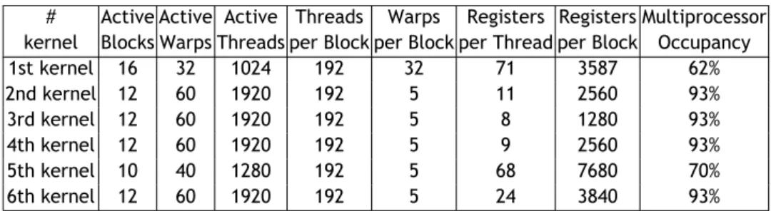
kernel) . . . 31 3.10.5 Computation of the triangle vertices (5-th kernel) . . . 31 3.10.6 Computation of normal vectors to the surface (6-th kernel) . . . . 32 3.10.7 Merging Partial Triangulations and Rendering . . . 32 3.11 Optimization of CUDA Kernels . . . 32 3.12 Results and Performance Evaluation . . . 35 3.13 Concluding Remarks . . . 39
4 GPU-Based Detection of Protein Cavities using Gaussian-like Implicit Surfaces 43 4.1 Introduction . . . 43 4.2 Related Work . . . 45 4.3 Background . . . 47 4.4 Cavity Detection Algorithm . . . 49 4.5 GPU Implementation . . . 49 4.5.1 Reading Atomic Centers from a PDB File . . . 50 4.5.2 Computation of the Bounding Box . . . 50 4.5.3 Voxelization of the Bounding Box . . . 50 4.5.4 Computation of the Scalar Field F . . . 51 4.5.5 Collecting Cavity Voxels . . . 51 4.5.6 Formation of Cavities . . . 51 4.6 Molecular Triangulation . . . 52 4.7 Results . . . 53 4.7.1 Hardware/Software Setup . . . 53 4.7.2 Time Performance . . . 53
4.7.3 Memory Space Complexity . . . 56 4.7.4 CUDA Code Optimization . . . 56 4.7.5 Comparison with other Geometric Algorithms . . . 58 4.7.5.1 Ground-Truth Dataset of Cavities . . . 58 4.7.5.2 Benchmarking Methods . . . 59 4.7.5.3 Benchmarking Metrics . . . 59 4.8 Concluding Remarks . . . 63
5 A Curvature-Based Cavity Detection Method 65
5.1 Introduction . . . 65 5.2 Related Work . . . 66 5.3 Theoretical Background . . . 68 5.3.1 Scalar Fields . . . 68 5.3.2 Gaussian Molecular Surface . . . 69 5.3.3 Critical Points . . . 69 5.4 Cavity Detection Algorithm . . . 71 5.4.1 Overview . . . 71 5.4.2 Voxelization . . . 71 5.4.3 Evaluation of the Discrete Scalar Field . . . 72 5.4.4 Computation of Critical Points . . . 72 5.4.5 Computation of the Number of Critical Points . . . 74 5.4.6 Clustering Critical Points into Cavities . . . 74 5.4.7 Surface Triangulation . . . 74 5.4.8 Rendering of Surface and its Pockets . . . 74 5.5 Results . . . 76 5.5.1 Hardware/Software . . . 76 5.5.2 CUDA Code Optimization . . . 77 5.5.3 Ground-Thruth Molecule Dataset . . . 79 5.5.4 Time Performance . . . 79 5.5.5 Memory Space Performance . . . 81 5.5.6 Accuracy Testing . . . 81 5.5.6.1 Benchmarking Methods and Metrics . . . 81
5.5.6.2 Benchmarking Metrics . . . 82 5.6 Concluding Remarks . . . 86
6 Conclusions and Future Work 87
6.1 The Revisited Research Plan . . . 87 6.2 Final Conclusions . . . 88 6.3 Future Work . . . 88
List of Figures
2.1 (a) Voronoi diagram of a molecule (i.e., set of spherical atoms); (b) convex hull of the atom centers, together with Delaunay triangulation; (c) alpha shape with triangles, edges, and vertices in black, where the empty triangles denote the existence of a cavity (taken and modified from [LWE98] [WPS07]). . . 11 2.2 (a) van der Waals surface in black, and inner blending surface as a
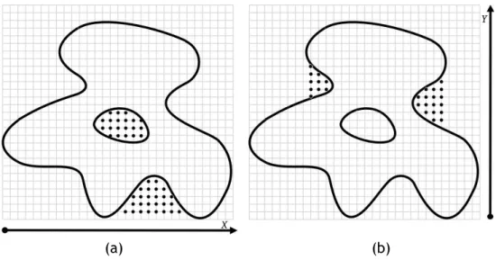
con-nected arrangement of blue and black spherical patches; (b) inner blend-ing mesh constructed from the atom enters and blendblend-ing surface; (c) outer blending surface as a connected arrangement of red and black spherical patches; (d) outer blending mesh as the convex hull of atom centers (taken from Kim et al.[KCC+08]). . . 12 2.3 Detecting cavities through POCKET: (a) in the x-direction; (b) in the
y-direction. . . 14 2.4 Detecting cavities through LIGSITE: (a) in the -45◦-direction; (b) in the
+45◦-direction. . . 15 2.5 Detecting cavities using the center of gravity of the molecule and
succes-sive nearest surface atoms (NSAs): (a) the first cavity; (b) the remaining four cavities (picture taken from [LJ06]). . . 16 2.6 Detecting a cavity through SURFNET: (a) Each probe sphere is placed at
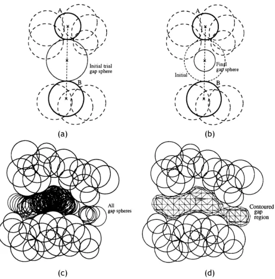
the midpoint of a pair of atoms (A, B); (b) but, if such probe sphere overlaps at least an atom (dashed spheres), its radius has to be reduced until it just has a tangential contact with the overlapped atom; (c) all probe spheres placed into cavity after considering all pairs of atoms; (d) the cavity surface enclosing all probe spheres of the cavity (pictures taken from [Las95]). . . 17 2.7 Detecting cavities through PASS: (a) coating the molecular surface with
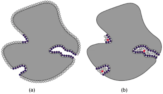
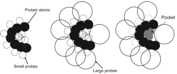
a first layer of probe spheres; (b) a second layer of probe spheres is, in this case, enough to find cavities on molecular surface (pictures taken from [WPS07], and inspired by [BS00]). . . 18 2.8 Detecting cavities through PHECOM: (a) Small probes are placed on the
Van der Waals surface; (b) large probes are placed on the Van der Waals surface; (c) small probes that overlap with the large ones are removed (taken from [KG07]). . . 19
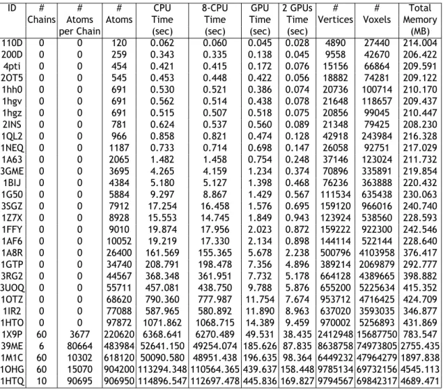
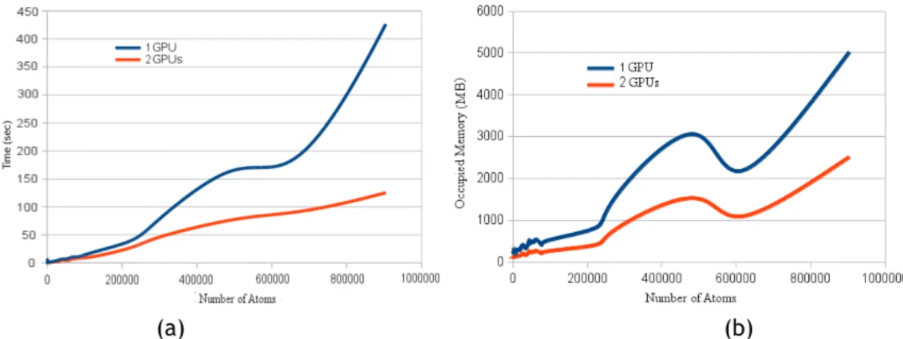
3.2 Practical complexity of GPU-based programs in terms of the number of atoms: (a) time complexity; (b) memory space complexity. . . 39 3.3 Examples of molecular surfaces displayed using GPU-based marching cubes
algorithm: (a) PDB id: 39ME; (b) PDB id: 1M1C; (c) PDB id: 1OHG. . . 41 3.4 Examples of molecular surfaces displayed using GPU-based marching cubes
algorithm: (d) PDB id: 1HTO, (e) PDB id: 1X9P, (f) PDB id: 2G34. . . 42
4.1 Molecular surfaces with the same blobiness β = 0.4 and distinct isovalues: (a) c = 1.0; (b) c = 1.2; (c) c = 1.4; (d) c = 1.6; (e) the previous four molecular surfaces (a)-(d) overlapping. . . 47 4.2 Molecular surfaces with the same isovalue c = 1.0 and distinct blobiness
values: (a) β = 0.34; (b) β = 0.32; (c) β = 0.30; (d) β = 0.28; (e) the previous four molecular surfaces (a)-(d) overlapping. . . 47 4.3 Molecular visualization of the 110D molecule: (a) cavities are not
de-picted on the surface; (b) cavities are dede-picted on the surface as deter-mined by our algorithm. The small spheres in blue indicate the location of cavities as calculated by MetaPocket [Hua09]. . . 52 4.4 (a) Time performance of CPU algorithm with (in red) and without (in blue)
surface triangulation; (b) Time performance of GPU algorithm with (in red) and without (in blue) surface triangulation. . . 53 4.5 (a) Memory space occupancy with respect to the number of atoms (n);
(b) the number of voxels in memory with respect to the number of atoms (n). . . 54 4.6 Histograms of hit performance of GaussianFinder in relation to LIGSITE,
PASS, SURFNET, POCASA, and fpocket: (left) numerical hit performance
ni in function of the dispersion of the number of cavities ∆i; (right)
posi-tional hit performance hi in function of the dispersion of the number of
cavities ∆i. . . 57
4.7 Cumulative cavity percentage of various detection methods in function of the distance d to ground-truth geometric centers: GaussianFinder, LIGSITE, PASS, SURFNET, POCASA, and fpocket for apo (left) and holo (right) structures. . . 60 4.8 Examples of 3 proteins and their cavities as detected by GaussianFinder
(left) and LIGSITE (right): (a) PDB id: 1NEQ (13 cavities); (b) PDB id: 1SVR (10 cavities); (c) PDB id: 2NWD (15 cavities). . . 64
5.1 Critical points as red balls considering: (a) the voxels outside or inter-secting the surface; (b) only the voxels interinter-secting the surface. . . 73
5.2 CriticalFinder (left) and LIGSITE (right) output the same number of cavi-ties on the same locations for two proteins: (a) PDB id: 2gqv (20 cavicavi-ties); (b) PDB id: 2nwd (18 cavities). . . 75 5.3 Time performance of the algorithm with (in red) and without (in blue)
the triangulation and rendering steps: (a) CPU implementation; (b) GPU implementation. . . 78 5.4 (a) Memory space occupancy with respect to the number of atoms (n);
(b) the number of voxels in memory with respect to the number of atoms (n). . . 78 5.5 Histograms of hit performance of CriticalFinder in relation to LIGSITE,
PASS, SURFNET, POCASA, and fpocket: (left) numerical hit performance
niin function of the dispersion of the number of cavities ∆i; (right)
posi-tional hit performance hi in function of the dispersion of the number of cavities ∆i. . . 82
5.6 Cumulative cavity percentage of various detection methods in function of the distance d to ground-truth geometric centers: CriticalFinder, LIGSITE, PASS, SURFNET, POCASA, and fpocket for apo (left) and holo (right) struc-tures. . . 85
List of Tables
3.1 Kepler GPU's memory and compute capabilities. . . 33 3.2 Performance data before optimizing the CUDA kernels. . . 34 3.3 Performance data after optimizing the CUDA kernels. . . . 34 3.4 Time performance/memory occupancy for a number of molecular
sur-faces before optimization. . . . 36 3.5 Time performance/memory occupancy for a number of molecular
sur-faces after optimization. . . 37 4.1 Kepler GPU's memory and compute capabilities. . . 55 4.2 Performance data before optimizing the CUDA kernels. . . 55 4.3 Performance data after optimizing the CUDA kernels. . . . 55 4.4 Hit performance (ni and hi) of GaussianFinder with respect to LIGSITE,
PASS, SURFNET, POCASA, and fpocket. . . 57 5.1 Kepler GPU's memory and compute capabilities. . . 76 5.2 Performance data before optimizing the five CUDA kernels (i.e., five steps
of CriticalFinder). . . 76 5.3 Performance data after optimizing the CUDA kernels. . . . 76 5.4 Hit performance (ni and hi) of CriticalFinder with respect to LIGSITE,
List of Abbreviations
3D Three dimensionalÅ Angstrom
ALU Arithmetic logic units
API Application programming Interface APROPOS Automatic Protein Pocket Search
ASPs Active site points CPU Central Processing Unit
CUDA Compute Unified Device Architecture CUDDP CUDA Data Parallel Primitives Library
GB Gigabytes
GPU Graphical processor unit HPC High-performance computing
MB Megabytes
MC Marching Cubes NBO Normal Buffer Object NSA Nearest surface atom OpenCL Open Computing Language OpenMP Open Multi-Processing
PDB Protein Data Bank PhD Philosophiae doctor
SAS Solvent-accessible surface
sec Seconds
SES Solvent-excluded surface SM Streaming Multiprocessors STL Standard Template Library VBO Vertex buffer object VDB Virus Data Bank VDW Van der Waals
Chapter 1
Introduction
This chapter briefly describes the rationale behind the research carried out during the PhD programme, which fits in the scope of geometric computing and molecular graphics.
1.1
Motivation
Most organisms in the world are made up of cells, which in turn are composed by molecules that are fundamental in biochemical processes that sustain life. Essentially, molecules interact with other molecules, so these interactions are the basis for almost cellular processes (e.g., DNA transcription).
Interactions between molecules occur on specific molecular regions, also called binding sites. But, not all binding sites of a molecule are compatible with the binding sites of another molecule. For example, the challenge in molecular docking is to know the compatible sites of two coupling molecules in advance. Note that the compatibilility between two molecules must occur at both geometric and chemical levels.
That requires a deep understanding of how a molecule binds to a specific surface region of another molecule, and how such information can be used to predict which molecules are supposed to bind to that region. Because of the structural variety and complexity of molecules, such an understanding seems to be a rather difficult task to accomplish. Aware of these difficulties, the scientific community has used to computational re-sources to make this problem more tractable and timely. However, most of the under-lying algorithms are not able to discriminate between correct results and false positives obtained in the simulation. In a way, this is so because the known shape descriptors are not able to output the (local and global) shape of a molecule uniquely, i.e., without ambiguities, not to say timely.
As known, shape plays a key role in bio-molecular recognition and function, in particular in the non-covalent interactions between molecules, as needed in applications such as drug design and protein engineering. Binding site recognition is the computational approach that allow us to locate or predict the location where cavities of a molecule are. The identification of protein binding sites has significant impact on understanding protein function. With the increasing number of protein datasets available across the web, predictive methods using geometric information only for protein interaction have drawn increasing interest. Currently, there are over 60 thousand protein 3D structures known, and this number is even increasing. One can use these data to predict the
binding sites in a protein structure of interest. Thus, the identification of those binding sites is often the first step to study protein functions and structure-based drug design. The focus of this work is to predict the molecular binding sites, that is, to find regions on the protein surface that can bind to other molecules (i.e., ligands), exclusively using geometric algorithms. Such protein surface regions are here called cavities, which include pockets, voids, tunnels, and so forth. But, as mentioned above, not all protein cavities are ligandable. That is, cavities are tentative binding sites of proteins.
1.2
Cavity Detection Methods: an Overview
In general, the computational algorithms to find cavities on a molecule divide in four categories: geometry-based, energy-based, evolutionary-based, combined approaches [VGGR10]. However, in this thesis, we are only interested in geometry-based algo-rithms. These geometric-based algorithms divide in three categories [KG07]:
• Grid-based • Sphere-based • Triangulation-based
Grid-based algorithms consist in mapping a protein onto an axis-aligned 3D grid, after
which we apply a scanning method to label each grid point as occupied or not by the protein, using then some geometric filtering method that outputs the non-occupied grid points that are deemed to be cavities or pockets. For example, Levitt and Banaszak determine which grid points lie in a pocket or a cavity by checking whether each of them is bracketed by two occupied points in either the x, y, or z directions [LB92]. Two significant drawbacks of grid-based methods is that they are not invariant to position and orientation of the molecule in 3D, i.e., the number of cavities and their locations may be distinct. The ambiguity of these methods becomes even more striking if one takes into account that it is not trivial to distinguish among all nodes outside the molecule those belonging to cavities. Besides, these methods are memory space-consuming because they have to hold the grid of nodes in memory.
The leading idea of sphere-based algorithms is to detect cavities, surface grooves and surface pockets by fitting probe spheres into them in some manner, defining then a surface around the resulting cluster of probe spheres. That is, these algorithms make usage of flood-filling procedure of probe spheres into cavities, grooves and pockets [Las95]. On average, sphere-based methods perform slower than grid-based methods in detecting of pockets, although they consume much less memory because it is not necessary to proceed to the voxelization of the 3D space surrounding the protein.
Triangulation-based algorithms usually focus on alpha shapes. An alpha shape is
possesses the same number of pockets, concavities, and tunnels [EM94]. More specifi-cally, an alpha shape can be defined as a subset of Delaunay tessellations of atom cen-ters, where the removal of edges longer than the sum of the radii of two atoms allows to detect pockets. Examples of algorithms that fall in this category are CAST [BNL03] and APROPOS (Automatic Protein Pocket Search) [PFF96]. Alpha shape methods for pre-diction of protein binding sites are also time-consuming, at least for large molecules. In general, the aforementioned geometric algorithms for the search of binding sites are very demanding in terms of mathematical computations, in particular when the number of atoms goes beyond some thousands. To keep the computational runtimes within reasonable limits, most algorithms only deal with small molecules. These circumstances get worse when we use 3D voxelizations of the space surrounding a molecule, as it the case of grid-based methods, since the computational complexity tends to be cubic, unless we take advantage of the parallel computing facilities (e.g., CUDA and OpenCL) of modern graphics cards.
1.3
Research Hypothesis
This thesis addresses a distinct approach to segment protein surfaces into clefts and knobs (i.e., concavities and convexities). We can even say that this approach leads to a new category of predicting methods for molecular cavities as tentative binding sites. Essentially, it is based on the theory of scalar fields, in particular scalar field topology. So the thesis statement behind the research described in this thesis can be stated as follows:
It is feasible to detect molecular cavities (as tentative binding sites) using shape descriptors based on the theory of scalar fields.
Particularly, we intend to investigate the relationships between critical points of the scalar field outside the molecular surface of the protein and its cavities. The computa-tion of the critical points allows us to identify the exact locacomputa-tion of pockets or clefts on the protein surface. Moreover, we believe that this can be done without using any 3D spatial grid or sphere-based coating of the molecular surface, or even any triangulation, but this has not been tried in the course of this thesis.
1.4
Research Plan
In the course of our research to prove the thesis statement mentioned above, we had to achieve a number of milestones, as described below:
• Molecular Surfaces and Electron Density Fields of Molecules. This step aimed at firstly finding a suited mathematical formulation for molecular surfaces. But,
we had also in mind an adequate mathematical formulation to describe the elec-tron density field generated by the molecule. As described in this thesis, we use Gaussian-like scalar fields to model both the electron density field and the surface of each molecule. This two-in-one solution is advantageous in many re-spects, in particular in respect to finding the critical points of electron density field of each molecule. Additionally, molecular interactions in physiochemical processes mean that molecules are not static and, consequently, their surfaces are not static either. Molecular surfaces changes over time depending on the interac-tions a molecule establishes with others locally. In atomic terms, this means that atoms change their positions within a molecule, which adopts different confor-mations depending on various factors such as temperature, hydrophobic effects, etc. Unlike those models found in the literature, our mathematical model keeps the spherical atoms and their surface envelope together. In this way we are able to directly associate each surface point to a single atom. Such a surface envelope of a molecule has been defined as a Gaussian-like surface, which can be easily triangulated and rendered using the marching cubes algorithm [LC87].
• Molecular Triangulation Algorithm. In order to visualize the results of our cavity detection algorithms, we primarily needed to develop a triangulation algorithm for Gaussian-like molecular surfaces, which was inspired in Blinn's formulation [Bli82]. The leading idea was to tackle the problem of triangulating and rendering surfaces of molecules with at least 0.5 million atoms in real-time. This real-time requirement led us to develop triangulation algorithms on GPU, in particular using CUDA.
• Intrinsic Shape Descriptors. An intrinsic shape descriptor is one that is invariant to geometric transformations, typically rotations and translations. These shape descriptors should take into account the local shape of the surface surrounding a given point, and also be robust to noise and sampling errors. Shape descrip-tors should also have a meaningful comparison function, one that scales roughly linearly with perceived shape change and is robust to noise. Example of shape descriptors with such characteristic are spherical harmonics algorithms, Laplace-Beltrami descriptors or surface curvature. However, in general, shape descriptors operate on the molecular surface only, not taking into account the surrounding space of the molecular surface. With such domain-extended analysis it is pos-sible to know the surface regions where the interactions are pospos-sible with other molecules. Thus, the analysis of protein cavities is of crucial importance to under-stand the biological processes in which proteins are involved in. Herein, we focus on the computational analysis of protein cavities, which is based on scalar field topology. This mathematical theory allows us to detect and recognize the loca-tion of different protein cavities, without spending much time and computaloca-tional resources.
developed protein cavity detection algorithms based on intrinsic shape descrip-tors. The first algorithm uses two implicit surfaces of a level set generated by a Gaussian-like scalar field. This technique solves the ambiguity problem of grid-based methods used in protein cavity detection. The second algorithm uses the same Gaussian-like scalar field, but the cavities are detected using scalar field topology, i.e., the critical points of the such Gaussian-like scalar field. These algorithms were also implemented on GPU via CUDA.
• Thesis Writing. The thesis was being written in agreement with the course of the PhD programme, and also with the pace of publishing scientific papers. So, the core chapters of this thesis have given place to papers published or under revision in journals and conference proceedings.
1.5
Contributions
Taking into account the thesis statement mentioned above, it can be said that the main contribution of the research work behind this thesis is the following:
• It is possible to detect cavities on the protein surface using scalar field topology, independently of the position and orientation of the protein. In other words, it is possible to identify the protein cavities without ambiguities.
As seen further ahead, such cavities correspond to particular critical points of the scalar field that describes both the molecular surface and the electron density field generated by the protein. As secondary contributions, we would list the following:
• A fully multi-GPU-based multi-threaded variant of the marching cubes algorithm to triangulate and render molecular surfaces, with all the particularities of com-puting the molecular surface locally in the neighborhood of each atom.
• An algorithm for detecting cavities on molecular surfaces through the Boolean difference of the interiors of two Gaussian molecular surfaces that approximate the same molecule.
• An algorithm based on an intrinsic shape descriptor taken from the scalar field topology to identify the molecular cavities without ambiguities.
These algorithms sustain themselves on grid-based methods, i.e., on the voxelization of the domain. Nevertheless, we believe that it is possible to implement the latter al-gorithm without voxelizing the domain; in particular using path minimization and max-imization in the domain (cf. [Gom14] for more details). This is an open issue for future work.
1.6
Publications
Within the scope of the research behind this thesis, we have produced the following articles, most of which already are published in journals and conference proceedings:
• Sérgio Dias and Abel Gomes. A Scalar Field Topology-Based Method for the De-tection of Protein Cavities. IEEE/ACM Transactions on Computational Biology and
Bioinformatics (submitted for publication).
• Sérgio Dias and Abel Gomes. GPU-Based Detection of Protein Cavities using Gaussian-like Implicit Surfaces. IEEE/ACM Transactions on Computational Biology and
Bioin-formatics (under 2nd revision).
• Sérgio Dias and Abel Gomes. Triangulating Gaussian-like Surfaces of Molecules with Millions of Atoms. Chapter in Walter Rocchia and Michela Spagnuolo (eds.), Computational Electrostatics for Biological Applications, Elsevier, Chapter 9, Springer-Verlag, pp.177-198, 2015.
• Sérgio Dias and Abel Gomes. Triangulating Molecular Surfaces over a LAN of GPU-Enabled Computers. Parallel Computing, Vol.42, pp.35-47, 2015.
• Sérgio Dias and Abel Gomes. Triangulating molecular surfaces on multiple GPUs. In Proceedings of the of the International Workshop on Parallelism in Bioinformatics (PBio'13), held as part of 20th European Conference on Message Passing Interface (EuroMPI'13), Madrid, Spain, September 15- 18, ACM Press, 2013.
• Sérgio Dias and Abel Gomes. Graphics processing unit-based triangulations of Blinn molecular surfaces. Concurrency & Computation: Practice & Experience, Vol.23, no.17, pp.2280-2291, 2011.
• Sérgio Dias and Abel Gomes. CUDA-based Triangulation of Convolution Molecular Surfaces. In Proceedings of the 5th International ACM Symposium on High Perfor-mance Distributed Computing (HPDC'10), Workshop on Emerging Computational Methods for the Life Sciences (ECMLS'10), Chicago, USA, June 21-25, ACM Press, 2010.
• Sérgio Dias and Abel Gomes. GPU-based Triangulations of the van der Waals Sur-face. In Proceedings of the IEEE International Conference on Bioinformatics & Biomedicine (BIBM'10), Hong Kong, December 18-21, IEEE Press, 2010.
1.7
Organization of the Thesis
This doctoral thesis aims at introducing new ways of understanding, representing, and detecting cavities on protein surfaces. In this context, the thesis is structured as fol-lows:
• Chapter 1: This chapter introduces the research work underlying the present PhD thesis. Here the rationale behind the research done during the PhD programme is presented, specially in the area of geometric representation and recognition methods of protein cavities, as needed, for example, in protein docking.
• Chapter 2: This chapter carries out the review of the literature concerning cavity detection methods. Therein, it is presented the general ideas behind these meth-ods, with a special emphasis on their leading ideas, advantages, and limitations. • Chapter 3: In this chapter a multi GPU-based triangulation algorithm for
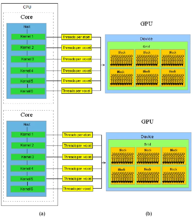
molec-ular surfaces is described in detail. It is a fast, scalable, parallel triangulation algorithm for molecular surfaces that takes advantage of multicore processors of CPUs and GPUs of modern hardware architectures, where each CPU core works as the master of a single GPU, being the processing burden distributed over the CPU cores available in a single computer or a cluster. The algorithm is based on a parallel version of the marching cubes algorithm that triangulates molecular sur-faces on multiple GPUs using CUDA and OpenMP. Besides it is carried out a study that compares a sequential version (CPU) to a parallel version (GPU) of well-know marching cubes (MC) algorithm to render molecular surfaces.
• Chapter 4: This chapter presents an algorithm that detects protein cavities through the analysis of the voxels located between two molecular surfaces. The novelty of the method lies in the fact that it makes usage of the scalar field of the protein in 3D space, from which we are able to define two distinct surfaces for the protein, being that the protein cavities are in the middle. Besides, as far as we know, this is the first geometric detection algorithm for protein cavity detection on GPU. • Chapter 5: This chapter proposes a new molecular shape descriptor to recognize
protein cavities through the topology (i.e., critical points) of the scalar field that features the electron density field of the protein. The approach is based on the study of the (normalized) Hessian matrix at points outside the molecular surfaces, not at points on the surface of the protein. Thus, the main novelty of the method relies in the use of the Morse theory as applicable to scalar fields. Furthermore, we compare our algorithm with others, in order to better assess the quality of the found cavities.
• Chapter 6: This chapter concludes this thesis, with important clues for future developments of this work.
As a marginal note, let us say that the target audience of this thesis is not only the com-munity of computer graphics and geometric computing, but primarily the comcom-munity of computational biology and bioinformatics, for whom we have designed the algorithms described herein.
Chapter 2
Cavity Detection Methods for Proteins: a Survey
The detection of protein cavities provides useful information about biological processes (e.g., protein docking). However to know the location of such regions it is necessary to collect information about the position, and the type of atoms that comprise a given molecule. Usually, this is done by X-ray crystallography, which is a technique based on the diffraction patterns of a X-ray irradiation in a crystal molecule. The result is then recorded in an appropriate plate that provides information about the position of each atom. Fortunately, for these molecules, their constituent atoms, and 3D atomic coordinates (x,y,z values) can be retrieved from a PDB file. With the three-dimensional structure of a protein, it is possible to find places where other molecules may bind. Such interactions generally happens in specific regions of the protein, called cavities, which usually correspond to pockets, clefts, inner cavities, or grooves on the surface of a given protein. In the literature, there are essentially three categories of computational algorithms to detect cavities on the protein surfaces: evolution-based, energy-based, and geometry-based. However, this chapter only surveys geometric algorithms.
2.1
Introduction
In 1894, Fischer conducted the initial studies on detection of protein cavities. From these studies, he concluded that the binding of a molecule to another is similar to the paradigm of inserting a key into a lock. In other words, this means that the affinity between two molecules exists if the shape of a molecule matches the shape of the other. However such model was considered very simplistic, because shape cannot be the only factor that influences the detection of protein cavities, since proteins are highly flexible and change shape over time. Generally, protein binding sites are specific large and deep clefts [LLST96]. However, protein shape can vary considerably, depending on the protein we have in hand. For example, the protein binding site of a ribonuclease is an extended rut, while the protein binding site of a endonuclease is a spherical cavity, and for a enzyme it is usually the largest cavity [LWE98].
In fact, a protein can bind many types of molecules, largely because of its non-negligible number of cavities. Many properties can be inferred from these molecular regions, helping us to understand how molecular interactions take place, as well as to be aware of important information on protein structures in the design of compounds, as usual in pharmaceutical and biotechnological domains. But, some of those protein zone targets are more receptive to bind with certain ligands than others. Those regions generally
have larger surface areas, and correspond to the concave, cleft or hole-shaped regions on a protein surface [KG07].
With molecular complexes involved in various molecular interactions, it is necessary to have adequate tools to characterize protein cavities as, for example, shape, size, and depth. But, a protein cavity only becomes a binding site if a number of factors like ambient temperature, pH, shape complementarity, electrostatics, hydrogen-bonding, and solvent interactions, combine gracefully. It is the conjugation of all of these factors that enable a ligand to identify the correct place to bind a given protein and induce its biological effect [HK06].
To make laboratory experiences easier it would be helpful to have computational ods capable of simulating such bio-chemical processes. However, computational meth-ods capable of simulating such processes are extremely difficult to recreate. The dif-ficulties behind that are related with the variety of admissible ligands, the variety of protein cavities, the protein shape variations themselves, and the physico-chemical factors that act on a cavity region.
It happens that such regions are usually located in pockets, clefts, inner cavities, and grooves on protein surfaces. Therefore, a better understanding of the process entan-gled in binding proteins requires the detection of cavities on the molecular surfaces. A computational estimate of the location of such protein regions, before initiating any ex-perimental work in the drug discovery process, may be instrumental in the improvement of the design of new drugs. For that purpose, a number of algorithms for predicting and identifying protein cavities have been develop so far. Such algorithms can be divided into three major categories:
• Evolutionary algorithms: They rely on multiple sequence alignments to find the location of cavities on a given protein surface.
• Energy-based algorithms: In this case, cavities are detected through the calcu-lation of the interaction energies between protein atoms and a small-molecule probe (e.g., Grid [Goo85], QSiteFinder [LJ05], and AutoLigand [HOG08]).
• Geometric algorithms: These algorithms are based on the analysis of geomet-ric properties of a molecular surface to detect cavities (e.g., SURFNET [Las95], LIGSITE [HRB97], and PocketDepth [KC08]).
However, each method has its own drawbacks. For example, geometric methods rely-ing on a grid are sensitive to protein position and grid spacrely-ing. Energy-based methods depend on their filtering procedures, force field parameterizations, and scoring func-tions. In turn, evolutionary-based methods depend on the quality of the alignment tool, and also on the number of available sequences. These problems show us that there is still a long way to go in this field, so that there is a need for further analysis of all the processes involved in the detection of binding sites of proteins [KG07]. This explains why the detection of molecular cavities still is a very active research area [HSAH+09].
2.2
Triangulation-Based Methods
The foundations of the triangulation-based methods lie in the field of computational geometry, in largely after the introduction of alpha shapes by Edelsbrunner and Mucke [EM94], in 1994. Edelsbrunner is a well-known computer scientist in the field of com-putational geometry. Interestingly, Edelsbrunner himself and colleagues [EFFL95] pub-lished a work on measuring proteins and voids in proteins in 1995.
In 1996, Peters et al. [PFF96] introduced new methods to detect molecular cavities through Voronoi diagrams, as in APROPOS program. The main objective was the devel-opment of an algorithm to identify cavities in proteins using only geometric criteria. In 1998, this technique was further developed by Liang et al. [LWE98], as illustrated in Fig. 2.1, which was called CAST. For that purpose, Liang and colleagues' algorithm firstly creates a Voronoi space decomposition from the atoms (atomic coordinates) of the molecule (Fig. 2.1(a)), from which one calculates the corresponding convex hull (i.e., Delaunay triangulation) (Fig. 2.1(b)), removing then the simplexes (e.g., trian-gles) that are not completely inside the molecule, resulting so in an alpha shape of the original molecule (Fig. 2.1(c)). The leading idea here is to get a triangulation with the same topological type as the original set of atoms that comprise the molecule, so that we can extract the cavities in a straightforward manner.
(a) (b) (c)
Figure 2.1: (a) Voronoi diagram of a molecule (i.e., set of spherical atoms); (b) convex hull of the atom centers, together with Delaunay triangulation; (c) alpha shape with triangles, edges, and vertices in black, where the empty triangles denote the existence of a cavity (taken and modified from [LWE98]
[WPS07]).
Another algorithm based on the same steps as the CAST algorithm was developed by Yaffe et al. [YFW+08], and called MolAxis. The only difference is in the type of surface that is used to detect cavities; CAST uses the molecular surface, while MolAxis uses the van der Waals surface. Also, SplitPocket developed by Tseng et al. [TDCL09], in 2009, has the same steps of CAST [LWE98], but SplitPocket has the capability of splitting a cavity if necessary through the analysis of atomic structure. In 2013, Sridharamurthy et al. [SDP+13] introduced a similar algorithm to CAST, with the difference that one uses
In 2007, Xie et al. [XB07] proposed an algorithm similar to CAST (cf. Fig. 2.1), but they introduced a new shape descriptor, the geometric potential, in order to distinguish lig-and binding sites from non-liglig-and binding sites. Following the same line of research, Kim et al. [KCC+08] built up a blending mesh of triangles derived from a surface gen-erated from blending atoms, as illustrated in Fig. 2.2. Then, they construct the convex hull from such a blending mesh. Cavities are found in places of the convex hull that are not occupied by the blending mesh. Interestingly, Kim et al. [KCC+08] use the Voronoi diagram of atoms, not the Voronoi diagram of atom centers, to easily calculate the molecular surface.
(a) (b)
(c) (d)
Figure 2.2: (a) van der Waals surface in black, and inner blending surface as a connected arrangement of blue and black spherical patches; (b) inner blending mesh constructed from the atom enters and blending surface; (c) outer blending surface as a connected arrangement of red and black spherical patches; (d)
outer blending mesh as the convex hull of atom centers (taken from Kim et al.[KCC+08]).
Guilloux et al. [LGST09] introduced Fpocket, which builds upon on the concept of al-pha spheres due to Liang et al. [LWE98]. For that purpose, one has to compute the Voronoi tessellation of atom centers, what is performed using the publicly available Qhull's source code on http://www.qhull.org, a well-known package that primarily calculates the convex hull of a set of points through the Quickhull algorithm. Recall that an alpha sphere is an external sphere that stands in contact with four boundary atoms of the molecule simultaneously, featuring thus the local curvature of molecular surface. Thus, cavities are located where we find alpha spheres; this obviously requires the use of some clustering of alpha spheres to form such cavities. This means that locating alpha spheres is equivalent to detect cavities on protein surfaces.
2.3
Grid-based algorithms
Grid-based algorithms essentially map any molecule onto an axis-aligned 3D grid, using then a specific geometric parameter to detect eventual cavities on the molecular sur-face. The most common geometric parameters are the following: distance, visibility, and depth.
2.3.1 Distance-Based Grid Algorithms
In respect to distance-based grid algorithms, let us mention FRODO, which is due to Voorintholt et al. [VKV+89], and is considered by many as the first cavity detection
algorithm. This algorithm assigns a real value to every single grid point, which depends on whether such point is inside the molecule, between the van der Waals (vdW) surface and the solvent accessible surface (SAS), or beyond SAS. Such real value assigned to each grid point is produced by a real function that depends on the distance of such grid point to the nearest surface atom, so that we end up having a distance map associated to the grid. It is clear that cavities are located between the vdW surface and SAS, but truly speaking FRODO does not actually detect cavities [GT94], having it been designed only for the visualisation of SAS. In fact, as noted by Ho and Marshall [HM90], although FRODO is effective in finding regions where cavities are located, it is not that easy to isolate and define the extent of each specific cavity. To overcome this lack of specificity, Ho and Marshall [HM90] introduced the CAVITYSEARCH, which implements a search function to isolate and delineate each cavity of interest starting from a seed point, through a filling procedure of such cavity, thereby producing a cast of the cavity.
Another well-known distance-based grid algorithm is due to Zhang and Bajaj [ZB07]. More specifically, they use a signed distance function from the molecular surface to de-termine its pockets. The extraction of pockets can be performed for any closed compact molecular surface (e.g., van der Waals surface, Gaussian isosurface, and SAS) embed-ded in a regular grid, being the signed distance function to the surface evaluated for grid points far away and outside from the surface, as well as for grid points of cubic cells intersecting the surface. The cavities (e.g., pockets, tunnels, and voids) correspond to grid points outside the molecular surface where the signed distance function is positive.
2.3.2 Visibility-Based Grid Algorithms
In respect to visibility-based grid algorithms, also known as scan-based grid algorithms, they are all built upon the idea of line of sight or visibility; a line of sight is a scanning direction. This idea of scanning cavities along one or more directions was first proposed by Levitt and Banaszak [LB92]; their algorithm is known as POCKET. As a grid-based algorithm, this algorithm firstly maps the molecule onto an axis-aligned grid of equally-spaced points. The detection of cavities is carried out by scanning them along x, y, and
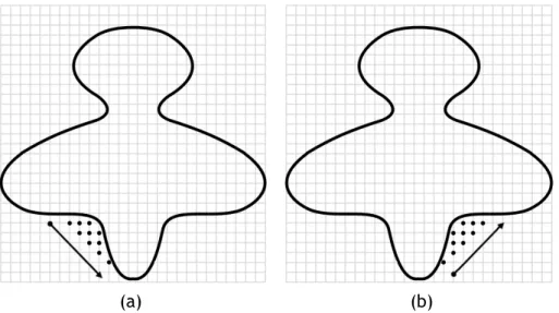
![Figure 2.5: Detecting cavities using the center of gravity of the molecule and successive nearest surface atoms (NSAs): (a) the first cavity; (b) the remaining four cavities (picture taken from [LJ06]).](https://thumb-eu.123doks.com/thumbv2/123dok_br/18663374.913319/48.892.111.752.106.391/figure-detecting-cavities-gravity-molecule-successive-remaining-cavities.webp)