Editor
Carla Nunes, FCT, Universidade do Algarve, Faro, Portugal
Editorial Board
Brion Duffy, Agroscope FAW Wadenswil Bacteriology, Switzerland Carla Nunes, FCT, Universidade do Algarve, Portugal
Christian Larrigaudiere, IRTA-Institut de Recerca i Tecnologia Agroalimentàries, Spain Josef Streif, Inst. Sonderkulturen & Produktsphysiologie, Hohenheim, Germany Maribela Pestana, FCT, Universidade do Algarve, Portugal
Maria Graça Barreiro, Instituto Nacional de Investigação Agrária, Portugal Maria Dulce Antunes, FCT, Universidade do Algarve, Portugal
Miguel Salazar, CICAE, Instituto Universitário Dom Afonso III, Portugal Mustafa Erkan, Akdeniz University, Turkey
Paolo Bertolini, Universita de Bologna, Italy Pol Tijskens, Wageningen University, Netherlands Shimshon Ben-Yehoshua, A.R.O. Volcani Centre, Israel Susan Lurie, A.R.O. Volcani Centre, Israel
The papers contained in this book report some of the peer reviewed Proceedings of the International Conference “Environmentally friendly and safe technologies for quality of fruit and vegetables”, but also other papers related with the subject were included. The manuscripts were reviewed by the Editor and Editorial Board, and only those papers judged suitable for publication were accepted. The Editor wish to thank to all the reviewers and authors for their contribution.
Proceedings of the International Conference “Environmentally friendly and safe
technologies for quality of fruit and vegetables”, held in Universidade do Algarve, Faro,
Portugal, on January 14-16, 2009. This Conference was a join activity with COST Action 924.Convener
Carla Nunes, Universidade do Algarve, Portugal
Scientific Committee
Carla Nunes, Universidade do Algarve, Portugal Amílcar Duarte, Universidade do Algarve, Portugal
Angelos Kanellis, Aristotle University of Thessaloniki, Greece Bart Nicolaï, Katholieke Universiteit Leuven, Belgium
Brion Duffy, Agroscope FAW Wadenswil Bacteriology, Switzerland
Christian Larrigaudiere, IRTA-Institut de Recerca i Tecnologia Agroalimentàries, Spain Domingos de Almeida, Universidade do Porto, Portugal
Josef Streif, Inst. Sonderkulturen & Produktsphysiologie Hohenheim, Germany Krzysztof Rutkowski, Research Inst. of Pomology and Floriculture, Poland Maria Dulce Antunes, Universidade do Algarve, Portugal
Maria da Graça Barreiro, Instituto Nacional de Investigações Agrárias, Portugal Mustafa Erkan, Akdeniz University, Turkey
Paolo Bertolini, Universita de Bologna, Italy Pol Tijskens, Wageningen University, Netherland Shimshon Ben-Yehoshua, A.R.O. Volcani Centre, Israel
Organizing Committee
Carla Nunes, Universidade do Algarve, Portugal Amílcar Duarte, Universidade do Algarve, Portugal Bart Nicolaï, Katholieke Universiteit Leuven, Belgium Maria Dulce Antunes, Universidade do Algarve, Portugal Maria Emília Costa, Universidade do Algarve, Portugal Maribela Pestana, Universidade do Algarve, Portugal
Miguel Salazar, Instituto Universitário Dom Afonso III, Portugal
Sponsors
COST, European Cooperation in the field of Scientific and Technical Research
Fundação para a Ciência e a Tecnologia
International Association of Students in Agriculture and Related Sciences, Faro
Serviço Técnico Pós-colheita do IRTA em Portugal Algarve.resorts.net
Câmara Municipal de Faro Câmara Municipal de Albufeira
Câmara Municipal de Aljezur Câmara Municipal de Lagos
Câmara Municipal de S. Brás de Alportel Crédito Agrícola, Caixa do Algarve A Farrobinha 80 g C.N. Kopke & Cª PrimeDrinks, S.A. Uniprofrutal Frutas Mourinho
Se c ti o n 3 . Q u a li ty m a n a g em en t o f f ru it a n d v eg et a bl eS
SECTION 3. QuALITy mANAgEmENT
OF FRuIT AND VEgETABLES
En v ir o n m En ta ll y F ri En d ly a n d S a FE tE ch n o lo g iES F o r Q u a li ty o F F ru it S a n d vE g Eta bl ES
16. DESERT PLANTS WITH mEDICINAL VALuE
Rivka Ofir
Dead Sea &Arava Science Center, Newe Zohar, 86910, Israel E-mail: rivir@bgu.ac.il
abstract
Treatment of cancer with chemotherapy has two main problems: toxicity to normal cells and failure to kill cancer cells. Cancer cells are characterized by uncontrolled cells proliferation and unlimited life span. Development of anti-cancer drug should involve the search for compounds capable of halting cell proliferation and/or leading to cell death. Combination of both types of drugs will make efficient chemotherapy. The compounds selected in this study are unique in their mode of action: they activate the protein procaspase-3, a critical enzyme in cell death process known as: apoptosis or programmed cell death. Although programmed cell death occurs naturally, too much or too little apoptosis cause diseases. Not enough apoptosis cause cancer. Apoptosis involves a cascade of enzymes (caspases) that are made as latent zymogens (pro-enzymes); procaspases activated following apoptotic death stimuli, lead to cleavage of cellular proteins, cleavage of DNA and cell death. The enzyme caspase-3 acts in a point of no return in this cascade. As such, compounds that will activate caspase-3 will be considered as potential anti-cancer drugs. In practice, for a compound to be considered as a potential lead drug, it should be a small molecule, stable, selective, and able to penetrate cellular membranes effectively. Following screening of plant extracts against several cancer models and through development of an assay that can detect compounds which activate caspase-3, several extracts capable of killing cancer cells by activating caspase-3 were identified.
Introduction
Cancer is one of the leading causes of death. In the United States there are 1.4 million cases of cancer per year and 0.5 million cases of death. Cancer of the prostate, breast, lung and colon are the most frequent type of cancers. onventional treatment work by killing the cancer cells with a dose of chemo-or radiotherapy. New generation of anti-cancer drug will be based on the knowledge regarding how cells kill themselves- a process called apoptosis or programmed cell death (Weil et al. 1996). Unlike cell death that occurs during and after tissue injury, apoptosis occurs in healthy tissue and does not trigger inflammation. Although programmed cell death occurs naturally, too much or too little cause disease. Excessive apoptosis may underlie the nerve damage in diseases such as Parkinson’s and Alzheimer’s. Not enough apoptosis, on the other hand, causes cancer. Too little apoptosis leads to too many cells; compounds capable of activating cell death will enable the reduction of cell number. The goal is a drug that triggers apoptosis in cancer cells but not in normal cells. At the heart of the conserved biochemical pathway that mediates the highly ordered process of apoptosis are a family of cysteine proteases, termed ‘caspases’ (cysteinyl aspartate-specific proteinases). Caspases are produced as precursor molecules that require processing into two subunits to produce a fully active enzyme (Thornberry & Lazebnik 1998). On the basis of primary structure, proapoptotic caspases can be divided into two classes, class I including caspases that contain a long amino-terminal prodomain, and class II with a short or absent prodomain. One of the key regulatory steps for apoptosis is the activation of caspases, leading to the characteristic morphological changes associated with apoptotic cells including chromatin condensation, DNA fragmentation into nucleosomal fragments, nuclear membrane break down, externalization of phosphatidylserine and formation of apoptotic bodies that are readily phagocytosed. The mechanism of caspase activation is poorly understood but recent studies demonstrate that procaspases can be activated through dimerization/oligomerization. Caspases are present as inactive pro-enzymes, most of which are activated following cleavage at a specific aspartate cleavage site and assembly of their active subunits. Caspase-8, caspase-9 and caspase-3 are situated at
Se c ti o n 3 . Q u a li ty m a n a g em en t o f f ru it a n d v eg et a bl eS
pivotal junction. Caspase-3 appears to amplify the caspase-8 and caspase-9 signal into a full commitment to disassembly. We have recently shown that p53-null T lymphoma cell lines, but not normal T cells, are highly sensitive to inhibition of gene/protein expression. These lymphoma cells seem to be primed for apoptosis but protected by an unknown labile protein (Ofir et al. 1999, and unpublished results). Some protein inhibit caspase activity: when they are not produced, apoptosis begins. The hope is to find drugs that will inactivate such inhibitors of apoptosis only in cancer cells.
Natural products research continue to be an invaluable source of leads, with examples such as antibiotics, analgestics such as epibatidine, the cholesterol-lowering drug mevinolin and the anticancer agent taxol. Purification of the active constituents of natural products enable to define their chemical structure and make it possible to chemically modify them to improve their efficacy and their safety. According to recent studies on apoptosis in plants it became clear that the process of apoptosis is conserved also in plants and as such it is reasonable to assume that there are activators/inhibitors of apoptosis in plants. It has been shown recently that natural compounds like resveratrol (Jang et al. 1997; Szende et al. 2000), wheat bran (Jenab & Thompson 2000), the Chinese herb Tripterygium Wilfordii hook (Lee et al. 1999), agents found in plant extracts like mistletoe and Semicarpus anacardium (Thatte et al. 2000) induce apoptosis. The progress in defining, at the molecular and cellular levels, the mechanisms of pathology in plants and the finding of caspase- like enzymes in plants (Korthout et al. 2000) make the search for caspase activating compounds in plant less dependent on serendipity than in earlier times.
In the present study we identified plant extracts capable of activating the protein procaspase-3. Two pathways for processing of procaspase-3 are described (Scheme 1): I. Cleavage between the large and small domains following by cleavage between the pro domain and the large domain. II. Cleavage between the pro domain and the large domain following by cleavage between the large and the small domains. Among the active extracts described in this project, one extract uses pathway II and three use pathway I for the activation of procaspase-3.
En v ir o n m En ta ll y F ri En d ly a n d S a FE tE ch n o lo g iES F o r Q u a li ty o F F ru it S a n d vE g Eta bl ES
results
In this section results will be presented showing that the selected active plant extract activate the enzymatic activity of caspase-3. The parameters tested were: enzymatic activity, DNA ladder, Western blotting and processing of procaspase-3 in situ (in the test tube) and in cellular extracts. In addition, the processing of procaspase-3 synthesized in reticulocytes lysate from the cDNA of procaspase-3 was also tested. The active plant extracts were identified by the following symbols: RO4 and RO39, are aqueous plant extracts; RO139, RO144 and RO197 are plant extracts prepared in ethanol.
Cytotoxicity of Plant Extracts in various tumor models
Cell lines representing various tumor models were incubated with plant extracts and cellular viability was measured; results are summerized in Table 1.
table 1. Viability of human tumor cell lines following incubation with plant extracts.
Plant Extract Human tumor models
Lymphoma Prostate cancer Melanoma Breasts cancer Ovarian
RO4 + + - -
-RO39 + - - -
-RO139 + + + +
-RO144 + + + -
-RO197 + + + +
-+ more than 50% killing; –less than 50% killing.
Viability was defined as cell survival above spontaneous death. Viability was measured with the reagant XTT ( XTT as a tetrazolium salt react with mitochondtrail enzymes that are active only in alive cells).
apoptosis of human and mouse hematopoietic Cell lines following Incubation with Plant Extracts
Cell were incubated with plant extracts and the apoptotic cells were identified by FACS analysis. The results are summerized in Table 2.
table 2. FACS analysis of leukemic cells following incubation with plant extracts. Plant Extract
Apoptotic cells (%)
Mouse cell line Human cell line
5 h 16 h 5 h 16 h Control 0.5 2 1 1 RO4 2 47 0 58 RO39 0.5 6 1 10 RO139 2 56 0 0 RO144 22 33 0 13 RO197 0 26 1 25
Caspase-3 activation following Incubation of tumor Cell lines with Plant Extracts
Initial screening for proapoptotic activity in plant extracts lead to the identification of 5 extracts containing agents capable of inducing caspase-3 enzymatic activity. The results of caspase-3 activity, as measured in cellular extracts following the incubation of mouse leukemic cells with plant extracts, are summarized in Table 3 and Fig 1.
Se c ti o n 3 . Q u a li ty m a n a g em en t o f f ru it a n d v eg et a bl eS
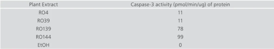
table 3. Caspase-3 activity following incubation of mouse leukemic cells with plant extracts*. Plant Extract Caspase-3 activity (pmol/min/ug) of protein
RO4 11
RO39 11
RO139 78
RO144 99
EtOH 0
*mouse leukemic cells were incubated with plant extracts for 6 h Caspase-3 activity was measured in cellular extracts using a colorimetric substrate according to the manufecturers instructions.
fig 1. The inhibitor DEVD-CHO inhibits Caspase-3 activity induced by plant extracts. Leukemic mouse cells were induced to undergo apoptosis by treatment with a plant extract. Cellular extracts prepared in lysis buffer (50 mM HEPES, 1 mM DTT, 0.1 mM EDTA, 0.1% CHAPS, pH 7.4) were incubated with the colorimetric substrate DEVD-pNA. Cleavage of the substrate by the active caspase-3 lead to changes in the absorbency at wavelength 405nm as measured along the experiment (every 15-30 min). Caspase-3 activity is expressed as arbitrary units of absorbance at 405 nm. In order to show the specificity of the cleavage, the enzymatic reaction was performed also in the presence of the caspase-3 specific inhibitor DEVD-CHO (0.1μM).
One of the hallmarks of apoptosis is the unique fragmentation of DNA into a ladder pattern. Caspase-3 activation following incubation of mouse leukemic cells with RO139, RO144 and RO197 resulted in DNA fragmentation (data not shown).
Processing of Procaspase-3 in Vitro following Incubation of Cellular Extract with Plant Extracts One of the goals of the project was to find a compound capable of directly activating procaspase-3. We developed an assay, which will enable us to follow in vitro the processing of the inactive procaspase-3 into
En v ir o n m En ta ll y F ri En d ly a n d S a FE tE ch n o lo g iES F o r Q u a li ty o F F ru it S a n d vE g Eta bl ES
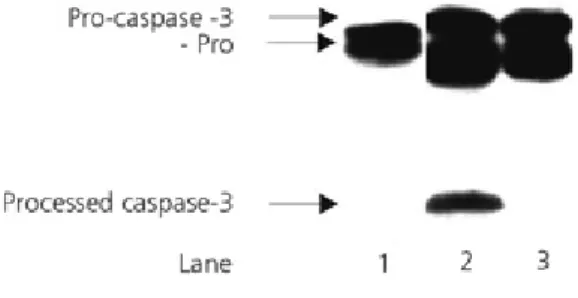
an active enzyme. A cellular lysate prepared from untreated cells was incubated with plant extract for 90 min at 37 ºC. According to the Western blotting shown in Fig 2, the extracts RO39, RO197, RO144 lead to the conventional processing of procaspase-3 (scheme 2 pathway I): 32kDa--->20kDa--->17kDa (lanes 1,3,4) while RO4 (lane 2) uses an alternative path (scheme2 pathway II): 32kDa--->29kDa--->17kDa. A similar processing mode is used by RO4 when incubated with procaspase-3 produced in a reticulocyte lysate (Fig 3 lane 1; the procaspase-3 protein is the translation product of the cDNA of procaspase-3). Heat inactivation of RO4 abolished its capability to process procaspase-3 (data not shown), indicating that RO4 contain an active protease.
fig 2. Activation of procaspase-3 following incubation of plant extracts with naïve cellular extract. Western blot analyses of the activation of procaspase-3. Procaspase-3 is detected in naïve cellular extract (extract prepared from untreated U937 cells) as 32kDa protein by antibody directed against the large subunit. The presence of large sububit indicates activation and is detected following treatment with RO39, RO4, RO197, and RO144, lanes 1,2,3,4, respectively. Treatment with RO39, RO197 and RO144 generated the intermediate pro+large subunit as well (lanes 1,3,and 4, respectively). Treatment with RO4 generate an intermediate consisit of large and small subunit (-pro).
fig 3. Procaspase-3 protein translated in reticulocytes lysate is processed following treatment with plant extracts. Western blot analyses of translation product of procaspase-3 cDNA incubated with caspase-3 activator plant extract C8 (lane 2) or with RO4 (lane 1). Detection was performed with anti caspase-3 antibodies.
Conclusion
The extracts RO4, RO39, RO139, RO144, RO197 directly induce caspase-3 processing and enzymatic activity. It is suggested that each extract contain at least one active compound that can be lead for anti cancer drug.
Se c ti o n 3 . Q u a li ty m a n a g em en t o f f ru it a n d v eg et a bl eS
references
Jang M, Cai L, Udeani GO, Slowing KV, Thomas CF, Beecher CWW, Fong HHS, Farnsworth NR, Kinghorn AD, Mehta RG, Moon RC, Pezzuto JM. 1997. Cancer chemotherapy activity of resveratrol, a natural product derived from grapes. Science 275:218-20
Jenab M, Thompson LU. 2000. Phytic acid in wheat bran affect colon morphology, cell differentiation and apoptosis. Carcinogenesis 21:1547-52
Korthout HA, Berecki G, Bruin W, van Duijn B, Wang M. 2000. The presence and subcellular localization of caspase 3-like proteinase in plant cells. FEBS Lett 475:139-44
Lee KY, Chang WT, Qiu D, Kao PN, Rosen GD. 1999. PG490 (Triptolide) cooperates with tumor mecrosis factor alfa to induce apoptosis in tumor cells. J Biol Chem 274:13451-5
Ofir R, Zhang LC, Adams JM. 1999. Interference with gene expression induces apid apoptosis in p53-null T lymphoma cells. Cell Differ 6:1216-21
Szende B, Tyihak E, Kiraly-Veghely Z. 2000. Dose-dependent effect of resveratrol on proliferation and apoptosis in endothelial and tumor cell cultures. Exp Mol Med 32:88-92
Thatte U, Bagadey S, Dahanukar S. 2000. Modulation of programmed cell death by medicinal plants. Cell Mol Biol 46:199-214
Thornberry NA, Lazebnik Y. 1998. Caspases: enemies within. Science 281:1312-6
Weil M, Jacobson MD, Coles HSR, Davis TJ, Gardner RL, Raff KD, Raff MC. 1996. Constitutive expression of the machinery for programmed cell death. J Cell Biol 133:1053-9