i FACULDADE DE CIÊNCIAS E TECNOLOGIA
Departamento de Ciências Biológicas e Biotecnologia
Identification of reliable reference genes for
quantitative gene expression in the Mozambique
tilapia, Oreochromis mossambicus.
Master’s Degree in Marine Biology
(especialização em aquacultura)
André Alçada Baptista Rebelo de Andrade
Supervisor: Professor Doctor Adelino CanárioCo-supervisor: Doctor Laurence Deloffre
ii
Acknowledgments
Gostaria de agradecer ao Professor Adelino Canário pela oportunidade que me concedeu de realizar este projecto sob a sua supervisão e pelas correcções. Queria também agradecer especialmente à Laurence por todos os preciosos ensinamentos, pela paciência nos meus piores momentos, por adaptar os horários do laboratório ao trabalho no cinema e pelas conversas divertidas. À Eduarda pelas correcções preciosas e a força transmitida.
À Alexandra por todos os qRT-PCRs e extracções que fizemos juntos, acho que formamos uma boa equipa.
À Rita e Ângela pela ajuda na histologia, e simpatia.
Ao Pedro e Nicolai pelos almoços, cafés e óptimas conversas
À Rute, Ricardo, Cristina e Marco, enfim a todo o pessoal que frequentava o 2.28 e que adorei conhecer.
Ao Peter pela disponibilidade do laboratório quando foi preciso dissecar os ovos naqueles infindáveis dias.
Especialmente à Débora, pela força, companheirismo e paciência quando eu não arranjava tempo para nada.
iii
I.
Abstract
Gene-expression analysis is increasingly important in biological research, with real-time reverse transcription polymerase chain reaction (qRT-PCR) becoming the method of choice for accurate expression profiling of selected genes. One or more appropriate reference genes must be selected to accurately compare mRNA across different samples and tissues. Although reference genes constitute the best normalizers, a major problem is that there is substantial evidence in the literature that most of the commonly used reference genes are regulated under some circumstances, such as at different stages of development or in response to different treatments, indicating that there may be no universal reference gene with a constant expression.
In this study we evaluated the expression of ten candidate reference genes (18S ribosomal RNA, β-actin, HPRT-1, GADPH, Tubulin A, TATA binding protein, Elongation factor 1 alpha, Beta-2 microglobulin, Cathepsin D, Cathepsin Z) by qRT-PCR in 13 distinct adult tissues and during five stages of oogenesis in Oreochromis mossambicus. Two different software tools (Genorm and Normfinder) were used to analyze the data given by qRT-PCR in order to validate the stability of the reference genes and to determinate of the most suitable one(s). The analysis revealed that the more stable genes in somatic tissues were 18S ribosomal RNA, Elongation factor 1 alpha, Beta-2 microglobulin and TATA binding protein while during oogenesis it were β-actin, Elongation factor 1 alpha and Cathepsin D. It was also observed that GADPH clearly had large variability in its expression in both cases tested in our study and is not recommended for normalization.
In order to test and to compare the sets of reference genes chosen by the two programs they were used in the normalization of the expression of two bone morphogenetic protein antagonists, BAMBI and Gremlin in the five groups of oocytes at different stages of development. The test of the reference genes revealed that the software geNorm had more satisfactory results then Normfinder software and that it should be used for best normalization the set of four reference genes CTSD, B-actin, EFa1 and CTSZ.
iv
II.
Resumo
A importância na análise de expressão genética tem vindo a crescer significativamente nas últimas década, sendo o real-time reverse transcription PCR (qRT-PCR) o método por excelência devido à sua grande eficiência e precisão. Um dos requisitos para medir a expressão genética através do qRT-PCR é utilização de um ou mais genes de referência, que funcionam como controlos internos para eliminar factores de variação inerentes a este método. Embora os genes de referência sejam a melhor ferramenta para a normalização, um dos grandes constrangimentos que se tem vindo a verificar é que cada vez mais estudos são reportados em que demonstram que muitos dos genes de referência até agora utilizados, são regulados em algumas circunstâncias tais como em diferentes estados de desenvolvimento ou em resposta a diferentes tratamentos. Indicando assim que não existe um gene de referência universal que tenha uma expressão constante em todas as células e diferentes situações.
Neste estudo avaliámos a expressão genética de 10 possiveis genes de referência (18S RNA ribossomal, β-actina, HPRT-1, GADPH, Tubulina A, proteína de ligação TATA, factor de alongamento alpha 1, Beta-2 microglobulina, Catepsina D e Catepsina Z) através de qRT-PCR, em 13 tecidos de indivíduos adultos da espécie Oreochromis mossambicus e em cinco grupos de ovocitos em desenvolvimento de fêmeas desta espécie. A selecção de genes de referência é considerado um problema circular, já que mesmo estes genes precisam de ser normalizados. Diversas ferramentas informáticas baseadas em algoritmos têm sido desenvolvidas para contornar este problema. Duas destas ferramentas são os programas geNorm e Normfinder, os quais foram utilizados neste trabalho para analisar os dados fornecidos pelo qRT-PCR com a finalidade de validar a estabilidade destes genes e para permitir a determinação do(s) mais adequado(s). A análise efectuada por estes programas revelou que os genes mais estáveis ao longo dos tecidos foram os genes 18S RNA ribossomal, factor de alongamento alpha 1, Beta-2 microglobulina e o proteína de ligação TATA, e que nos cinco grupos de ovocitos em desenvolvimento foram os genes β-actina, factor de alongamento alpha 1 e Catepsina D. Outra conclusão obtida neste estudo foi o facto de o gene GADPH, muito usualmente usado como gene de
v referência revelar-se pouco recomendado para a normalização no nosso caso, já que foi o gene que revelou uma maior variabilidade genética quer nos tecidos como nos ovocitos.
Para testar e comparar o(s) gene(s) de referência apontados por estes dois programas, estes foram utilizados para a normalização da expressão genética de duas proteínas morfogenéticas ósseas antagonistas, BAMBI e Gremlin, nos cinco grupos de ovocitos em desenvolvimento. O teste revelou que o programa geNorm apresentou resultados mais satisfatórios que o programa Normfinder e que se deverá utilizar para a melhor normalização o conjunto de quatro genes CTSD, B-actin, EFa1 e CTSZ .
vi
III.
Abbreviations
18S -18S Ribosomal RNA B2m - Beta-2 microglobulin B-act - Βeta-actin
BAMBI - Bone morphogenetic protein and activin membrane-bound inhibitor BMP - Bone morphogenetic protein
Bp - base pairs
CAS - Cortical alveoli stage cDNA - Complementary DNA Cq – Quantification cycle
CNS - Chromatin nucleolar stage CSTD - Cathepsin D
CSTZ - Cathepsin Z Ct – Threshold cycle
DEPC - Diethylpyrocarbonate DNA - Deoxyribonucleic Acid dsDNA – Double stranded DNA
dNTPs - Deoxynucleotides triphosphates EF1a - Elongation factor- 1 alpha
FSH - Follicle stimulating hormone
GADPH - Glyceraldehyde 3-phosphate dehydrogenase GSI – Gonodosomatic Index
GVBD - Germinal vesicle breakdown Gre – Gremlin
HPRT1 - Hypoxanthine phosphoribosyltransferase 1 MIS - Maturation-inducing steroids
mRNA - Messenger RNA MVB - Multivesicular bodies MS - Maturation stage
PCR - Polymerase chain reaction PGC - Primordial germ cells PS - Perinuclear stage
qRT-PCR – Real Time Quantitative PCR
RACE-PCR - Rapid amplification of cDNA ends PCR rDNAse - RNAse free DNAse
RNA - Ribonucleic acid
RT-PCR - Reverse transcriptase PCR SD - Standard deviation
ssDNA – Single stranded DNA PGCs - Primordial germ-cells TBP - TATA Binding Protein Tub A - Tubulin, alpha 1 Ta – Annealing temperature Tm – Melting temperature UTR - Untranslated region VS - Vitellogenic stage Vtgs - Vitellogenins
vii
IV.
Table of contents
I. ABSTRACT III
II. RESUMO IV
III. ABBREVIATIONS VI
IV. TABLE OF CONTENTS VII
1. INTRODUCTION 1
1.1 REAL-TIME QUANTITATIVE PCR(QRT-PCR) 4
1.1.1 FLUORESCENT MARKERS 6
1.1.2 ONE-STEP VS TWO-STEP REACTION 7
1.1.3 QUANTITATION OF RESULTS 8
1.2 REPRODUCTION AND FOLLICLE DEVELOPMENT IN TELEOSTS 9
1.2.1 SEX DETERMINATION AND REPRODUCTION STRATEGIES 10
1.2.2 OVARY 11
1.2.3 FOLLICLE DEVELOPMENT 11
1.3 TILAPIA AS A MODEL 15
1.3.1 THE TILAPIAS 15
1.4 OBJECTIVES 17
2. MATERIAL AND METHODS 18
2.1 CLONING OF PUTATIVE REFERENCE GENES 18
2.1.1 CDNA SYNTHESIS 18
2.1.2 POLYMERASE CHAIN REACTION 19
2.1.3 CLONING 20
2.2 CLONING OF TWO ANTAGONISTS OF BMPS,GREMLIN AND BAMBI 22
2.2.1 PARTIAL CDNA CLONING OF GREMLIN 22
2.2.2 FULL LENGTH CDNA CLONING OF BAMBI 23
2.2.3 MULTISEQUENCE ALIGNMENT AND IDENTIFICATION OF CONSERVED DOMAINS 25
2.3 TISSUE COLLECTION 25
2.4 DISSECTION OF OOCYTES AT DIFFERENT STAGES 26
2.5 HISTOLOGY 27
2.6 QUANTITATIVE-POLYMERASE CHAIN REACTION 27
2.6.1 PRIMER DESIGN 27
2.6.2 QUANTITATIVE RT-PCR ON ADULT TISSUES 28
viii
3. RESULTS 39
3.1 HISTOLOGY 39
3.2 QUANTITATIVE RT-PCR 41
TABLE 8–ACCESSION NUMBER OF ALL GENES CLONED IN THIS STUDY. 41
3.2.1 QUANTITATIVE PCR ON ADULT TISSUES 42
3.2.2 QUANTITATIVE RT-PCR ON OOCYTES DURING DEVELOPMENT 48
3.2.3 BAMBI AND GREMLIN EXPRESSION IN OOCYTES 55
4. DISCUSSION 60
5. REFERENCES 68
ANNEX 1: DNA LADDER A
ANNEX 2: P-GEM-T EASY VECTOR RESTRICTION MAP B
ANNEX 3: CALCIUM CHLORIDE METHOD C
ANNEX 4: SOLUTIONS D
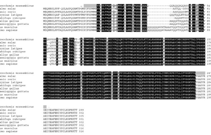
ANNEX 5 – MULTISEQUENCE ALIGNMENT FOR ALL PUTATIVE REFERENCE GENES G
ANNEX 6 – MULTISEQUENCE ALIGNMENT, MATRIX OF IDENTITIES AND PHILOGENETIC
TREES FOR BAMBI AND GREMLIN GENES P
1
1.
I
NTRODUCTIONUnderstanding the patterns of gene expression is expected to provide insight into complex regulatory networks and will most probably lead to the identification of the genes relevant to a biological process. In fact, the importance of gene-expression analysis is increasing in many fields of biological research. The real-time reverse transcription quantitative polymerase chain reaction (qRT-PCR) technique is one of the tools of choice to analyze gene expression by measuring transcript abundance and has gained much popularity in recent years [3].
This method is well suited for validation of microarray expression screening results and for studies of a selected number of candidate genes or pathway constituents in an experimental setup (biopsies, treated cell cultures or any other sample collection) [30]. qRT-PCR can now be performed on 384-well block thermal cyclers that allow gene expression analysis of even higher number of genes and samples (1 to 48 samples for 48 to 3072 different genes) [30].
Any measured variation in gene expression is caused by two sources. One is the true biological variation, and the other is non-specific variation which includes mainly factors relative to template input quantity and quality, pipetting errors, inhibitory compounds, yields of the extraction process and the enzymatic reactions (reverse transcription and polymerase chain reaction amplification). One of the major difficulties in obtaining reliable expression patterns is the removal of this experimentally induced non-biological variation from the true biological variation. This can be done through normalization by controlling as many of the variables as possible [30].
There are several strategies to remove experimentally induced variation but most of these methods cannot completely reduce all sources of variation. That is why it is crucial to standardize each step of the work, if not, variation will be introduced in the results that cannot be eliminated by applying the final normalization. Indeed it has been shown to be very important to try to control all sources of variation along the entire workflow of PCR based gene expression analysis [31].
2 It is generally agreed that the use of reference genes, also called housekeeping genes, is the preferred way of normalizing qRT-PCR data. This method is particularly attractive because the reference genes are internal controls that are affected by all sources of variation during the experimental workflow in the same way as the genes of interest [30]. The reference genes are expressed in the cells, and their mRNAs are present during nucleic acid extraction, storage, and any enzymatic processes such as DNase treatment and reverse transcription [17].
The ideal reference gene is a gene which expression occurs at a constant level in all tissues, independently of experimental conditions and developmental stages. These genes are generally well conserved throughout evolution and play key roles in cell survival, and their synthesis occurs in all nucleated cells types [7] [8].
Although reference genes constitute the best normalizers, a major problem is that there is substantial evidence in the literature that most of the commonly used reference genes are regulated under some circumstances, such as at different stages of development or in response to different treatments, indicating that there may be no universal reference gene with a constant expression [1] [24].
Genes frequently used, and considered as classical reference genes, are highly expressed genes such as glyceraldehyde-3-phosphate dehydrogenase, Beta-actin and ribosomal protein 18S that due to their key role in metabolism, cytoskeleton and ribosome structures, respectively, it was generally assumed that their expression levels were constant [1]. However it has been reported that these so called classical reference genes are also susceptible to regulation in certain circumstances [40]. Because of this, many other genes have been investigated as potential reference genes, including TATA binding protein, beta-2-microglobulin, tubulin alpha, L3 ribosomal protein and hypoxanthine phosphoribosyl transferase 1, but they all present the same problems of expression variation as the classical reference genes and are therefore of limited value as universal quantitative references [6]. These variations can partially be explained by the fact that some of the proteins encoded by the reference genes can participate in functions other than basal cell metabolism [7].
3 Therefore it is crucial to validate for each experimental situation if a candidate reference gene is suitable as reference gene for normalization. If unrecognized, unexpected changes in reference gene expression can result in erroneous conclusions about real biological effects [30].
In order to identify the most suitable reference gene a set of potential reference genes should be analyzed and validated for each different condition in a specific experimental study [8]. Typical experiments are performed by measuring the relative gene expression of 10 candidate genes in 10 representative samples from each tissue or sample group. The relative gene expression values are then imported to appropriate software and ranked according to their expression stability, choosing among the set the ones who have minimal variability. It has been reported that using a single non-validated reference gene may result in a significant bias and typically between 2 and 5 reference genes are required for accurate normalization [8].
Selection and validation of a reference gene could be a circular problem because the expression of this target gene itself also needs to be standardized. In order to try to circumvent this paradox and to analyze expression stability, several algorithms and software tools have been developed. Genorm [3] or Normfinder [47] are examples of such software that can be used to determine the most adequate reference genes to select.
This thesis is included as part of a more extensive science project. The objective of this work is to select suitable reference genes for qRT-PCR to be used in normalization of gene expression in Oreochromis mossambicus. Because there is evidence that classical reference genes may have expression variations at different stages of development, it is also important to evaluate the reference genes suitability in ovary follicules at different stages. During ovogenesis great structural and functional changes happened, regulated by gene expression. It is expected that the most suitable reference genes for follicles will be different from those of somatic tissues. If successful in recognizing the most suitable reference genes, this work will be a useful tool to apply in normalization of gene expression in studies with this species.
4
1.1
Real-Time quantitative PCR (qRT-PCR)The qRT-PCR has become one of the most widely used methods for gene expression analysis due to its large dynamic range, sensitivity, as well as accurate quantification of specific gene expression [2].
The process of qRT-PCR is similar to traditional PCR except that the quantification of PCR product is measured after each cycle of amplification (along the reaction) instead of an end-point quantification which is not necessarily proportional to the amount of target present in the samples [26] [24]. PCR is an in vitro process that increases the amount of a specific DNA template starting with a small amount of the template in a three-step cycling process.
For the PCR amplification to occur the essential components are 2 synthetic oligonucleotide primers, complementary to the regions that flank the target DNA sequence on the opposite strands and that presents the 3’ hydroxyl end oriented toward each other, a target sequence in the DNA sample, a thermostable DNA polymerase that can withstand multiple cycles of heating and dNTPs (19).
The PCR cycling process begins with the thermal denaturation of the DNA sample by raising the temperature to approximately 95ºC. In the second step, renaturation or annealing, the primers present in the reaction, in a vast molar excess, ligate to their complementary DNA sequences as the temperature is slowly reduced to near 55ºC (depending on the primer). The step of synthesis or extension is the third and final step and the temperature is raised up to 72ºC, the optimal temperature for the catalytic function of the Taq DNA polymerase for the extension of the sequences initiated in the primers, thus making copies of the target sequence[19].
For quantification to occur in qRT-PCR a fluorescent marker is commonly used. This marker or reporter produces a fluorescent signal measured by a camera or detector and during the reaction the reporter generates a signal that reflects the amount of product formed. In the initial cycles the signal is weak (small amount of product) and during the remaining of the reaction, as the amount of product accumulates, the signal increases exponentially, until it reaches saturation [24].
5 The fluorescent signal readings during a qRT-PCR can be analyzed as follows (Figure 1): linear ground phase, the beginning of the PCR reaction, in which the fluorescence emission at each cycle has not yet risen above the background; the early exponential phase, where the fluorescence emission will be statistically significantly higher than the background or baseline levels and is in this phase that the fluorescence of each reaction crosses the threshold level. The cycle in which it occurs is the threshold cycle (CT) or quantitative cycle (Cq), and is a parameter that characterizes each individual reaction. The Cq value is inversely correlated with the logarithm of initial copy number of template and is used for the calculation of the experimental results, log-linear phase follows the early exponential phase and corresponds to the phase when the PCR reaction reaches its optimal amplification and is expected that the amplification curves for different reactions to be parallel in this phase. The final PCR phase is the plateau stage, and is when the amplification rate drops to near zero and little more product is synthesized as the limiting reagent of the reaction is completely used [2].
Figure 1 - Phases of the PCR amplification curve. PCR amplification curve charts the accumulation of fluorescent emission at each reaction cycle. The curve can be divided in four different phases: the linear ground, early exponential, log-linear, and plateau phases. Data gathered from these phases are important for calculating background signal, cycle threshold (Ct), and amplification efficiency. Rn is the intensity of fluorescent emission of the reporter dye divided by the intensity of fluorescent emission of the passive dye (a reference dye incorporated into the PCR master mix to control for differences in master mix volume). ΔRn is calculated as the difference in Rn values of a sample and either no template control or background, and thus represents the magnitude of signal generated during PCR. This graph was generated with ABI Prism SDS version 1.9 software (Applied Biosystems) (in Wong et al, 2005 [2]).
6 1.1.1 Fluorescent markers
Currently, several detection chemistries are used in real-time PCR but the most common are TaqMan® probes and the SYBR Green dye. When TaqMan® probes are used specific primers and probes for the amplicon are added to the reaction. The probes - synthetic oligonucleotides with a fluorescent reporter dye attached to the 5’ end - are designed to hybridize to the cDNA and to be incorporated to the internal region of the PCR product. When the polymerase replicates the template, the probe is incorporated and the fluoropher and quencher are cleaved, thus resulting in the increase of the emitted fluorescence (Figure 2). The use of TaqMan® probes is very specific to the target amplicon, requires less optimizations in qRT-PCR method compared to other markers used, although it can be considerably expensive [4]. On the other hand, SYBR Green dye is not as specific as TaqMan probes as it binds to any double stranded DNA present in the reaction and upon excitation emits light (Figure 2). As this dye does not bind in a sequence-specific manner, the assay is prone to false positives. A strategy that can be used to discard the presence of false positives is the dissociation curve analysis, where the presence of different PCR products is reflected on the melting peaks [2] Melting peaks or melt curves allow a comparison of the melting temperatures of amplification products and, to be produced, the final PCR products are exposed to a temperature gradient from about 50 °C up to 95 °C while the fluorescence signal is continuously measured. Products of different lengths and sequences will melt at different temperatures and are observed as distinct peaks [23].
The use of SYBR Green dye requires more optimization, nevertheless is the simplest, flexible and most economical format used for detecting and quantifying PCR products in real-time PCR reactions [4].
Nonetheless, a good reporter should have low background fluorescence, emit a high signal upon amplicon formation and high target specificity [24].
7
Figure 2 – Schematic representation of the functioning of the SYBR Green Dye and TaqMan probe. SYBR Green binds to the dsDNA as it is being synthesized, and starts to emit fluorescence. The TaqMan probe binds to the DNA with the primers and as the fluorophore (green) is excited, it transfers the energy to a quencher molecule (orange) present in the probe; as the extension occurs, the probe is broken down, the fluorophe is released emitting fluorescence. (Taken from Velden et al 2003 [18]).
1.1.2 One-step VS two-step reaction
For the qRT-PCR to be applied in gene expression analysis, the messenger RNA (mRNA) must be copied into complementary DNA (cDNA) by reverse transcription. This is a crucial step as it has to reflect in the cDNA the amounts of the target present in the mRNA [24]. qRT-PCR can be performed in one-step or two-step reactions.
One-step reaction is characterized by cDNA synthesis and PCR amplification in a single tube, while in the two-step reaction both reactions are done separately. The one-step reaction is thought to minimize experimental variation but it may not be suitable in situations where the sample is assayed for several times and is less sensitive than the two-step reaction. On the other hand the two-step reaction, by initially doing the reverse transcription, allows the use of the same amount of cDNA in several different assays by using dilutions of the original cDNA template [2].
8 1.1.3 Quantitation of results
Depending on the level of quantitation needed to the experiment, several approaches can be adopted in order to quantify the amount of template. Among the different methods used for quantification are included the absolute standard method, the relative standard method and the comparative threshold (Ct) method.
1.1.3.1 Absolute standard method
In this method the input copy numbers of the transcript of interest are determined usually by relating the PCR signal to a standard curve [27]. This curve is constructed from a serial dilution of cRNA, ssDNA or dsDNA of known concentrations [2] and is used as a reference standard for extrapolating quantitative information for mRNA targets of unknown concentrations [27]. The external standard used for the curve construction should have the same primer binding sites of the target sequence in order to eliminate potential differences in quantification due to annealing. Ideally, it should contain sequences that are the same as those in the target sequence or which vary slightly [2].
A DNA standard must be a pure species and can be synthesized by cloning the target sequence into a plasmid, purifying a conventional PCR product, or directly synthesizing the target nucleic acid [2]. Spectrophotometric measurements at 260nm are normally used to determine the concentrations of these DNAs.
For correct application of this method the amplification efficiencies of the standard and the target gene should be the same. Normalization to a housekeeping gene or reference gene should be performed in order to control variations in the efficiency of the reverse transcription step and also to correct the variation introduced to variable RNA inputs [4].
1.1.3.2 Relative standard method
In the relative standard method, the quantity of each experimental sample is first determined using a standard curve and then expressed relative to a single
9 calibrator sample [2]. The target quantity is determined from the standard curve and divided by the target quantity of the calibrator to generate relative expression levels. The calibrator is designated as unity, and all the other quantities are expressed as an n-fold difference relative to the calibrator [27].
This method is often used also when the amplification efficiencies of the reference and target genes are not equal and results should also be normalized by endogenous controls, usually housekeeping genes.
1.1.3.3 Comparative threshold (Ct) method
The comparative threshold method is a mathematical model that calculates changes in gene expression as a relative fold difference by comparing Ct values between experimental samples and a control or calibrator such as a non-treated sample or RNA from normal tissue [2] [4]. The Ct values of both the calibrator and the samples of interest are normalized to an appropriate endogenous housekeeping or reference gene [4].
The comparative Ct method is also known as the 2-[delta][delta]Ct method, where [delta][delta]Ct = [delta]Ct,sample – [delta]Ct,reference and [delta]Ct,sample is the Ct value for any sample normalized to the endogenous housekeeping gene and [delta]Ct,reference is the Ct value for the calibrator also normalized to the endogenous housekeeping gene [4].
For the [delta][delta]Ct calculation to be valid, the amplification efficiencies of the target and the endogenous reference must be approximately the same. This can be established by looking at how [delta]Ct varies with template dilution. If the plot of cDNA dilution versus delta Ct is close to zero, it implies that the efficiencies of the target and housekeeping genes are very similar [4].
1.2 Reproduction and follicle development in Teleosts
The infraclass Teleostei, commonly referred to as teleosts or bony fishes, embraces a large number of species distributed in a great variety of aquatic environments, and is the most representative group among vertebrates [9]. This diversity leads to a wide range of reproductive adaptations, or “reproductive
10 strategies”, of interest in comparative biology. Also the numerous biological models that comprise this group can be particularly convenient for the acquisition of physiological or genetic knowledge [12], as the knowledge in the molecular pathways involved in reproduction processes in vertebrates, even in mammalian models is still limited [9].
Among several different aspects involving teleost reproduction, ovarian follicle development is a crucial step for their success on larvae recruitment. Studies on this development process can provide useful information not only for basic research, but also for aquaculture, fisheries management, as well as environmental and biomedical sciences. Increasing the knowledge in molecular pathways involved in this process may turn very useful to improve the industry of aquaculture since one of their biggest concerns is to produce “high-quality eggs”.
1.2.1 Sex determination and reproduction strategies
Teleosts have a great plasticity in respect of sex determination with a range of gonochoristic species - where individuals are either male (M) or female (F) - or species in which gender change may occur during their lifetime (protogynous F→M; protandrous M→F) [11].
Reproductive behavior is also much diversified among teleosts depending on the species, from spawning without egg care, until nest building and complex parental care [12].
There are three main types of ovarian development among teleosts, the synchronous, where all oocytes develop and ovulate at the same time, the “group-synchronous”, where at least two different stages of oocyte development are present in the ovary, and asynchronous, where all stages of oocytes are present without a dominant population. Group synchronous fishes spawn in a single episode or in a short period of time, while the asynchronous have several spawning batches during the spawning season. The reproductive effort can be indicated by the gonadosomic index (GSI) - the ratio between the gonad weight and total weight of the fish [13].
11 1.2.2 Ovary
The ovary of teleosts shows a similar general structure. In almost all the cases they usually appear as paired elongated organs oriented longitudinally within the abdominal cavity, under the kidney (and, if present, the swim bladder). An oviduct is situated at the posterior part of each ovary and is connected to the genital papilla [12].
Ovaries are compartmentalized by numerous septa formed by folds of the germinal epithelium projected into the ovarian lumen. These septa are called ovigerous lamella and contain nests of oogonia and oocytes at early stages, and also follicules at various stages of oocyte maturation. In adult teleost females the oogonia keep on proliferating contrarily to mammals, renewing the stocks of young oocytes and follicles. At ovulation, mature oocytes are released from their follicle into the ovarian cavity, before being laid through the oviduct and the genital papilla [12].
1.2.3 Follicle development
The formation of a mature egg in all teleosts undergo the same basic pattern (Figure 3): formation of primordial germ-cells (PGCs); the transformation of PGCs into oogonia; transition of oogonia into oocytes (onset of meiosis); followed by vitellogenesis (meiotic arrest at the end of prophase I); maturation with resumption of meiosis and germinal vesicle breakdown (GVBD); finishing with ovulation where mature eggs are expelled from its follicule [11].
1.2.3.1 Primordial germ cells and primary growth
The first step of oogenesis is the formation of PGCs that are produced from germ line cells developed by association with the accumulation of maternal RNA [11]. PGCs have a distinct nucleus membrane and one or two prominent nucleoli, they are of relatively large size and have large nuclei.
12
Figure 3 - Oocyte growth, oocyte maturation, and meiosis. Oocytes produced by the entry of mitotically proliferating oogonia into meiosis stop their meiotic cell cycle at prophase I, during which they grow by the accumulation of yolk (vitellogenesis). The prophase I-arrested oocytes are immature. Upon hormonal stimulation, the immature oocytes resume meiosis and proceed to metaphase II, at which stage they mature. (taken from [25]).
The PGCs are transformed into oogonia through structural changes and then oogonia multiply by mitotic divisions forming oogonial nests.
At this stage each oogonium becomes surrounded by a monolayer of somatic granulosa cells that secrete a basement lamina. Outside the basement lamina it is formed the theca which is a monolayer of somatic cells associated with blood vessels [11]. The oocyte with its surrounding granulosa cells, basement lamina and theca somatic layer constitutes the ovarian follicle and forms the primary oocyte. The transition from oogonium to a primary oocyte is also characterized by the initiation of the first meiotic division, before leaving the oogonial nest [11]. The primary oocyte growth encompasses the meiotic chromatin-nucleolus stage until early cortical stage and is involved with the development of the follicle layers surrounding the oocyte (Figure 4). It is also known that at this stage the organelles and molecules required for later stages are synthesized [11].
13
Figure 4 - Diagrammatic representation of the distinction between an oogonium and an intact ovarian follicle oocyte (adapted from [12]).
Oocyte enlargement occurs associated to the appearance of cortical alveoli at the periphery of oocytes that characterizes the so called “primary vittelogenesis” stage. At this stage the cortical alveoli multiplies, increasing the oocyte size by filling the cytoplasm with vesicles which differ in composition among species. At the end of this stage the content of cortical alveoli that are displaced to the periphery of the oocyte, is released to the egg surface leading to the formation of the chorion by restructuring of the egg envelop proteins [11].
1.2.3.2 Vitellogenesis
Vittelogenesis, in almost all teleosts, is the stage in which the oocyte shows a massive growth and may account as much as 95% of the final egg size and GSI increasing between 50 and 100 fold [13]. This growth is due to the incorporation by the oocyte of mainly of vitellogenins (Vtgs), but also vitamins and carbohydrates, all carried from the blood stream. These components will be crucial for the future development of the embryo. Vtg is a glycophospholipoprotein composed of 79% protein and 19% lipid. It is synthesized by the liver in response to estrogenic stimulation and its cleavage into yolk proteins is probably caused by the proteolytic activity of the enzyme
14 cathepsin D. Vtg enters in the oocyte passing through thecal capillaries until the granulosa layer and then through pore canals of the zona radiata into the oocyte membrane. At the membrane it binds to specific receptors in the endocytotic clathrin-coated pits of the vesicles. These vesicles then move into the peripheral ooplasm and fuse with lysosomes forming multivesicular bodies (MVB) [20].
1.2.3.3 Final Maturation and Ovulation
Oocyte final maturation processes are characterized by the reduction or the end of endocytosis, resumption of meiosis, germinal vesicle breakdown (GVBD), the formation of a monolayer of cortical alveoli under the oolemma and yolk platelet dissolution. The first meiotic division gives rise to two cells differing in size, the small cell with first polar body degenerates and the large mature oocyte is formed.
Numerous studies have shown that the transition of vitellogenesis into maturation is associated with an increase of plasma LH levels and increased expression of the LH receptor, leading to the production of maturation-inducing steroids (MIS) regulated by a LH driven switch [22]. The MIS is a derivative of progesterone and binds to oocyte-specific receptors to activate the maturation promoting factor in the ooplasm, which leads to the dissolution of the germinal vesicle and reinitiates meiosis. The stage of final maturation is also characterized by yolk clarification, oil droplet fusion and an increase of the oocyte volume. This increase is due to the intake of water and it happens mainly on marine species as the embryos need to have a water reservoir, in order to survive in hyper-osmotic seawater and for flotation [11].
Ovulation is the process of release of the mature oocytes from its follicle into the ovarian or abdominal cavity. The mature oocytes are released by separation from the granulosa layer and follicle layer. For some species, the stimulation by MIS leads not only to maturation but also to ovulation, in other species the ovulation is stimulated by arachidonic acid and its metabolites including prostaglandins [11].
15 1.3 Tilapia as a model
The complexity of tilapias, along with their economic interest worldwide makes them an important object of study. The relative ease of handling and rearing make them also a good study model for researchers. Recent research on tilapia has been mainly to define the mechanisms of the female gonad and development organization [28]. Among tilapia species, the Nile tilapia (Oreocheromis niloticus) has been used in the majority of studies, although, Mozambique tilapia (Oreocheromis mossambicus) (Figure 5) shares several common features that also make this species a suitable model for research in various fields.
Figure 5 - Oreochromis mossambicus (Peters, 1852). http://www.fao.org/fishery/species/2408: accessed 8 March 2011.
1.3.1 The Tilapias
Tilapias is the common name given to a group of subtropical to tropical freshwater fish of the family Cichlidae (Order Perciformes) that are native to Africa and the south-western Middle East [14]. Tilapias are grouped into three genera, according to parental care patterns: Oreochromis (arena-spawning maternal mouthbrooders); Sarotherodon (paternal or biparental mouthbrooders); Tilapia (substrate spawners) [14] [15].
Many species of Tilapia have been dispersed almost worldwide primarily for use in biological control, as bait fish, ornamental fish and for food supply by
16 farming in aquaculture systems [14]. Tilapias are now the second group of fish most farmed worldwide (behind carps) with an annual production of 2.5 million tons [29]. The species more representative in this farming industry are Mozambique tilapia (Oreochromis mossambicus), Nile tilapia (O. niloticus) and Blue tilapia (O. aureus). These species are well-suited for aquaculture production mainly because they are fast growers and tolerant of a wide range of environmental conditions, such as salinity and oxygen. Also the fact that they are omnivorous and, in some cases, disease resistant and overcrowding tolerant, makes them good targets to use in aquaculture [14]. Farming is based on all male monosex populations due to higher growth rates of males compared to females. This is explained because females of these species become sexually mature very early , which, associated with time spending in elaborated parental care, requires a large energy effort that reduces their growth rates. The monosex populations are normally achieved by androgen treatments and despite the success of its use, there is a preference for genetic control which is more suited to avoid environmental contaminations by hormones [15].
All the previously described characteristics made this group a case of success not only in aquaculture farming but also a successful invasive species since they are probably the most widely distributed group of exotic fish in the world [14], which is another strong argument to better understand this species in the attempt to minimize their destructive impact on the invasion of new habitats.
1.3.1.1 Reproduction
Tilapia species are sexually mature when they are 8-10cm and 2-3 months old. Adult fish can live until 6 to 8 years and in some cases even 12 years. The spawning season of tilapia usually starts in the spring months in temperate regions when the temperature rises, and continues throughout the year as long as water temperatures are above 22 ºC [16].
Tilapias make nests in the form of shallow pits in the substrate where courting and spawning happens. In most cases, males are territorial and develop aggressive behavior [16]. Female release her eggs in the nest and, in most cases, after fertilization, parents collect them into their mouth to undergo incubation. After hatching parents can maintain their young in the mouth for a period of time
17 for protection [16]. Females have multiple spawns due to their asynchronous ovaries and can produce a large variation in the number of eggs, depending on the species, body size and age [16].
1.3.1.2 Determination of sex
Tilapias are heterogametic (XX/XY). “All male populations” can be achieved genetically through the development off YY “supermales” and it has been shown that sex determination on tilapia species is more complex than a simple XX/XY monofactorial system [15].
Although sex determination is strongly determined by chromosomes, it is now clear that other factors such as the environment are also acting on sex. It has been demonstrated in various tilapia species that temperature can have a strong effect on sex determination. As an example, Baroiller et al., in 1995 masculinised XX progenies (100% females) with functional male phenotypes by raising water temperature above 32 ºC [15].
1.4 Objectives
The main objective of this thesis is the selection of suitable reference gene(s) for qRT -PCR in the Mozambique tilapia.
The study will start by cloning of ten potential reference genes (Table 1), followed by qRT-PCR and statistical analysis to determine their suitability.
The method of qRT-PCR will be used to measure the mRNA transcript level in different somatic tissues (brain, esophagus, stomach, anterior intestine, medium intestine, posterior intestine, liver, kidney, head kidney, spleen, heart, gill, and gonad) and during follicle development.
Gene expression levels will be compared among tissues according to sex and between oocytes maturation stages. Two different software tools (Genorm (version 3.5) http://medgen.ugent.be/~jvdesomp/genorm [3] and Normfinder http://www.mdl.dk/publicationsnormfinder.htm [47]) will be used to validate the stability of the reference genes and to allow the determination of the most suitable one(s).
18 After validation, the reference genes chosen will be tested in the analysis of the expression of two bone morphogenetic protein antagonists, BAMBI and Gremlin. The coding region of the two antagonists will be cloned and expression levels analyzed by qRT-PCR.
Table 1
Candidate reference genes which were evaluated.
Abbreviation. Gene name Function
18S 18S Ribossomal RNA Ribosome structure
β-act Βeta-actin Cytoskeletal structural protein
GADPH Glyceraldehyde 3-phosphate dehydrogenase
Glycolytic enzyme
HPRT1 Hypoxanthine
phosphoribosyltransferase 1
Purine nucleotide synthesis
TBP TATA Binding Protein Transcription
B2m Beta-2 microglobulin Major histocompatibility complex
TubA Tubulin, alpha 1 Cytoskeletal structural protein
EF1a Elongation factor- 1 alpha Protein synthesis
CSTD Cathepsin D Lipoprotein uptake and yolk processing
CSTZ Cathepsin Z Lipoprotein uptake and yolk processing
2.
Material and methods
2.1 Cloning of putative reference genes
2.1.1 cDNA synthesis
Complementary DNA (cDNA) is a single stranded DNA molecule that is synthesized by the action of a reverse transcriptase enzyme from a template of single-stranded RNA. This molecule is an important tool for molecular biology, providing the information from the RNA, in a stable form.
Total RNA from O. mossambicus ovary, treated with DNase I, was available in the laboratory and was used for this task. Three micrograms of total RNA were denaturated at 65ºC for 10 min. Reverse transcription was carried out in a 40 µl
19 reaction volume, 40 U of MMLV reverse transcriptase (Promega), 8 U of Ribolock RNase inhibitor (Fermentas), 0.2 mM dNTPs and 10 pmol oligo(dT) adaptor (5’-CGAGTCGACATCGATCGT(T)16-3’). A negative control without reverse transcriptase was used. Synthesis reactions were incubated for 1 hour at 37ºC.
2.1.2 Polymerase Chain Reaction
The polymerase chain reaction or PCR is a method for selective replication of a particular DNA fragment. It uses DNA polymerase that catalyzes the replication of a DNA template in a buffered reaction with an excess of a pair of short oligonucleotides primers and four deoxynucleoside triphosphates (dNTPs) to make millions of copies of the target sequence.
2.1.2.1 Primer design
To ensure primer efficiency different parameters were controlled with the software following the common established rules for primer design. The percentage of nucleotides G and C were between 40% and 60%, melting temperature in the range between 54 and 68ºC (2ºC for each A or T base and 4ºC for each G or C base) and differing no more than 5ºC between forward and reverse primers, size between 20 and 30 base pairs.
For each of the putative reference genes with the exception of the 18S ribosomal RNA, for which the sequence is available in the non-redundant nucleotide database (nr) at the National Center for Biotechnology Information (NCBI), a mutisequence alignment was carried out using available vertebrate
nucleotide sequences for that particular gene
(http://multalin.toulouse.inra.fr/multalin/multalin.html). Primer pairs were designed in the conserved region of the sequences. Properties of the primers were controlled using the online software, Oligonucleotide Properties Calculator (http://www.basic.northwestern.edu/biotools/oligocalc.html). All primers were synthesized by Sigma-Aldrich and are presented on table 2 at the end of methods section.
20 2.1.2.2 PCR reactions
The optimization of PCR reactions conditions determined the optimal annealing temperature and MgCl2 concentration for each pair of primers.
Frequently the Ta estimated by the software or given by the primer manufacturer may not produce the best results with the protocol used in the laboratory. To determinate the optimal Ta a gradient of temperature was tested.
Magnesium is an important DNA polymerase co-factor and excessive concentration used in the reaction will result in specificity loss. To optimize this parameter, different concentrations were tested in parallel to the temperature gradients.
PCR reactions (50 µl volume) were performed using ~ 20 ng of cDNA, appropriate concentration of MgCl2, 0.1 mM of dNTPs, 50 pmol of each sense and antisense primers and 0.6 U of DreamTaqTM DNA polymerase (Fermentas). All reactions were carried out on a MyCycler thermocycler (Biorad), using the following conditions: initial denaturation at 94ºC for 5 min, 30 cycles at 94ºC for 45 sec, adapted annealing temperature (table 3) for 1min, 72ºC for 1 min and final extension at 72ºC for 10 min.
The PCR products were analyzed in a 1.5% agarose gel electrophoresis. PCR products with the expected size were purified on columns with the GeneJET™ Gel Extraction Kit (Fermentas, USA) according to the manufacturer’s instructions. The purified PCR product was eluted on 30 µl of water, quantified by a nanodrop ND-1000 spectrophometer.
2.1.3 Cloning
The pGEM®-T Easy (Promega, Spain) (Annexe 2) was used as cloning vector. It is a linearized plasmid with a single 3´-terminal thymidine at both ends which allows the direct cloning of PCR products since they have 3’-deoxyadenosine (A) residues at the extremities, as a result of the activity of DreamTaqTM DNA polymerase used in this work. This vector has multiple cloning sites within the α-peptide coding region of the enzyme β-galactosidase. This enzyme is responsible for X-Gal
(5-bromo-4-chlor-3-indolyl-beta-D-21 galactopyranoside) degradation; as a result it produces blue colored colonies of bacteria. When a DNA fragment is ligated into the vector, the α-peptide is inactivated resulting in white colonies. This test is called the blue/white screen.
2.1.3.1 Ligation
To ligate the purified and quantified PCR products into the pGEM®-T Easy vector, the mass ratio insert:vector of 3:1 was used. The reaction was done as following: ~ 50 ng of PCR product were added to 15 ng of pGEM®-T Easy (Promega, Spain), 1.5 weiss U of T4 ligase (Promega, Spain), 1 μl of 10x ligation buffer and sterile water up to 10 μl. The mixture of ligation reaction was incubated overnight at 4ºC and then used for the transformation of competent bacteria.
2.1.3.2 Transformation
For transformation, 5 μl of ligation reaction were added to 100 μl of competent XL-1 Blue MRF` cells previously prepared for competence by the calcium chloride method (Annex 3). After incubation on ice for 20 min, the competent cells were heat shocked at 42ºC for 2 min. Transformed bacteria were plated on LB/ampicillin/IPTG/X-Gal plates and incubated overnight at 37ºC to allow bacteria to grow.
2.1.3.3 Colony PCR
Twelve of the white colonies were picked from each plate and put to grow in 96 well plates for 4 hours with 75 μl of LB Broth plus 50 μg/ml of ampicillin. After growing, colony PCR was performed, in 12.5 µl final volume, with 1.5 μl of growth colony, 2 mM MgCl2, 0.1 mM of dNTPs, 50 pmol of each T7 and SP6 primers and 0.25 U of DreamTaqTM DNA polymerase (Fermentas). PCR conditions were: 94ºC for 10 min to allow cell breakage and DNA denaturation, 30 cycles of 30 sec at 94ºC to denature DNA into single strands on every cycle, 30 sec at 48ºC for primer annealing and 30 sec to 1 min at 72ºC, variable depending on expected size of the amplicon. PCR products were run on 1.5% agarose gel electrophoresis stained by ethidium bromide.
22 2.1.3.4 Plasmid extraction
Extractions of the bacteria plasmids were performed by the alkaline lysis method, which uses an alkaline solution (NaOH) and a detergent (sodium dodecyl sulphate) to disrupt the bacterial cell wall and dissolve the cell membrane for releasing the plasmids to the solution.
Positive colonies were grown overnight in 3 ml of LB/ampicilin at 37ºC and constant agitation. Bacterial cultures were transferred to 1.5ml fresh tubes and centrifuged for 5 min at 12000 g. The supernatant was discarded and the pellet resuspended in 100 μl of cold GTE (Annex 4), in which RNase was added (50 μg/ml), and incubated 5 min at room temperature. Two hundred microliters of lysis buffer were added and the tubes were mixed by inversion and kept on ice until the solutions clarified (maximum of 5 min to avoid DNA degradation). Neutralization of the lysis reaction was done by addition of 150 μl of cold KAc 3M, pH 4.8. Tubes were mixed by inversion until flocculation and were incubated 15 min on ice. After centrifugation at 12000 g at 4ºC for 15 min, supernatants were recovered into fresh tubes and 450 μl of cold isopropanol were added to precipitate the plasmids. After mixing, the tubes were left 20 min at -20ºC and centrifuged at 12000 g at 4ºC for 20 min. Supernatants were discarded and the pellets were washed with ethanol at 75%. Aftercentrifugation at 12000 g for 5 min, ethanol was discarded and the pellets were air dried at room temperature. Pellets were resuspended in 30 μl of sterile water. Plasmids quality was analyzed on a 1.5% agarose gel. After quantification, they were sequenced at the CCMAR sequencing facilities and stored at -20ºC. The sequences obtained were compared by similarity searches against the non-redundant nucleotide database at NCBI using BLASTn (http://blast.ncbi.nlm.nih.gov/).
2.2 Cloning of two antagonists of BMPs, Gremlin and BAMBI
2.2.1 Partial cDNA cloning of Gremlin
To carry out cloning of Gremlin, the strategy was similar to the one described previously for the putative reference genes.
23 Briefly, primers were designed based on conserved regions determined by a multisequence alignment of published nucleotide sequences. Two pairs of primers were used for the cloning: the second pair allows the nesting of the PCR product obtained with the first pair. Primers sequence and localization on the predicted cDNA are presented in table 4. Oligo(dT) adaptor primed cDNA was synthesized using DNA free total RNA from mature ovary.
The first PCR was carried (50 µl volume) with ~ 20 ng of cDNA, 2mM of MgCl2, 0.1 mM of dNTPs, 50 pmol of each sense and antisense primers and 0.6 U of DreamTaqTM DNA polymerase (Fermentas). All reactions were performed using the following conditions: initial denaturation at 94ºC for 5 min, 35 cycles at 94ºC for 45 sec, 58°C for 45 sec, 72ºC for 45 sec and final extension at 72ºC for 10 min. After control by nested PCR, products were purified, cloned into pGemT-easy plasmid (Promega) and sequenced.
2.2.2 Full length cDNA cloning of BAMBI
A mature mRNA molecule is composed a 5’-UTR, a coding region (initiate by a methionine and terminate by a stop codon), a 3’UTR and poly(A) tail (Figure 7). Rapid amplification of cDNA ends (RACE) is a polymerase chain reaction-based technique, which facilitates the cloning of full-length cDNA sequences when only a partial cDNA sequence is available.
The 5’ RACE which allows the cloning of the initial methionine and the 5’ UTR and the 3’ RACE, from which the objective is to obtained the stop codon and the 3’ UTR with the poly-adenylation signal.
A large cDNA fragment of the coding region of BAMBI had been previously cloned which was used to obtain the full length cDNA.
2.2.2.1 5’- RACE-PCR
Classical techniques designed to clone the 5’ region of cDNAs are based on the addition of an adaptor to the 5’ extremity of the cDNA. The added adaptor, for which the sequence is known, will be used as site to anchor a primer during the PCR. This addition is done by template switching, extension of a poly(A) tail in the 5’ of the cDNA or ligation of the adapter on the 5’ end of the RNA after deletion of the protective CAP.
24 We have experimented an unconventional protocol in which the addition of the adaptor on the 5’ extremity is not necessary. Figure 7 outlines this technique. Briefly, after cDNA synthesis using a gene-specific primer, a first PCR is done with a unique reverse primer designed upstream of the available sequence. A second PCR is done with this reverse primer and a primer designed to be complementary to the first one. During the PCR, some of primers will hybridize together but the majority anneals to the cDNA and allows the amplification of the 5’ of the cDNA.
A cDNA was synthesized using a BAMBI-specific primer designed from available sequences (Table 5). Three micrograms of total RNA were denaturated at 65ºC for 10 min. Reverse transcription was carried out in a 40 µl reaction volume containing 40 U of MMLV reverse transcriptase (Promega), 8 U of Ribolock RNase inhibitor (Fermentas), 0.2 mM dNTPs and 10 pmol BAMBI cDNA primer. A negative control without reverse transcriptase was used. Synthesis reactions were incubated for 1 hour at 37ºC.
For the first PCR, ~ 20 ng of cDNA, 2 mM of MgCl2, 0.1 mM of dNTPs, 50 pmol of BAMBI-5’-nor primer and 0.6 U of DreamTaqTM DNA polymerase (Fermentas) were used in a 50 µl reaction volume. PCR conditions were the following: initial denaturation at 94ºC for 5 min, 35 cycles at 94ºC for 45 sec, 55°C for 45 sec, 72ºC for 30 sec and final extension at 72°C for 10 min.
Two µl PCR of the product obtained was used as template for the second PCR and the conditions were the same as described in PCR-1 with the exception of the addition of 50 pmol of BAMBI-5’-opp primer. The PCR product was run on a 1.5% agarose gel before purification, ligation in pGemT-easy plasmid and sequencing.
2.2.2.2 3’- RACE-PCR
For the 3’-RACE-PCR, PCR reaction was done using an oligodT primed ovary cDNA as template. Two forward primers were designed based on the available sequence and are described in table 5.
The reaction volume was 50 µl and contained ~ 20 ng of cDNA mixed to 2mM of MgCl2, 0.1 mM of dNTPs, 50 pmol of BAMBI-3’ primer, 50 pmol of oligo(dT) adaptor and 0.6 U of DreamTaqTM DNA polymerase (Fermentas).
25 Reactions were performed using the following conditions: initial denaturation at 94ºC for 5 min, 35 cycles at 94ºC for 45 sec, 58°C for 45 sec, 72ºC for 45 sec and final extension at 72ºC for 10 min. After control by nested PCR with BAMBI-3’-nest primer, the PCR product was purified, inserted in plasmid and sequenced as previously described. The sequences were submitted to the ncbi public database. (http://blast.ncbi.nlm.nih.gov/).
2.2.3 Multisequence alignment and identification of conserved domains
Alignments were performed using ClustalW [33]. Phylogenetic and molecular evolutionary analyses were conducted using MEGA version 4 [34].
To identify conserved domains in the BAMBI protein sequence, the sequence was compared to the NCBI’s Conserved domain database (CDD; http://www.ncbi.nlm.nih.gov/Structure/cdd/cdd.shtml). The existence of BMP receptors transmembrane domains in BAMBI protein was determined using the online tool, SOSUI (http://bp.nuap.nagoya-u.ac.jp/sosui/sosui_submit.html). The main objective of this program is to determinate whether the protein is a soluble or transmembrane protein by prediction of helices in the amino acid sequences.
2.3 Tissue collection
Tilapias were obtained from a broodstock reared at the University of Algarve. Fish were maintained in re-circulating freshwater tanks under a simulated natural photoperiod and fed once a day with commercial cichlid food (Nutrafin basix®; Rolf C. Hagen Inc.).
All animals were euthanized with MS-222 (3-aminobenzoic acidethyl ester; Sigma-Aldrich), administrated in a concentration of 1 g/L. Animals were then decapitated and dissected.
Samples were collected from 13 different tissues (brain, esophagus, stomach, anterior intestine, medium intestine, posterior intestine, liver, kidney, head kidney, spleen, heart, gill and gonad) of three males and three females. Fish were measured and weighted. Gonadosomatic index was calculated as
26 GSI=gonad weight/body weight*100. Immediately after dissection all tissues were flash frozen in liquid nitrogen and later stored at -80ºC until RNA extraction. Portions of the ovaries were separated previously to be frozen and fixed in Bouin-Hollande (Annex 4) to confirm maturation by histology.
2.4 Dissection of oocytes at different stages
Five sexually mature ovaries were collected from five females. Fish length, body weight and ovary weight were recorded. A portion of the ovary was fixed in Bouin-Hollande for posterior confirmation of the maturation stage by histological analysis. Gonads were dissected manually using a binocular microscope (Olympus SZX7) in Cortland medium and follicles were separated into 5 different groups according to their maturation stage: chromatin nucleolar stage (CNS), perinuclear stage (PS), cortical alveoli stage (CAS), vitellogenic stage (VS) and final maturation stage (MS) (Figure 6). For the first 4 stages, ~ 50 eggs were pooled while for the last stage, groups of 25 eggs were pooled and the yolk emptied. After dissection all samples were immersed in liquid nitrogen and stored at -80ºC until RNA extraction.
Figure 6 – Schematic drawing of zebrafish folliculogenesis. In the primary growth stage (stage I), follicles appear translucent and rangefrom 7–140 µm in size. Stage II follicles (0.15–0.34 mm) have cortical alveoli, typically surrounding the germinal vesicle. Vitellogenesis takes place in stage III follicles, whereas oocyte maturation occurs in stage IV. During oocyte maturation, germinal vesicle (GV) migrates from center to the periphery and the GV membrane breaks down. Stage V is the mature egg. (From Clelland et al., 2007 [32]).
27 2.5 Histology
Histological analysis was carried out on three ovaries. After 24 hours in Bouin-Hollande’s, tissues were washed twice with a solution of 1x PBS/20% sucrose overnight. Ovaries were treated in a semi-enclosed tissue processor Leica TP1020 starting with dehydration in increasing ethanol gradient: 70% (10min); 95% (30 min x2); 100% (1 hour x2) - followed by a mixture of ethanol:xylene (1:1) (1 hour), xylene 100% (1 hour and 1.5 hour), a mixture of xylene:paraffin (1:1) (2 hours) and paraffin 100% (2 hours). Paraffin embedded tissues were set in paraffin blocks using the thermal, dispensing and cryo consoles (Miles Scientific).
Serial sections of 7µm were obtained using a Leica RM 2135 microtome and mounted on poly-L-lysine-coated slides. Sections were dried overnight and stained with haematoxylin-eosin (Annex 4). For the staining, paraffin was removed from the sections by immersion in xilol (2x 15min) followed by hydration through a graded ethanol series: 100% (10 min); 95% (5 min); 70% (5 min), and distilled water (5 min). Sections were immersed in haematoxylin for 5 min and washed with running water and distilled water. Sections were then immersed in eosin Y for 2 min and washed with distilled water with a few drops of acetic acid and with distilled water. Dehydration was preformed through an increasing series of ethanol (70%, 95% and 100%, 5 min each), followed by immersion in xylene (2x 15 min) and mounted with DPX.
2.6 Quantitative-Polymerase Chain Reaction
2.6.1 Primer design
Different factors were taken into account following the established common guidelines to design primers for the qRT-PCR method. To prevent the reducing of annealing efficiency it was avoid runs of 4 or more bases. Amplicon sizes were kept short, ideally between 50-200 bases in length, since this allows for faster and more efficient reactions and increased consistency of results. The choice of forward and reverse primers that span exon-exon junctions, can avoid amplification of potential contaminating genomic DNA in cDNA targets. To
28 reduce the occurrence of secondary sequences such as hairpins, self-dimers and cross-dimers, complementary sequences between the primers were avoided. The percentage of G/C in primers was kept between 40 to 60%.
Primers were designed for each gene based on the cloned sequences and using the software Beacon Designer 7.7 (Premier biosoft international) (Table 3). Primers were tested and optimized on ovary cDNA in a MyCycler gradient thermocycler, with different temperatures (50 to 60ºC). PCR products were ran on a 3% gel and sequenced to confirm primer specificity. PCR efficiencies and coefficients of determination (Table 3) were determined with duplicated standard curves generated from 10-fold dilution series (from 1 ng to 1 fg) of purified PCR fragments as templates and with an IQ-5 real-time PCR detection system using SsoFastTM Evagreen® supermix (Biorad).
The PCR products used for making the standard curve were prepared as the following: PCR were performed as described on section 2.1.2 for all target genes using as templates the correspondent plasmid DNA extracted (see section 2.1.3). It was used 2 µl of plasmids samples (diluted to have the concentration of 100 ng/ul) in reactions of 100 µl. PCR products were purified on columns with GeneJETTM gel extraction kit (Fermentas) and after quantified using nanodrop ND-1000 spectrophotometer.
2.6.2 Quantitative RT-PCR on adult tissues
2.6.2.1 RNA extraction and removal of genomic contamination
Total RNA extraction was performed using 50 mg of each tissue and the Maxwell® 16 Instrument (Promega) with the respective kit and following the protocol provided by the manufacturer.
To each tissue sample, 500 µl of Lysis buffer previously prepared was added and the tissue was immediately disrupted using an ultra Turrax® homogenizer (IKA). Homogenized samples were then incubated on ice for 10 min to ensure complete lysis and transferred to fresh tubes. Blue RNA dilution buffer (835 µl) was added to the lysate followed by 125 µl of clearing agent. The tubes were mixed thoroughly, and incubated at 70ºC for 3 min. After vortexing
29 for 30 s; tubes were newly incubated for 5 min at room temperature, transferred to clearing columns and centrifuged at 12000 g for 2 min. The flow trough was transferred to the Maxwell® 16 RNA cartridge rack. Elution tubes were prepared by the addition of 300µl nuclease-free water. At the end of the program, eluted samples were transferred into fresh tubes.
RNAs were concentrated by precipitation. To each tube, 2.5 volumes of absolute ethanol and 0.1 volumes of NaAc (5 M, pH =8) were added. The solution was mixed thoroughly and incubated at -20ºC overnight. Following incubation, RNA was precipitated by centrifugation at the maximum speed at 4ºC during 20 min. The pellet was washed in 75% ethanol (in DEPC treated water), air dried and resuspended in 10 or 20 µl of nuclease free water depending on the total RNA pellet size. RNAs were ran in an agarose gel to check the quality and quantified by spectrophotometer.
Tissue total RNA extracted was treated with DNA-freeTM kit (Ambion, USA), to remove potential genomic DNA (gDNA) contamination. The removal of the gDNA was carried out using 8 µg of RNA that were transferred into fresh tubes and to which 5 µl of rDNAse I 10x Buffer, 2 U of rDNAse I and nuclease free water were added, up to a final volume of 50 µl. Samples were incubated at 37ºC for 30 min. After addition of 5 µl of inactivation buffer to terminate the reaction, the mix was incubated 5 min at room temperature. The tubes were centrifuged at 10000 g for 1.5 min to pellet the DNase inactivation reagent. To avoid any inhibition during the cDNA synthesis related to the inactivation buffer, only 40 µl of the reaction was transferred in a new tube and stored at -20ºC.
Before cDNA synthesis, the control of DNase treatment efficiency was performed by PCR amplification with primers designed against the housekeeping gene beta-actin (β-actin) (Wenjing Ruan, Mao de Lai, 2007).
PCR was performed, in 25 µl final volume, with ~ 10 ng of cDNA, 2 mM MgCl2, 0.1 mM of dNTPs, 50 pmol of each sense and antisense primers (sense, 5’-GGCCGCGACCTCACAGACTAC-3’ and antisense, 5’-ACCGAGGAAGGATGGCTGGAA-3’) and 0.5 U of DreamTaqTM DNA polymerase (Fermentas). PCR conditions were: initial denaturation at 94ºC for 5 min, 30 cycles at 94ºC for 30 sec, 48ºC for 30
![Figure 4 - Diagrammatic representation of the distinction between an oogonium and an intact ovarian follicle oocyte (adapted from [12])](https://thumb-eu.123doks.com/thumbv2/123dok_br/18651244.912489/21.892.274.672.113.488/figure-diagrammatic-representation-distinction-oogonium-ovarian-follicle-adapted.webp)