RESUMO.- [Aumento da concentração plasmática de óxido nítrico em cães com doença renal crônica natu-ralmente adquirida.] A determinação de óxido nítrico no plasma em cães clinicamente estáveis em diferentes
estágios da doença renal crônica (DRC) não foi estudada, constituindo este o objetivo do presente estudo. Foram estudados cinco grupos de cães, com idade variando en-tre quatro a 18 anos, compreendendo o grupo controle, composto por animais sadios (controle, n=17), grupo com DRC estágio 1 (DRC-1, n=12), grupo com DRC estágio 2 (DRC-2, n=10), grupo com DRC estágio 3 (DRC-3, n=13) e grupo com DRC estágio 4 (DRC-4, n=10). Os cães com DRC estavam com o quadro clínico estável e sem receber qual-quer tipo de tratamento. Foram estudados cinco grupo de cães, com idade variando entre quatro a 18 anos, compre-endendo o grupo controle, composto por animais sadios (controle, n=17), grupo com DRC estágio 1 (DRC-1, n=12), grupo com DRC estágio 2 (DRC-2, n=10), grupo com DRC estágio 3 (DRC-3, n=13) e grupo com DRC estágio 4 (DRC-4, n=10). Os animais sadios ou com DRC foram submetidos a
Increased nitric oxide plasma concentration in dogs with
naturally acquired chronic renal disease
1André B. Galvão2*, Marileda B. Carvalho2, Luciane G. Batalhão3, Juliana C.B. Silva4,
Marcelo Batalhão5 and Evelin C. Carnio5
ABSTRACT.- Galvão A.B., Carvalho M.B., Batalhão L.G., Silva J.C.B., Batalhão M. & Carnio E.C. 2017. Increased of nitric oxide plasma concentration in dogs with naturally ac-quired chronic renal disease. Pesquisa Veterinária Brasileira 37(8):847-852. Medicina Veterinária, Faculdade de Ciências Agrárias e Medicina Veterinária, Universidade Estadual Paulista, Via de Acesso Prof. Paulo Donato Castellane s/n, Jaboticabal, SP 14884-900, Bra-zil. E-mail: [email protected]
This study aimed to determine the amount of plasma nitric oxide in clinically stable dogs at different stages of chronic kidney disease (CKD). Five groups of dogs were studied, aged from 4 to 18, comprising of a control group composed of healthy animals (control n=17), group CKD stage 1 (DRC-1, n=12), group CKD stage 2 (CKD-2, n=10) group, CKD sta-ges 3 (CRD-3, n=13) and Group CKD stage 4 (DRC-4, n=10). Dogs with CKD were clinically stable and received no treatment. Two blood samples were collected at 24 hours inter-vals (repeated measures) to obtain serum and plasma. The serum creatinine values were used to classify dogs as CG, CKD-1, CKD-2, CKD-3 and CKD-4, and were (1.02±0.02mg/dL), (1.07±0.04mg/dL), (1.81±0.03mg/dL), (3.40±0.15mg/dL) and (6.00±0.20mg/dL) respec-tively. The determination of nitric oxide (NO) was performed by dosing nitrate/nitrite indi-rectly, and used for measurement of nitrate according to the NO/ozone chemiluminescence. The data were submitted to ANOVA for nonparametric analysis(Kruskal-Wallis)(P<0.05).
The concentration of plasmatic NO did not differ significantly among GC (10.81±0.51µM), CKD-1 (15.49±1.97µM) and CKD-2 (19.83±3.31µM) groups. The plasma concentration of CKD-3 (17.02±1.73µM) and CKD-4 (83.56±13.63µM) was significantly higher compared
with healthy dogs. In conclusion, the NO plasma concentration can increase in dogs with
CKD and become significantly higher in stage 3 and 4 dogs.
INDEX TERMS: Nitric oxide plasma concentration, dog, chronic renal disease, azotemia, creatinine, proteinuria, hypertension.
1 Received on May 26, 2016.
Accepted for publication on September 12, 2016.
2 Programa de Pós-Graduação em Medicina Veterinária, Departamento
de Clínica e Cirurgia Veterinária, Faculdade de Ciências Agrárias e Medi-cina Veterinária (FCAV), Universidade Estadual Paulista (Unesp), Via de acesso Prof. Paulo Donato Castellane s/n, Jaboticabal, SP 14884-900, Bra-zil. *Corresponding author: [email protected]
3 Departamento de Morfologia e Fisiologia Animal, FCAV/Unesp, Via de
acesso Prof. Paulo Donato Castellane s/n, Jaboticabal, SP 14884-900, Brazil.
4 Embrapa Pantanal, Rua Vinte e Um de Setembro, Nossa Senhora de
Fatima, Corumbá, MS 79320-900, Brazil.
5 Escola de Enfermagem de Ribeirão Preto, Universidade de São Paulo
duas coletas de sangue, com intervalo de 24 horas (amos-tras repetidas), para obtenção de soro e plasma. Os valores
de creatinina sérica, que definiram a classificação dos pa -cientes do controle, DRC-1, DRC-2, DRC-3 e DRC-4, que fo-ram 1,02±0,02mg/dL; 1,06±0,05mg/dL; 1,80±0,03mg/dL; 3,39±0,21mg/dL e 6,00±0,28mg/dL, respectivamente. A determinação plasmática indireta de óxido nítrico (NO) foi realizada por meio da dosagem de nitrato/nitrito, através da técnia de quimioluminescência NO / ozono. Os dados foram submetidos à ANOVA para análise não paramétrica (Kruskal-Wallis) (P <0,05). Os resultados das
concentra-ções plasmáticas de NO não diferiram significativamente quando comparados os dados do controle (10,81±0,51µM), DRC-1 (15,49±1,97µM), DRC-2 (19,82±3,31µM). No en
-tanto, o NO plasmático do grupo DRC-3 (17,01±1,73µM) e DRC-4 (83,55±13,63µM), foi significativamente maior, em
relação às médias dos cães sadios. Concluímos que a con-centração plasmática de NO pode aumentar em cães com
DRC e torna-se significativamente mais elevada nos está -gios 3 e 4 da doença.
TERMOS DE INDEXAÇÃO: Concentração plasmática, óxido nítri-co, cães, doença renal crônica, azotemia, creatinina, proteinuria, hipertensão.
INTRODUCTION
Chronic kidney disease (CKD) is defined as the presence
of functional or structural abnormalities in the kidneys; it
implies a 75% reduction in the glomerular filtration rate
(GFR) that typically corresponds to a loss of substantially more than 75% of the functional nephrons. Regardless of the cause or causes of nephron loss, CKD is characterized by irreversible structural lesions. Patients with CKD can be categorized into stages along a continuum of progres-sive kidney disease. The value of staging CKD is to facilitate application of appropriate clinical practice guidelines for diagnosis, prognosis and treatment. The IRIS (2013) has proposed a system for staging CKD in dogs and in cats.
Re-garding the renal condition, dogs can be classified as stage
0, patients at risk of developing CKD. Patients with CKD can be categorized into four distinct stages according to clinical
and laboratory data. For dogs in the first stage, the patient is
not azotemic (serum creatinine <1.4 mg/dL); in the second stage the patient is characterized by mild renal azotemia
(serum creatinine 1.4 ≥2.0mg/dL) and clinical signs are
usually mild or absent; in the third stage there is moderate
renal azotemia (serum creatinine ≥2.1-5.0mg/dL) and clin -ical signs may be present; and in the fourth stage, the pa-tient has severe renal azotemia (serum creatinine >5.0mg/ dL) and clinical signs are usually present. It is also possible
to establish subcategories within each stage, based on uri-nary protein excretion and arterial blood pressure. Patients are considered proteinuric when their protein/creatinine
ratio (U-P/C) exceeds 0.5. Patients may be classified as hy -pertensive with no complications when their systolic blood pressure (SBP) exceeds 160mmHg (Polzin et al. 2005, IRIS 2013).
Renal injury may be caused by activation of the re-nin-angiotensin, endothelin, and sympathetic nervous systems and a lack of vasodilatory action of nitric oxide
(NO). The potent vasoconstrictor effect of NO deficiency
causes decline in renal function and the
non-hemodynam-ic actions of NO defnon-hemodynam-iciency include permissive mesangial
growth and matrix overproduction, promoting glomeru-lar sclerosis (Aiello et al. 1997). The NO pathway has been implicated in many physiological functions in the kidney, including regulation of glomerular hemodynamics, medi-ation of pressure-natriuresis, maintenance of medullary perfusion, blunting of tubuloglomerular feedback, inhi-bition of tubular sodium reabsorption and modulation of renal sympathetic nerve activity (Polzin et al. 2005). These
findings have clinical relevance, given the evidence that NO deficiency occurs in rats and humans as result of CKD. The deficiency the NO has been implicated in the hypertension
and proteinuria in CKD (Aiello et al. 1997). However, de-termination of plasma in the NO in clinically stable dogs at different stages of naturally acquired CKD has not been studied, thus establishing the scope of this study.
MATERIALS AND METHODS
Ethics statement. The experiments were performed in ac-cordance to the guidelines and practices of animal welfare esta-blished by the Animal Ethics Committee of Colégio Brasileiro de Experimentação Animal (CBEA) (Protocol 013690/11).
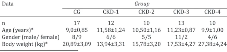
Animals. In this study, 62 adult dogs were evaluated from diverse races (28 females and 34 males). The patients were di-vided into 5 groups; the first group included 17 healthy dogs, which were included in the control group (CG) (serum creatinine 1.02±0.02mg/dL). The second group included 12 dogs (serum cre-atinine 1.07±0.04mg/dL) in the CKD stage 1 group (CKD-1). The third group included 10 patients (serum creatinine 1.81±0.03mg/ dL) in the CKD stage 2 group (CKD-2). The fourth group included 13 dogs (serum creatinine 3.40±0.15mg/dL) in the CKD stage 3 group; and the fifth group included 10 dogs (serum creatinine 6.00±0.20mg/dL) in the CKD stage 4 group. The 45 dogs (19 fe-males and 26 fe-males) with clinically stable CKD and no treatment have been distributed according to the stage of CKD established by the IRIS (2013) as summarized in Table 1. These five groups were formed by selected CKD adult dogs, without distinction of sex or breed, and no other unrelated primary concurrent disorder.
The exclusion criteria were: (a) diagnosis of acute kidney
Table 1. Characteristics of healthy dogs in control group and with CKD
Data Group
CG CKD-1 CKD-2 CKD-3 CKD-4
n 17 12 10 13 10
Age (years)* 9,0±0,85 11,58±1,24 10,50±1,16 11,23±0,87 9,9±1,00
Gender (male/ female) 8/9 6/6 5/5 11/2 4/6
disease or lower urinary tract disease, (b) patient uremic crisis requiring immediate treatment or (c) insufficient sample volume obtained for all tests.
Experimental protocol. Once eligible for composing groups, healthy or CKD dogs were submitted to two assessment sessions with 24-hour intervals. Blood and urine samples were collected for analysis. Systolic blood pressure (SBP) was measured by Doppler vascular an indirect method (Doppler vascular DV10 microrem).
The blood samples were extracted from the right jugular vein to parameter determination of red blood cell count (RBC), hema-tocrit (HCT), mean corpuscular volume (MCV), mean corpuscular hemoglobin concentration (MCHC), platelet count (PLT) and whi-te blood cell count (WBC), by a hematology automawhi-ted analyzer (Coulter model ABC T8). At the same time the serum samples bio-chemical indices analysis were centrifuged at 2.000g for 5 min, and samples from the jugular vein collected in heparinized tube to analyze the nitric oxide levels, were centrifuged at 2.000g for 20 min at 4oC. The plasma was transferred to another tube and
immediately frozen and stored at -70oC.
The urine sample was collected and analyzed for protein-cre-atinine ration (U-P/C).
Serum samples were processed for the determination of cre-atinine (modified Jaffe method), urea (enzymatic method), total calcium (cresolphthalein complex one method) and phosphorus (method the phosphomolybdate). The urine samples were used for the determination of creatinine (modified Jaffe) and total protein (pyrogallol red method). All biochemical analysis were carried out with System Labtest sets of reagents. Semi-automatic spectrophotometer was used for the readings.
Serum sodium and potassium were measured by the method of ion-selective electrode.
Nitric oxide levels in plasma of clinically stabled dogs at diffe-rent stages of naturally acquired CKD were evaluated indirectly by nitrite/nitrate quantification according to the technique des -cribed by Archer (1993). To determine plasma NO, the NO/ozone chemiluminescence technique was used, through a Sievers NO Analyzer 280 (GE Analytical Instrument, Boulder, CO, USA) and NO levels were expressed in µM.
Statistical analysis. Statistical comparisons of differences in the responses were analyzed by ANOVA test followed by nonpara-metric analysis (Kruskal-Wallis). Differences in the mean values were considered significant at (P<0.05), using the GraphPad Pris -ma program.version 6.02 for Windows, GraphPad Software, La Jolla California USA.
RESULTS
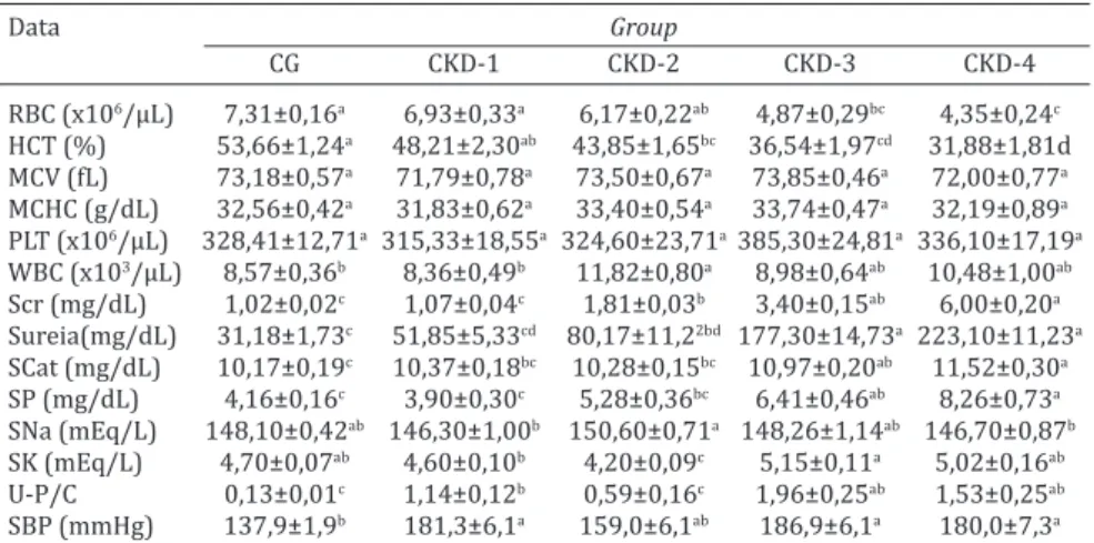
Renal dysfunction was demonstrated by increased serum creatinine and urea levels, as well as increases on UPC mean of CKD groups, when compared to healthy dogs. On the other hand, the PLT count did not differ between CKD groups, when compared to healthy dogs. CKD dogs showed a
signi-ficant rise in SBP compared to normal renal function dogs.
RBC and HCT noticeably decreased in renal dysfunction dogs in comparison to normal renal functional dogs (Table 2).
Nitric oxide levels
The NO levels did not differ significantly (P>0.05) be -tween groups GC, CKD-1 and CKD-2. Nevertheless, the
plas-matic group CKD-3 and CKD-4 was significantly (P<0.05)
higher compared to the mean of healthy dogs and CKD sta-ges 1, 2 and control group (Table 3).
DISCUSSION
The L-arginine, traditionally considered a nutritionally dispensable (nonessential) amino acid, serves multiple functions, among them; it is the precursor of NO (Lau et al. 2000). It is formed from citrulline, which is a product of amino acid metabolism in the gut wall. L-arginine is con-verted from L-citrulline via argininosuccinate synthetase and argininosuccinate lyase predominantly in the proximal tubules of the kidney (Modlinger et al. 2004). There are
many likely causes of NO deficiency in CKD, amongst them,
limitations on substrate (L-Arginine) availability, probably
Table 2. Laboratory dates and systolic blood pressure (SBP) of comparison of healthy dogs in control group, and dogs CKD. The values are presented as the
mean±standard error of the mean
Data Group
CG CKD-1 CKD-2 CKD-3 CKD-4 RBC (x106/µL) 7,31±0,16a 6,93±0,33a 6,17±0,22ab 4,87±0,29bc 4,35±0,24c
HCT (%) 53,66±1,24a 48,21±2,30ab 43,85±1,65bc 36,54±1,97cd 31,88±1,81d
MCV (fL) 73,18±0,57a 71,79±0,78a 73,50±0,67a 73,85±0,46a 72,00±0,77a
MCHC (g/dL) 32,56±0,42a 31,83±0,62a 33,40±0,54a 33,74±0,47a 32,19±0,89a
PLT (x106/µL) 328,41±12,71a 315,33±18,55a 324,60±23,71a 385,30±24,81a 336,10±17,19a
WBC (x103/µL) 8,57±0,36b 8,36±0,49b 11,82±0,80a 8,98±0,64ab 10,48±1,00ab
Scr (mg/dL) 1,02±0,02c 1,07±0,04c 1,81±0,03b 3,40±0,15ab 6,00±0,20a
Sureia(mg/dL) 31,18±1,73c 51,85±5,33cd 80,17±11,22bd 177,30±14,73a 223,10±11,23a
SCat (mg/dL) 10,17±0,19c 10,37±0,18bc 10,28±0,15bc 10,97±0,20ab 11,52±0,30a
SP (mg/dL) 4,16±0,16c 3,90±0,30c 5,28±0,36bc 6,41±0,46ab 8,26±0,73a
SNa (mEq/L) 148,10±0,42ab 146,30±1,00b 150,60±0,71a 148,26±1,14ab 146,70±0,87b
SK (mEq/L) 4,70±0,07ab 4,60±0,10b 4,20±0,09c 5,15±0,11a 5,02±0,16ab
U-P/C 0,13±0,01c 1,14±0,12b 0,59±0,16c 1,96±0,25ab 1,53±0,25ab
SBP (mmHg) 137,9±1,9b 181,3±6,1a 159,0±6,1ab 186,9±6,1a 180,0±7,3a
Averages in the same row, followed by at least one letter in common are not statistically different from each other (Kruskal-Wallis, P< 0.05).
due to impaired renal L-Arginine biosynthesis, decreased transport of L-Arginine into endothelial cells and possible competition between NO synthase (NOS) and competing metabolic pathways, such as arginase (Archer 1993, Le-villan et al. 2008). Surprisingly, total L-arginine synthesis is reportedly preserved in patients in end-stage renal disease.
The plasma levels of L-arginine did not change significantly
in rats with subtotal nephrectomy compared with normal rats (Levillan et al. 2008). This suggests that there was in-creased synthesis of L-arginine per nephron in the remai-ning renal mass and/or increased synthesis/ release of this amino acid at extra renal sites (Levillan et al. 2008). The remnant kidney undergoes hypertrophy after subtotal re-nal ablation. This may result in increased synthesis of L-ar-ginine per nephron, an adaptive mechanism that attempts to maintain renal production of L-arginine at levels seen in rats with intact kidneys (Modlinger et al. 2004). The kidney is not the only organ of L-arginine synthesis. Experiments with dogs show that renal arginine synthesis accounts for the greater part of whole body arginine synthesis, but it is clear that ~40% of this occurs outside the kidney (Mo-dlinger et al. 2004). The sources of this extra renal arginine synthesis certainly includes arginine regeneration from ci-trulline produced in macrophages, intestine and endothe-lial cells (Yu et al. 1996).
In experimentally induced CKD caused by renal mass reduction, the combination of increased plasma citrulline concentration and proximal tubular hypertrophy can com-pensate and maintain renal L-arginine production (Modlin-ger et al. 2004, Levillan et al. 2008). Appreciable quantities of arginine are synthesized in the kidney from citrulline produced by the intestine (Levillan et al. 2008).
The quantitative importance of renal arginine synthe-sis in the humans was demonstrated by Tiazenello et al.
(1980), who measured the net flux of amino acids across the kidney in normal subjects and in that chronic renal insuffi -ciency. In normal subjects, renal arginine synthesis amount-ed to ~1.75g/d. Curiously, in the patients with chronic renal
insufficiency, even GFR was reduced to 13% of normal, the
kidneys still produced ~0.7g arginine/d. The reason for this is probably related to the elevation in plasma citrulline
con-centration seen in chronic renal insufficiency. The dog me -tabolism does not strongly differ from that of humans and might constitute an excellent experimental model when hu-man kidneys are not available (Levillan et al. 2008). Yu et al. (1996) and Levillain et al. (2008) demonstrated that the dog arterial plasma exhibited appropriate levels of L-citrulline. Levillain et al. (2008) demonstrated that the dog produc-tion of L-arginine is proporproduc-tional to the level of L-citrulline,
the plasma concentrations of citrulline in dogs are sufficient
to sustain a significant production of L-arginine for body
needs as known in other species. These studies show that compensatory mechanisms can maintain adequate produc-tion of L-arginine and L-citrulline in dogs and in men, and may preserve NO production. Lau et al. (2000) studied the plasma levels of L-arginine and L-citrulline and examined the rate of whole body NO synthesis, using the stoichiomet-ric reaction involving conversion of [15N
2] arginine to [ 15N] citrulline, in human patients with initial diagnosis of hyper-tension and end-stage renal disease clinically stable with no acute complications. As a result, it was demonstrated that the level of whole body L-arginine synthesis is maintained in human patients in end-stage renal disease, possibly due to an increase in the availability and adaptive volume citrul-line. Furthermore, NO production in human patients in
end-stage renal disease was significantly increased when com -pared to healthy adults. Similar results were found, where all patients were clinically stable and without intercurrent
acute illness; there was a significant increase in the plasma
concentration of NO in dogs with end-stage renal disease compared with healthy dogs. Nevertheless, Wever et al. (1999) using L-[guanidine-15N
2] arginine in a tracer para-digm comparable to the one used here for determination of NO production, reported a reduced rate of NO production in human patients with chronic renal failure who were not on hemodialysis and blood pressure controlled by antihy-pertensive therapy, NO production was monitored by mea-suring the isotopic enrichment of [15N]-citrulline in plasma during intravenous infusion of [15N
2]-arginine. Tatematsu et al. (2007) induced CKD through heminepherctomy and nephrectomy in male dogs and studied the nitrite/nitrate as a way to estimate the plasma NO concentration, and re-ported that plasma nitrite/nitrate levels decreased in the studied dogs, as well as the plasma NO even under the con-dition of TFG reduction. In this study, the authors attribute the plasma NO decrease to the fact that the dogs had not developed hypertension. Aiello et al. (1997) studied rats with CKD induced by renal mass reduction and demonstrat-ed that unlike the kidney, the synthesis of NO increasdemonstrat-ed in
the systemic circulation, as reflected by higher than normal
plasma nitrite/nitrate concentrations. In this study, an in-crease in the expression of iNOS and eNOS in endothelial cells of large vessels was found. However, the eNOS expres-sion increase was not restricted to the endothelium but it was also detected in smooth muscle of large vessels. The authors believe that the probable cause for the increase in the production of NO in these sites is the increase in the
blood flux tension force exerted in the vessels endothelium,
which promotes mechanic-elastic alterations in the vessel and stimulates the eNOS expression. In this study the rats Table 3. The nitric oxide (NO) levels of comparison of in dogs healthy
controls, and dogs CKD. The values are presented as the mean ± standard error of the mean
Data Group
CG CKD-1 CKD-2 CKD-3 CKD-4
NO (µM) 10.81±0.51c 15.49±1.97bc 19,83±3,31bc 17,02±1,73b 83,56±13,63a
were hypertensive and as we know, in the HAS there is a pulsatile stretching of the arteries wall, which favors the NO synthesis. In addition to that, the rats were uremic which probably stimulated the iNOS expression through the
ac-tion of inflammatory mediators such as TNFα (Aiello et al.
1997). Nava et al. (1998) studied the plasma NO concentra-tion in the cardiovascular system of rats with spontaneous HAS and reported that the expression. of heart NOS and the micro vessels eNOS increased the NO production in such a
way that the plasma nitrate concentration was significantly
higher in hypertensive rats, when compared to healthy rats. Nevertheless, even with the NO increase, it is not
vasoac-tive to stimulate an adequate vasodilation. These findings
could justify the raised plasma levels of nitrate of our study, since our dogs with naturally acquired CKD in stage 1, 3 and 4 were hypertensive. Nevertheless, Yokokawa et al. (1995) demonstrated increased production of NO, determined by plasma nitrite and nitrate concentrations in human patients with end-stage renal disease and hemodialysis-induced hy-potensive episodes. Komeno et al. (2004) studied the role of NO in hemodialysis-related hypotension in experimental renal dysfunction dog model, demonstrated in renal dys-function dogs; a continuous hypotension occurred with a gradual increase in the plasma NO concentration. These re-sults suggest that an excessive production of NO suppresses the sympathetic nerve activity in renal dysfunction dogs, according to the authors (Komeno et al. 2004).
Previous studies showed elevated plasma nitrite/ni-trate levels in patients with end-stage renal disease, which was probably due to decreased excretion of nitrite/nitrate (Schmidt & Baylis 2000). Blum et al. (1998) studied three groups of patients with different kidney function and con-clude that renal nitrite/nitrate correlated with creatinine clearance. However, plasma nitrite/nitrate levels were hi-gher than in healthy controls. The investigators conclude
that end-stage renal disease is a state NO deficiency, but
that NO2 and NO3 accumulate in the plasma because of de-creased renal excretion. In our study, certainly the renal excretion of our patients was decreased, which might also justify the increased plasma concentration of NO.
Plasma L-arginine concentration is normal in patients with CKD although orotic acid increases which is indicative
of net L-arginine deficiency. If L-arginine transport into en -dothelial cells was impaired in CKD, this would reduce the
rate of removal of L-arginine from plasma and camouflage a
reduction in intracellular L-arginine availability (Wagner et
al. 2001). Studies with artificial solutions contenting high
concentrations of urea suggest that urea contributes and that the effect is not osmotically mediated or acutely rever-sible with excess arginine and exhibits an “all-or-nothing” response, switching on when the concentration in the me-dium exceeds ~ 15mmmol/L or ~45mg/100mL (Xiao et al. 2001). Furthermore, urea has to enter the endothelial cell to act, since in the presence of phloretin, a urea transport inhibitor, the inhibitory effect of urea on arginine transport was abolished. After 7 days of urea-induced inhibition of L-arginine transport, we observed decreased endothelial NOS (eNOS) activity in cultured endothelial cells, in the absence of changes in eNOS protein abundance, suggesting
that cumulative substrate deficiency might lead to limita -tion of NO produc-tion (Xiao et al. 2001). However, in study by Aiello et al. (1997) in a rat model of CKD induced renal mass reduction found that, although there was decreased renal production of NO, there was an increase in total body. These investigations suggested that this up-regulation of vascular NOS activity might be caused by guanidinosucci-nate, which is a toxin that accumulates in CKD humans and animals, and can increase NO production in cultured
endo-thelial cells (Aiello et al. 1997). Such a finding also could
account for the platelet dysfunction (Aiello et al. 1997). The authors suggest, that up-regulation of NO-forming en-zymes might be an early defense mechanism against hyper-tension of uremia (Aiello et al. 1997). The patients in our study were at a condition of uremia, a condition that also may have promoted the enhanced levels of plasma NO.
Jankowski et al. (2007) and Lautner et al. (2013) have discovered a new component of the renin angiotensin sys-tem, named alamandina a peptide that increases with pro-gression of CKD in rats, mice and humans. This peptide has vasodilating action by the increase of NO. In the present study, all patients were clinically stable, thus the increase of NO may be a defensive mechanism due to the various hemodynamic disorders caused by CKD. Research has de-monstrated that the ability to convert nitrate/nitrite to NO, and the use of oral supplementation of sodium nitrate in humans resulted in decreased risk of developing cardio-vascular disease, improvement in endothelial function, and vasodilatation (Carlström et al. 2011, Gilchrist et al. 2009).
CONCLUSION
The dogs with naturally acquired CKD presented higher NO plasma concentration concerning the values observed in
healthy dogs. However, the differences are significant only
in dogs in stages 3 and 4.
Acknowledgments.- This study was supported by FAPESP (Fundação de Amparo à Pesquisa do Estado de São Paulo, Proc. no 2011/08767-3).
Conflict of interest statement.- The authors have no competing interests.
REFERENCES
Aiello S., Noris M., Todeschini M., Zapella S., Foglieni G., Benigni A., Corna D.Z., Cavalloti D. & Remuzzi G. 1997. Renal and systemic nitric oxide syn-thesis in rats with renal mass reduction. Kidney Int. 52:171-181. Archer S. 1993. Measurement of nitric oxide in biological models. FASEB
J. 1:349-360.
Blum M., Yachnin T., Wollman Y., Chernihovsky T., Peer G., Grosskopf I., Ka-plan E., Silverberg D., Cabili S. & Iaina A. 1998. Low nitric oxide produc-tion in patients with chronic renal failure. Nephron 79:265-268. Carlström M., Persson A.E.G., Larsson E., Hezel M., Scheffer P.G., Teerlink T.,
Weitzberg E. & Lundberg J.O. 2011. Dietary nitrate attenuates oxidative stress, prevents cardiac and renal injuries, and reduces blood pressure in salt-induced hypertension. Cardiovasc. Res. 89:574-585.
Gilchrist M., Winyard P.G. & Benjamin N. 2009. Dietary nitrate: good or bad? Nitric Oxide 22:104-109.
IRIS 2013. International Renal Interest Society. Staging Chronic Kidney Disease (CKD) 2013. <http://www.irisiriskidney.com/pdf/IRISiris%20 A4%20Poster.pdf> Accessed Jan. 10, 2016.
Wiedon A., Beyermann M., Bader M., Todiras M. & Jankowski J. 2007.
Mass-spectrometric identification of a novel angiotensin peptide in hu -man plasma.Arterioscler. Thromb. Vasc. Biol, 27:297-302.
Komeno M., Akimoto A., Fujita T., Aramaki T., Aoki M., Shimada T. & Ohashi F. 2004. Role of nitric oxide in hemodialysis – related hypotension in an experimental renal dysfunction dog model. J. Vet. Med.Sci.66:53-57. Lau T., Owen W., Yu Y. M., Novishi N., Lyons J., Zurakowsky D., Tsay R., Ajami A., Young Y. & Castilho L. 2000. Arginine, citruline, and nitric oxide me-tabolism in end-stage disease patients. J. Clin. Invest. 105:1217-1225. Lautner R.Q., Villela D.C., Fraga-Silva R., Silva N., Verano-Graga V.,
Costa-Fra-ga F., Jankowski V., Souza F.S., Alzamora A., Soares E., Kjedsen C.B., Olivei-ra A., BOlivei-raga G., Savergninin S., Maia G., Peluso A., Passos-Silva D., FerreiOlivei-ra AF., Alves F., Martins A., Raizada M., Paula R., Motta-Santos D., Pimenta F.K.A., Alenina N., Sinisterra R., Bader M., Campagnole-Santos M.L. & San-tos R.A.S. 2013. Discovery and characterization of Alamandine – A novel component of the renin-angiotensin system. Cir. Res. 112:1104-1111. Levillan O., Rabier D., Duclo B., Gaudreau P. & Vinay P. 2008. L-argnine
metabolism in dog kidney and isolated nephron segments. Metabolism 57:9-23.
Modlinger P.S., Wilcox C.S. & Aslam S. 2004. Nitric oxide, oxidative stress, and progression of chronic renal failure. Semin. Nephrol. 24:354-365. Nava E., Farre A., Moreno C.A., Santos C., Pierre M., Francesco C. & Lüscher
T.F. 1998. Alterations to the nitric oxide pathway in the spontaneously hypertensive rat. J. Hypertens. 16:609-615.
Polzin D.J., Osborne C.A. & Ross S. 2005. Chronic renal failure, p.1756-1785. In: Ettnger S.J. & Feldman E.C. (Eds), Textbook of Veterinary Inter-nal Medicine. 6th ed. W.B. Saunders, Philadelphia.
Schmidt R.J. & Baylis C. 2000. Total nitric oxide production is low in pa-tients with chronic renal disease. Kidney Int, 58:1261-1266.
Tatematsu S., Wakino S., Kanda T., Homa K., Yoshioka K., Hasegawa K., Na-oki S., Kimoto M., Saruta T. & Hayachi K. 2007. Role of nitric oxide pro-ducing and degrading pathaways in coronary endothelial dysfunction in chronic kidney disease. J. Am. Soc. Nephrol.18:741-749.
Tiazenello A., Ferrari G., Gariboto G., Gurreri C. & Robaudo C. 1980. Renal metabolism of amino acid and ammonia in subjects with normal renal
function and in patients with chornic renal insufficiency.J. Clin. Invest. 65:1162-1173.
Wagner L., Klein J.D., Sands J.M. & Baylis C. 2001. Urea transports are dis-tributed in endothelial cells and mediate inhibition of L-arginine trans-port. Am. J. Physiol., Renal Physiol. 283:F578-F582.
Wever R., Boer P., Hijmering M., Stroes E., Verharr M., Kastelein J., Versluis K., Lagerwerf F., Herman R., Koomans H. & Rabelink T. 1999. Nitric oxide production is reduced in patients with chronic renal failure. Arterio-scler.Thromb. Vasc. Biol. 19:1168-1172.
Xiao S., Wagner L., Mahaney J. & Baylis C. 2001. Uremic levels of urea inhib-it L-arginine transport in cultered endothelial cells. Am. J. Physiol., Renal Physiol. 280:F989-F995.
Yokokawa K., Mankus R., Saklaven M.G., Kohno M., Yasunari K., Minami M., Kano H., Horio T., Takeda T. & Mandel A.K. 1995. Increased nitric oxide production in patients with hypotension during hemodialysis. Ann. In-tern. Med. 1:35-37.