RESUMO.-
[
Identificação de espécies e perfil de suscep
-tibilidade antimicrobiano de bactérias causadoras de
mastite subclínica em búfalos
.
] Microrganismos
causa-dores de mastites subclínicas em búfalas foram isolados
desde 20 amostras de leite de búfalos de quatro granjas
lei-teiras localizadas na região central do Estado de São Paulo,
Brasil, através dos testes contagem de células somáticas
(CCS), contagem padrão em placas (CPP), provas
bioquí-micas, reações de PCR e perfil antimicrobiano. A CCS apre
-sentou uma mediana de 721.000 cel/mL no leite, indicando
presença de mastite subclínica. A média geral de CPP foi de
1,8x10
4UFC/mL. Os microrganismos com maior frequên
-cia de isolamento segundo os testes bioquímicos foram:
Staphylococcus epidermidis
(17%),
Staphylococcus aureus
(15%),
Bacillus
spp. (14%),
Acinetobacter
spp. (12,5%);
frequência intermediaria:
Pseudomonas aeruginosa
(9,5%);
Shigella flexneri (7,0%),
Streptococcus
spp. (5,5%),
Cory-nebacterium
spp. (5,0%),
Escherichia coli
(4,5%),
Serratia
Species identification and antimicrobial susceptibility
profile of bacteria causing subclinical mastitis in buffalo
1Andrea Vásquez-García
2, Thaysa dos Santos Silva
3, Sabrina R. de
Almeida-Queiroz
4, Silvia H.S. Godoy
4, Andrezza M. Fernandes
4, Ricardo L.M. Sousa
4and Raul Franzolin
5*
ABSTRACT.-
Vásquez-García A., Silva T.S., Almeida-Queiroz S.R., Godoy S.H.S., Fernandes
A.M., Sousa R.L.M. & Franzolin R. 2017.
Species identification and antimicrobial pro
-file of bacteria causing subclinical mastitis in buffalo.
Pesquisa Veterinária
Brasilei-ra 37(5):447-452
. Departamento de Zootecnia, Faculdade de Zootecnia e Engenharia de
Alimentos, Universidade de São Paulo, Av. Duque de Caxias Norte 225, Pirassununga, SP
13635-900, Brazil. E-mail: rfranzol@usp.br
Microorganisms causing subclinical mastitis in water buffalo were isolated from 20
buffalo milk samples at four dairy farms located in central region of São Paulo State,
Bra-zil, through testing of somatic cell count (SCC), standard plate count (SPC), biochemical,
PCR assays and antimicrobial profile. The SCC showed average of 721,000 cells/mL in the
milk, indicating the presence of subclinical mastitis. The overall average for SPC was 1.8
x 10
4CFU/mL. The microorganism most frequently isolation according to biochemical
tests were:
Staphylococcus epidermidis
(17%),
Staphylococcus aureus
(15%),
Bacillus
spp.
(14%),
Acinetobacter
spp. (12.5%); with intermediate frequency:
Pseudomonas aeruginosa
(9.5%);
Shigella flexneri
(7.0%),
Streptococcus
spp. (5.5%),
Corynebacterium spp
. (5.0%),
Escherichia
coli
(4.5%),
Serratia marcescens
(4.0%),
Stenotrophomonas maltophilia
(4.0%),
and low incidence:
Klebsiella rhinoscleromatis
(0.5%),
Klebsiella ozaenae
(0.5%),
Tatumella
ptyseos
(0.5%),
Enterobacter cloacae
(0.5%). The molecular analysis indicated that sam
-ples positive by culture method of the genera
Staphylococcus
,
Streptococcus
and
E. coli
were
positive by PCR. Para
S. aureus
and
S. epidermidis
the highest percentages of observed
sen-sitivity were gentamicin (100%) and vancomycin (100%); for the genus
Streptococcus
to
gentamicin and oxacillin and
E. coli
to Ampicilin. These findings may help in the control and
treatment of subclinical mastitis in buffaloes and contribute to improving the efficiency
and quality of the milk produced.
INDEX TERMS: Antimicrobialprofile, mastitis, buffalo, antibiotics, bacteria, somatic cell count, milk.
1 Received on December 11, 2015
Accepted for publication on September 1, 2016.
2 Programa de Pós-Graduação em Ciências, Departamento de Enge -nharia de Alimentos, Faculdade de Zootecnia e Enge-nharia de Alimentos (FZEA), Universidade de São Paulo (USP), Av. Duque de Caxias Norte 225, Pirassununga, SP 13635-900, Brazil.
3 Programa de Pós-Graduação em Zootecnia, Departamento de Zootecnia, FZEA-USP, Av. Duque de Caxias Norte 225, Pirassununga, SP 13635-900, Brazil.
4 Departamento de Zootecnia e Medicina Veterinária, FZEA-USP, Av. Du -que de Caxias Norte 225, Pirassununga, SP 13635-900, Brazil.
marcescens
(4,0%),
Stenotrophomonas maltophilia
(4,0%),
e baixa incidência:
Klebsiella rhinoscleromatis
(0,5%),
Klebsiella ozaenae
(0,5%),
Tatumella ptyseos
(0,5%),
Ente-robacter cloacae
(0,5%). A análise molecular indicou que
as amostras positivas pelo método de cultura dos gêneros
Staphylococcus, Streptococcus
e
Escherichia coli
foram
po-sitivas por PCR. Para
S. aureus
e
S. epidermidis
os maiores
percentuais de sensibilidade observados foram
gentamici-na (100%) e vancomicigentamici-na (100%); para o gênero
Strepto-coccus
à gentamicina e oxacilina e para
E. coli
à ampicilina.
Este resultados podem ajudar no controle e tratamento da
mastite subclínica em búfalos e contribuir para melhorar a
eficiência e qualidade do leite produzido.
TERMOS PARA INDEXAÇÃO: Susceptibilidade antimicrobiano, bactérias, mastite subclínica, búfalos, antibióticos, contagem de células somáticas, leite.
INTRODUCTION
The buffalo milk production is a highly important
activi-ty in many countries, among which are highlighted Asian
countries, Italy and Brazil (Bernardes 2007). In Brazil, the
buffalo has been exploited for milk and meat, but the main
economic activity is the dairy industry, especially the
mo-zzarella cheese produced exclusively with buffalo milk (An
-drighetto et al. 2005, Bernardes 2007, Araujo et al. 2012).
Buffalo milk has a great potential for commercial
produc-tion due mainly contain particular physicochemical
charac-teristics with high total solids, fat and protein (Amaral et
al. 2005).
The importance of identifying the aspects related to the
health of the mammary gland and milk products of
buffalo-es has been highlighted in the world literature as in Brazil
(Medeiros et al. 2011), India (Tiwari et al. 2011), Pakistan
(Hussain et al. 2013), Nepal (Dhakal et al. 2007), Italy (Fa
-giolo & Lai 2007) and Germany (Braun & Preuss 2007).
Mastitis is an inflammation of the mammary gland paren
-chyma due to an infectious process predominantly caused
by many microorganisms, particularly bacteria, and may
also be involved fungi and yeast (Baloch et al. 2011). The
effective and accurate diagnosis is extremely important to
control this severe disease in buffalo (Viana et al. 2010).
However, the absence of macroscopic changes in the
tis-sues or secretions in cases of subclinical mastitis, does not
allow the identification of infected mammary quarters be
-fore milking, once routine diagnostic methods include only
physical examination, and fluid secretion (Bonini Pardo et
al. 2007). Several factors have been identified as predispo
-sing to subclinical mastitis in buffalo, as level of milk
pro-duction, body weight, calving period, udder type and
hygie-ne conditions for milking (Hussain et al. 2013).
Along the lack of information on the buffalo species,
the same animal management techniques for cattle milk
production are used for the control of mastitis in
buffa-lo milk production, resulting in lack of success since they
have peculiar habits of each ruminant species (Carvalho
et al. 2007, Medeiros et al. 2011). Indeed, most of mastitis
prevalence was found in cow’s milk (32%) than in buffalo
milk (22%) (Khan et al. 2013). Thus, subclinical mastitis
unfortunately has not been diagnosed with frequency and
consequently its etiology has not been widely investigated
(Fagiolo & Lai 2007). In addition, Brazilian literature has
presented a considerable number of publications about
the buffalo mastitis, but when compared to the number of
papers on bovine mastitis is small. A fact that needs more
research efforts because buffalo has attracted growing
in-terest of breeders and research institutions as an
alternati-ve for dairy farming (Langoni et al. 2001, Jorge et al. 2005,
Medeiros et al. 2011).
The objective of this work was the isolation and
phe-notypic characterization of the main microorganisms that
cause mastitis subclinical in buffaloes (
Bubalus bubalis
)
raised in four dairy farms located in central region of Sao
Paulo state, Brazil, as well as the molecular
characteriza-tion and evaluacharacteriza-tion of bacterial sensitivity profile for the
isolated species.
MATERIALS AND METHODS
This study was carried out at four commercial buffalo farms in the central region of São Paulo State, Brazil, using animals reared in a loose housing system with supplementation of concentrated ration according to the stage of lactation. Water and mineral sup -plementation were available ad libitum. The procedures involved
in this experiment were approved by Comissão de Ética no Uso de Animais (CEUA/FZEA) (Protocol #13.1.2338.74.7). In order to select buffaloes more prone to subclinical mastitis, sanitary con-trol data carried out by the property were evaluated and Somatic Cell Count test (SCC) of all of the female buffaloes in the herd at previous month were taken in account. Thus, milk samples were selected from twenty animals that showed SCC values above to 200,000 cells/mL (Dhakal 2006). Selected animals were dairy bu -ffaloes of Murrah breed, in the second or third stage of lactation, with average production of 7 kg of milk/animal/day, in two daily milking. After the physical examination of the mammary gland, twenty milk samples were collected by combining the four quar-ters of the mammary in duplicate with all necessary hygiene, pro-perly identified and packed in insulated boxes with ice packs.
One of the samples, collected in a bottle containing Bromopol (Microtabs®) was designed to determine the SCC by flow cytome -try using Somacount 500 equipment. The second sample was col -lected aseptically for microbiological analysis and antimicrobial susceptibility testing. For the Standard Plate Count (SPC), the milk samples were diluted in sterile peptone water solution (Hime-dia®, India) at 0.1% (w/v). Aliquot 0.1 mL of each dilution (10-1, 10-2, 10-3, 10-4 and 10-5) was inoculated into Brain Heart Infusion agar (BHI - Difco, USA) and incubated at 37°C under aerobic con -ditions for 48 hours. Then, the read of the plates was carried out after the incubation. Ten colonies with different characteristics were picked up randomly per animal and seeded by exhaustion for the isolation of colonies in different culture media: blood agar (Acumedia®, USA), Eosin Methylene Blue Agar (Himedia®, India), Mannitol Agar (Himedia ®, India) and MacConkey Agar (Himedia®, India). The microorganism pure of the respective boards were identified based on Gram stain, morphology and macroscopic characteristics.
Biochemical tests were subsequently carried out in accor-dance with bacterial groups identified in previous tests. Strains of Staphylococcus spp. were submitted to the free coagulase tests
(aerobiosis) and mannitol (aerobiosis and anaerobiosis) were performed according (Macfaddin 1980) and isolates classified in accordance with (Baird-Parker 1990).
Gram negative bacteria isolated were identified using Bactray Kit I, II and III® of biochemical identification (Laborclin, Brazil). The genus Bacillus was featured on nutrient agar by the formation of colonies rounded, smooth and irregular border with creamy consistency, gram stain and rod shape with catalase and oxidase test. The bacteria whose colonies had become small, round, whit -ish or creamy, with rough surface, measuring 1 to 2 mm in diam-eter, gram-positive rods, absence of hemolysis on blood agar, cata-lase production, were classified as Corynebacterium spp. Tests for Streptococcus genus identification included absence of catalase
production, growth in Bile Agar Esculin (Himedia®, India), type of hemolysis on blood agar (α, β and γ hemolytic) with 5% defibri -nated blood sheep and tolerance to tellurite.
The molecular characterization of isolated Staphylococcus
spp., Streptococcus spp. and E. coli were performed according to Shome et al. (2011) with some modifications. Isolated microor -ganisms of cultures incubated for 18 hours at 37°C in Brain Heart Infusion agar (BHI - Difco, USA) were replicated in one mL of broth in BHI and incubated at 37°C under stirring for 24 hours. After centrifugation at 10,000 x g for 10 min, the supernatant was discarded and the pellet was solubilized in 100 uL of MilliQ water, homogenized and incubated at 95°C for 10 min. Then the wells were centrifuged at 12,000 x g for 2 min at room temperature, and the supernatant was used as a substrate for PCR reactions (Fang & Hedin 2003). The concentration of genomic DNA was determined using genequant pro RNA/DNA calculator, GE Healthcare, EUA and stored at 20°C until use. Six pairs of primers were selected for amplification of genomic fragments of bacterial strains belonging to the genera Staphylococcus (23S rRNA, sodA, rdr and gap genes), three pairs of primers to the genera Streptococcus (16S rRNA and cpn60 genes), and one pair of primers to the E. coli (phoA gene) (Shome et al. 2011) (Table 1).
For the PCR reactions were used GoTaq® Green Master Mix kit (Promega Corporation, USA) according to the manufacturer’s recommendations. Briefly, the PCR reaction has consisted of a solution containing around 200ng of DNA; 12.5µL of GoTaq® Colorless Master Mix 2X; 0.4µM of each specific sense primer; 0.4µM of each specific antisense primer and 9.5μL of
nuclease-free water (GE Healthcare, USA) totaling 25.0μL. The thermo -cycling protocol (Swift® MaxPro Thermal Cycler, Esco Technolo-gies Inc., USA) was: initial denaturation at 94°C for 5 min, and 30 cycles of 94°C for 30 sec, 60°C for 30 sec and 72°C for 45 sec and final extension at 72°C for 10 min (Shome et al. 2011). The re -sulting amplicons were subjected to electrophoresis in agarose gel 2% in Tris-acetate/EDTA buffer (TAE 1X) in 8mL volume per sample, adding a 2mL of a solution containing 10mM Tris- HCl (pH 7.5), 50mM EDTA (pH 8.0), 0.03% (w/v) bromophenol blue, 0.03% xylene cyanol FF and 15% Ficoll® 400 (Blue / Orange Loading Dye, 6X, Promega, USA).
Subsequently, the gel was subjected to staining solution of SYBR® Gold nucleic acid gel stain (Life Technologies, USA) and ob -served under UV light, using a photo documentation system L-Pix ST and L-PixImage software (Loccus Biotechnology, Brazil). The size of the fragments was determined by comparison of the pat-tern of electrophoretic migration of a molecular weight marker 100pb (GE Healthcare, USA). The standard strains Staphylococ-cus aureus ATCC 29213, Staphylococcus epidermidis ATCC 12228, Escherichia coli ATCC 43895 were used as controls of the reac -tions.
The antimicrobial profile was determined using eight iso -lates of S. aureus, S. epidermidis, S. agalactiae and E. coli previ-ously identified in biochemical and molecular tests. After grow -ing in BHI incubated at 37 °C for 24 hours, the bacterial cultures were plated on Mueller Hinton Agar (Himedia®, India) for car -rying out the antibiograms, through the simple method disk, ac-cording to the technique described by Bauer (1966). The follow -ing antibiotics and dosages for Staphylococcus and Streptococcus
genera were used: cefepime (30μg) clindamycin (2μg), eryth -romycin (15μg), gentamicin (10μg), oxacillin (1μg), penicillin G (10μg), rifampicin (34μg), sulphazotrim (25 μg), tetracycline (30μg) and vancomycin (30μg) and for E. coli: ampicillin (10μg),
amoxicillin + clavulanate (30μg), ceftazidime (30μg), cefepime (30μg), cefoxitin (30μg), cefuroxime (30μg), gentamicin (10μg), meropenem (10μg), cephalothin (30μg) and Trimethoprim + sulphazotrim (25μg). The plates were incubated for 24 hours at 37°C. After reading the halos formed around the discs, we deter -mined the sensitivity profile and resistance of isolated accord -ing to the manual for antibiogram diffusion in Kirby-Bauer disk (Laborclin, Brazil).
Table 1. Sequence of primers used in the confirmation of the most frequent microorganisms in the isolation of subclinical mastitis in buffaloes
Microorganism Primer Gene Orientation Sequence of primer 5´-3´ Product (pb)
Staphylococcus aureus SAS2F 23S sense AGCGAGTCTGAATAGGGCGTTT 894
SAS2R rRNA antisense CCCATCACAGCTCAGCCTTAAC
Staphylococcus chromogenes SCHS1F sodA sense GCGTACCAGAAGATAAACAAACTC 222
SCHS1R antisense CATTATTTACAACGAGCCATGC
Staphylococcus haemolyticus SHS1F sodA sense CAAATTAAATTCTGCAGTTGAGG 214
SHS1R antisense AGAGCCCATTGTTCTTTGA
Staphylococcus epidermidis SERF rdr sense AAGAGCGTGGAGAAAAGTATCAAG 130
SERR antisense TCGATACCATCAAAAAGTTGG
Staphylococcus sciuri SSCGF gap sense GATTCCGCGTAAACGGTAGAG 306
SSCGR antisense CATCATTTAATACTTTAGCCATTG
Staphylococcus simulans SSMF gap sense AGCTTCGTTTACTTCTTCGATTGT 472
SMR antisense AAAAGCACACAAGCTCACATTGAC
Streptococcus agalactiae STAGF 16S sense GCTAATACCGCATAAGAGTAATTAAC 317
STAGR rRNA antisense GGTAGATTTTCCACTCCTACCAA
Streptococcus dysgalactiae STDGF 16S sense GGGAGTGGAAAATCCACCAT 572
STAGR rRNA antisense AAGGGAAAGCCTATCTCTAGACC
Streptococcus uberis STUBF cpn60 sense TCGCGGTATTGAAAAAGCAACAT 400
STUBR antisense TGCAATAATGAGAAGGGGACGAC
Escherichia coli ECPF phoA sense GGTAACGTTTCTACCGCAGAGTTG 468
RESULTS
The average Somatic cell count was 721,000 cells/mL of
milk (minimum: 205,000, maximum: 2.264 million), in
-dicating the presence of subclinical mastitis. All positive
-samples by culture method were also positive by PCR that
confirmed the identity of the
Staphylococcus aureus, S.
epi-dermidis
and
E.
coli
species with amplimers electrophoretic
pattern compatible with the described species. Two isola
-tes have showed an amplification product (500 bp) specific
to
Streptococcus dysgalactiae
and eight isolates have
sho-wed an amplification product of 300pb in PCR for
Strepto-coccus. agalactiae
.
All twenty samples showed bacterial growth in the BHI
agar. The overall average of standard plate count obtained
in this study was 1.8x10
4CFU/mL. Two hundred isolates
recovered from milk samples culture were submitted to
phenotypic and biochemical characterization (Table 2).
S. epidermidis
(17%) was the most frequently isolated
or-ganism, followed by
S. aureus
(15%). As further relates to
Gram-positive pathogens, there was high isolation
Bacillus
spp. in buffalo milk (14%). However, bacteria of the ge
-nus
Streptococcus
spp. had a lower frequency of isolation
(5.5%) as well as the gram negative bacteria
E. coli
(4.5%).
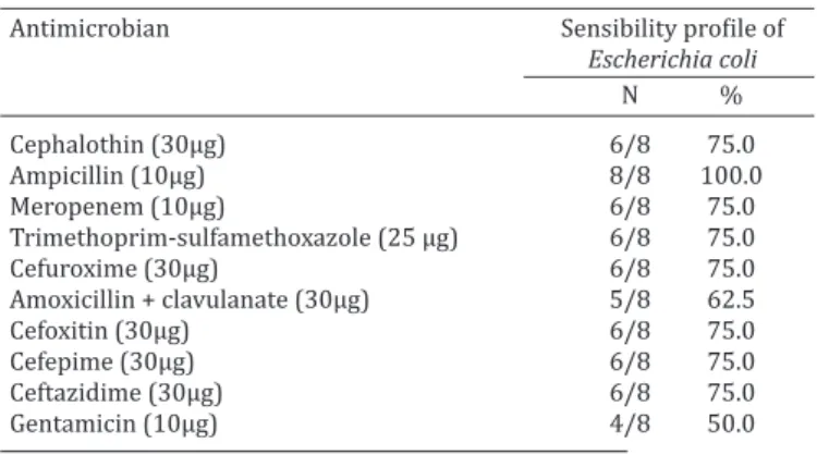
The study of bacterial sensitivity (Table 3) has showed
that
S. aureus
and
S. epidermidis
bacteria were more
sen-sitive to antibiotics gentamicin (100%) and vancomycin
(100%). For the genus
Streptococcus
, gentamicin and
oxa-cillin were the best action antibiotics, followed by
sulpha-zotrim. In contrast, gentamicin was a less effective antimi
-crobial (50%) for
Escherichia coli
isolates (Table 4).
DISCUSSION
The higher average obtained from the SCC (721,000 cells/
mL) was expected, since the sampling was restricted to bu
-ffaloes that had high SCC in order to obtain samples with
microorganisms that may cause subclinical mastitis. It was
high compared to average values of 63,000 cells/mL ob
-served in 2693 buffalo milk samples belonging to a single
herd from state of Sao Paulo (Cerón-Muñoz et al. 2002) and
63,380 cells/mL mean value identified in lactating buffa
-loes also in Brazil (Jorge et al. 2005). Subclinical mastitis
in buffalo milk samples was previously described with SCC
greater than 200,000 cells/mL and positive bacterial gro
-wth in culture in 21.7% (52/200) of the evaluated animals
(Dhakal 2006). High SCC has been obtained in crossbred
Murrah buffaloes, with values of 171,000 cells/mL for ani
-mals without mastitis, 799,000 cells/mL for ani-mals with
subclinical mastitis and 6,039,000 cells/mL with clinical
mastitis (Dhakal et al. 2008).
The most often agents isolated in the samples
Staphylo-coccus epidermidis
(17%) and
S. aureus
(15%) (Table 2)
were similar to those obtained in 49 adult Murrah buffalo
at different stages of lactation in herds from state of
Per-nambuco, Brazil (Oliveira et al. 2004). In that study, the
Staphylococcus
genus was considered the main etiological
agent, being of great epidemiological importance in buffalo
mastitis. Indeed, the genus
Staphylococcus
has been
pre-dominantly isolated in samples from buffalo Murrah
cros-sbred with subclinical mastitis (Dhakal et al. 2008) and
clinical mastitis (Pizauro et al. 2014). Khan and Muham
-mad (2005) obtained similar prevalence for
microbiolo-gical analysis in milk samples of buffalos from Pakistan:
Staphylococcus
spp. (45%),
Streptococcus
spp. (23%) and
Bacillus
spp. (14%). However, Khan et al. (2013) observed a
greater prevalence of
S. aureus
in cow milk samples (90%)
than in buffalo milk (84%). Langoni et al. (2001) observed
Table 2. Bacteria number and frequency of isolates (%) inbuffalo milk samples with subclinical mastitis
Microrganism Number Frequency (%)
Staphylococcus epidermidis 34 (17.0%)
Staphylococcus aureus 30 (15.0%)
Bacillus spp. 28 (14.0%)
Acinetobacter spp. 25 (12.5%)
Pseudomonas aeruginosa 19 (9.5%)
Shigella flexneri 14 (7.0%)
Streptococcus spp. 11 (5.5%)
Corynebacterium spp. 10 (5.0%)
Escherichia coli 9 (4.5%)
Serratia marcescens 8 (4.0%)
Stenotrophomonas maltophilia 8 (4.0%)
Klebsiella rhinoscleromatis 1 (0.5%)
Klebsiella ozaenae 1 (0.5%)
Tatumella ptyseos 1 (0.5%)
Enterobacter cloacae 1(0.5%)
Isolation total 200
Table 3. In vitro susceptibility profile of Staphylococcus aureus, Staphylococcus epidermidis and Streptococcus spp. isolated from buffalo milk samples with subclinical mastitis Antimicrobian Sensibility profile
Staphylococcus Staphylococcus Streptococcus
aureus epidermidis spp.
N̽ % N % N %
Gentamicin (10μg ) 8/8 100.0 8/8 100.0 8/8 100.0 Vancomycin (30μg) 8/8 100.0 8/8 100.0 2/8 25.0 Clindamycin (2μg) 4/8 50.0 4/8 50.0 2/8 25.0 Erythromycin (15μg) 4/8 50.0 4/8 50.0 2/8 25.0 Penicillin G (10μg) 2/8 25.0 3/8 37.5 1/8 12.5 Sulphazotrim (25 μg) 4/8 50.0 4/8 50.0 6/8 75.0 Oxacillin (1μg) 4/8 50.0 4/8 50.0 8/8 100.0 Cefepime (30μg) 4/8 50.0 4/8 50.0 2/8 25.0 Tetracycline (30μg) 2/8 25.0 3/8 37.5 - Rifampicin (34μg) 4/8 50.0 4/8 50.0 2/8 25.0 N = evaluated number of microorganisms.
Table 4. In vitro susceptibility profile of Escherichia coli isolated from buffalo milk samples with subclinical mastitis
Antimicrobian Sensibility profile of
Escherichia coli
N %
Cephalothin (30μg) 6/8 75.0
Ampicillin (10μg) 8/8 100.0
Meropenem (10μg) 6/8 75.0
Trimethoprim-sulfamethoxazole (25 μg) 6/8 75.0
Cefuroxime (30μg) 6/8 75.0
Amoxicillin + clavulanate (30μg) 5/8 62.5
Cefoxitin (30μg) 6/8 75.0
Cefepime (30μg) 6/8 75.0
Ceftazidime (30μg) 6/8 75.0
Gentamicin (10μg) 4/8 50.0
prevalence of
S. epidermidis
(30.1%) and
S. aureus
(4.8%)
in buffaloes with mastitis in São Paulo, Brazil. Other stu
-dies have also identified a high prevalence of
S. epidermidis
(Tenhagen et al. 2009, Sampimon et al. 2009, Pankaj et al.
2013), indicating the importance of this group of
microor-ganisms which are commonly described as lower
prevalen-ce of pathogens in the case of mastitis. In addition,
S.
epi-dermidis
is a component of the normal flora of the skin of
the udder and its prevalence, as observed in this study may
be a result of poor hygienic practices for milking.
Acinetobacter
was isolated with a frequency also
con-sidered high of 12.5% in this study. Similar results were
found in cows with clinical mastitis and milk samples with
mastitis in Korea (Nam et al. 2009, 2010, Gurung et al.
2013). The association of this bacterium with subclinical
mastitis in buffaloes presents a new challenge for the
treat-ment and control of disease. There is need for further stu
-dies to evaluate its role as a pathogen potential and identify
possible sources of contamination.
The percentage of
Pseudomonas aeruginosa
isolated,
can be related to the occurrence of the contamination in
milk during and/or after milking, such as contaminated
water used for washing the teat, cleaning and during
in-tramammary therapy (Fernandes et al. 2009, Langoni et al.
2009).
The incidence of
Bacillus
spp. observed (14%) is found
within a wide occurrence in buffalo milk samples ranging
from 2.4 to 32.4% (Langoni et al. 2001, Oliveira et al. 2004,
Dhakal et al. 2008). This level of detection may differ ac
-cording to the type of sample of milk, herd and region. This
kind of bacteria is widely distributed in soil, water, air, feces
and vegetation.
Although
Streptococcus
are considered less frequent
agents of mastitis, as also obtained in this study, these
op-portunistic pathogens are widespread in the environment
and can be found on the ground, water and manure. Howe
-ver, some studies have identified the
Streptococcus
spp. as
the major etiologic agent isolated from buffalo subclinical
mastitis (Fagiolo & Lai 2007).
Low frequency of
E. coli
was observed in this study
(4.5%) being in agreement with the data obtained by Saini
et al. (1994) that observed lower frequency of microorga
-nisms
E. coli
(12.9%) in buffalo milk samples from Punjab,
India. Moreover, Kumar (2009) observed buffalo milk sam
-ples with high incidence of mastitis
E. coli
(30%).
Other species of bacteria as
Lactococcus garvieae
and
Enterococcus gallinarum
have been isolated from milk
sam-ples of buffalo subclinical mastitis (Vianni & Lazaro 2003).
Arcobacter
species also has considered an important
sour-ce of bacteria in cow and buffalo milk with risk to public
health (Yesilmen et al. 2014).
Conventional bacterial culture is relatively slow
perfor-mance, since incubation of primary cultures often requires
48 hours (or 72 hours) to be completed, and additional
confirmatory tests are relatively time-consuming. The PCR
assay for the identification of microorganisms in milk sam
-ples with mastitis require an analysis time of 3 to 4 hours.
In the case of subclinical mastitis such quick results may
allow the identification of animals with this disease and
indicate the treatment, while optimizing the results with
appropriate use of antibiotics and reduce the
indiscrimina-te usage. Moreover, PCR has been shown to be more sen
-sitive and specific for the diagnosis of microorganisms in
subclinical mastitis than conventional culturing technique
(Shahzad et al. 2013).
Gentamicin was the antibiotic of choice for
Staphylococ-cus
and
Streptococcus
bacteria (Table 3) partially agreeing
with the findings of Cunha et al. (2006) who observed a
wide gentamicin action with high antimicrobial efficacy
(97.98%) on various bacteria and association including
Staphylococcus
spp.,
Streptococcus
spp. and
E. coli
. Howe
-ver, gentamicin was one of the less effective antimicrobial
(50%) for
E. coli
isolates (Table 4). But, this finding agrees
with the results of Costa (2008) who observed that
amino-glycosides and sulfonamides were the antimicrobials less
effective for
E. coli
, highlighting the sensitivity percentage
of 46.2% and 41.8% for gentamicin and neomycin, respec
-tively.
For the genus
Streptococcus
, gentamicin and oxacillin
were the best action antibiotics, followed by sulphazotrim
(Table 3), confirming the data of Langoni et al. (2001) who
have demonstrated a better efficiency of gentamicin (96%)
and oxacillin (95%) on the isolated agents with more
fre-quency:
Corynebacterium bovis, Staphylococcus epidermidis
and
Streptococcus agalactiae
.
The somatic cell count and standard plate count can
perform an assessment of the health status of the buffalo
mammary gland with subclinical mastitis and emphasize
the importance of a normative specific for buffalo that allo
-ws the control and inspection of milk, since the parameters
used for cattle may not be suitable for monitoring mastitis
in buffalo herds.
CONCLUSION
This study highlights the importance of some pathogens
in-volved subclinical mastitis in buffaloes and the possibility
of specific antimicrobial use in the control and treatment of
this serious problem, promoting increased quality and milk
production with effective reduction of production costs
and improvement in health foods.
Acknowledgements.- Credit to Coordenação de Aperfeiçoamento de Pes -soal de Nível Superior (CAPES-Brazil) for financial support and to farmers of buffalo for their interest in scientific research to improve the quality of milk products.
Conflict of interest statement.- The authors have no competing interests.
REFERENCES
Amaral F.R., Carvalho L.B., Silva N. & Brito J.R.F. 2005. Qualidade do leite de búfalas: composição. Revta Bras. Reprod. Anim. 29:106-110.
Andrighetto C., Jorge A.M., Gomes M.I.F.V., Hoch A. & Piccinin A. 2005. Ef -fect of monensin on milk production and composition, production of
mozzarela cheese and body condition score of Murrah buffalo cows. Re
-vta Bras. Zootec. 34:641-649.
Araujo K.B.S., Rangel A.H.N., Fonseca F.C.E., Aguiar E.M., Simplicio A.A., Novaes L.P. & Lima Júnior D.M. 2012. Influence of the year and calving
season on production, composition and mozzarella cheese yield of
Baird-Parker A.C. 1990. The staphylococci: an introduction. J. Appl. Micro
-biol. Symp. Suppl. 69:1S-8S.
Baloch H., Rind R., Kalhoro D.H. & Kalhoro A.B. 2011. Study on the inci
-dence of mastitis in buffaloes caused by bacterial species. Pak. J. Agri., Agril. Engg., Vet. Sci. 27:83-93.
Bauer A.W., Kirby E., Sherris E.M. & Turk M. 1966. Antibiotic by stan
-darized single disk method. Am. J. Clin. Pathol. 45:493-496.
Braun P.G. & Preuss S.E. 2007. Microbial quality of water buffalo milk and milk products in Germany. Milchwissenschaft 62:276-278.
Bernardes O. 2007. Buffaloes breeding in Brasil. Ital. J. Anim. Sci. 6:162-167.
Bonini Pardo R., Mendoza-Sánchez G., Nader Filho A., Santos T.A.B., Lango
-ni H., Tonhati H., Ferreir E.B.S., Ravena D.L., Oliveira M.E.A. & Sturion D.J. 2007. Microbiological evaluation of milk samples positive to California
Mastitis Test in dairy buffalo cows (Buballus bubalis). Ital. J. Anim. Sci.
6:884-887.
Carvalho L.B., Amaral F.R., Brito M.A.V.P., Lange C.C., Brito J.R.F. & Leite R.C. 2007. Contagem de células somáticas e isolamento de agentes causado -res de mastite em búfalas (Bubalus bubalis). Arq. Bras. Med. Vet. Zootec.
59:242-245.
Cerón-Muñoz M., Tonhati H., Duarte J., Oliveira J., Muñoz-Berrocal M. & Jurado-Gámez H. 2002. Factors affecting somatic cell counts and their relations with milk and milk constituent yield in buffaloes. J. Dairy Sci. 85:2885-2889.
Costa G.M. 2008. Mamite bovina em rebanhos leiteiros da região sul do Estado de Minas Gerais. Tese de Doutorado, Escola de Veterinária, Uni
-versidade Federal de Minas Gerais, Belo Horizonte, MG. 123p.
Cunha A.P., Silva L.B.G., Pinheiro Júnior J.W., Silva D.R., Oliveira A.A.F., Silva K.P.C. & Mota R.A. 2006. Perfil de sensibilidade antimicrobiana de agen -tes contagiosos e ambientais isolados de mastite clínica e subclínica de
búfalas. Arqs Inst. Biológico, São Paulo, 73:17-21.
Dhakal I.P. 2006. Normal somatic cell count and subclinical mastitis in Murrah buffaloes. J. Vet. Med. B 53:81-86.
Dhakal I.P., Dhakal P., Koshihara T. & Nagahata H. 2007. Epidemiological and bacteriological survey of buffalo mastitis in Nepal. J. Vet. Med. Sci. 69:1241-1245.
Dhakal I.P., Neupane M. & Nagahata H. 2008. Evaluation of direct and in
-direct measures of quarter milk from crossbred buffaloes. Anim. Sci. J. 79:628-633.
Fagiolo A. & Lai O. 2007. Mastitis in buffalo. Ital. J. Anim. Sci. 6:200-206. Fang H. & Hedin G. 2003. Rapid screening and identification of methi
-cillin-resistant Staphylococcus aureus from clinical samples by
selec-tive-broth and real-time PCR assay. J. Clin. Microbiol. 41:2894-2899. Fernandes M.C., Ribeiro M.G., Siqueira A.K., Salerno T., Lara G.H.B. & Listoni
F.J.P. 2009. Surto de mastite bovina causada por linhagens de Pseudomo-nas aeruginosa multirresistentes aos antimicrobianos. Arq. Bras. Med.
Vet. Zootec. 61:745-748.
Gurung M., Nam H.M., Tamang M.D., Chae MH., Jang G.C., Jung S.C. & Lim S.K. 2013. Prevalence and antimicrobial susceptibility of Acinetobacter
from raw bulk tank milk in Korea. J. Dairy Sci. 96:1997-2002.
Hussain R., Javed M.T., Khan A. & Muhammad G. 2013. Risks factors associ
-ated with subclinical mastitis in water buffaloes in Pakistan. Trop. Anim. Health Prod. 45:1723-1729.
Jorge A.M., Andrighetto C., Strazza M.R.B., Correa R.C., Kasburgo D.G., Picci
-nin A., Victoria C. & Domingues P.F. 2005. Correlation between the Cali -fornia Mastitis Test (CMT) and somatic cells count on milk from Murrah
buffalo cows. Revta Bras. Zootec. 34:2039-2045.
Khan J.M., Rasool M.H., Arshad M., Rahman S.U., Tahir M.F., Aslam B., Jing W., Jun Z. & Ghani M. 2013. Comparative Evaluation of Leukotoxic Activ -ities of Indigenous Staphylococcus aureus Isolates from Subclinical and
Clinical Mastitic Milk Samples of Buffalo and Cattle. Open Vet. J. 7:24-27. Khan A.Z. & Muhammad G. 2005. Quarter-wise comparative prevalence of
mastitis in buffaloes and crossbred cows. Pakistan Vet. J. 25:9-12. Kumar P.A. 2009. Evaluation of PCR test for detecting major pathogens of
bubaline mastitis directly from mastitis milk samples of buffaloes. Trop. Anim. Health Prod. 41:1643-1651.
Langoni H., Sakiyama D.T.P., Guimarães F.F., Menozzi B.D. & Da Silva R.C. 2009. Aspectos citológicos e microbiológicos do leite em propriedades no sistema orgânico de produção. Pesq. Vet. Bras. 29:881-886. Langoni H., Domingues P.F., Molero Filho J.R. & Baldini S. 2001. Etiologia e
sensibilidade bacteriana da mastite subclínica em búfalos (Bubalus bu-balis). Ars Vet. 17:213-217.
Macfaddin J.F. 1980. Biochemical Tests for Identification of Medical Bacte
-ria. Williams and Wilkins, Baltimore, USA.
Medeiros E.S., Freitas M.F.L., Saukas T.N., Azevedo S.S., Pinheiro Junior J.W., Brandespim D.F., Neto O.L.D. & Mota R.A. 2011. Risk factors associated with buffalo mastitis in the Brazilian Northeast. Pesq. Vet. Bras. 31:499-504.
Nam H.M., Lim S.K., Kang H.M., Kim J.M., Moon J.S., Jang K.C., Kim J.M., Joo Y.S. & Jung S.C. 2009. Prevalence and antimicrobial susceptibility of
gram-negative bacteria isolated from bovine mastitis between 2003 and
2008 in Korea. J. Dairy Sci. 92:2020-2026.
Nam H.M., Lim S.K., Kim J.M., Joo Y.S., Jang K.C. & Jung S.C. 2010. In vitro ac -tivities of antimicrobials against six important species of gram-negative
bacteria isolated from raw milk samples in Korea. Foodborne Pathog. Dis. 7:221-224.
Oliveira M.V.V., Mota R.A., Oliveira A.A.F., Meirelles F.S. & Silva F.F. 2004. Utilização do whiteside modificado e california mastites test no diag -nóstico da mastite subclínica em búfalas e sua relação com o exame
mi-crobiológico. Ciência Animal. 14:39-45.
Pankaj A.S., Chhabra R. & Sindhu N. 2013. Sub-clinical mastitis in Mur -rah buffaloes with special reference to prevalence, etiology and
anti-biogram. Buffalo Bull. 32:107-113.
Pizauro L.J.L., Silva D.G., Santana A.M., Clemente V., Lara G.H.B., Listoni F.J.P., Vaz A.C.N., Vidal-Martins A.M.C., Ribeiro M.G. & Fagliari J.J. 2014.
Prevalence and etiology of buffalo mastitis and milk somatic cell count
in dry and rainy seasons in a buffalo herd from Analandia, São Paulo State, Brazil. Arq. Bras. Med. Vet. Zootec. 66:1703-1710.
Saini S.S., Sharma J.K. & Kwatra M.S. 1994. Prevalence and etiology of sub
-clinical mastitis among crossbreed cows and buffalos in Punjab. Indian J. Dairy Sci. 47:103-107.
Sampimon O., Barkema H.W., Berends I., Sol J. & Lam T. 2009. Prevalence of intramammary infection in Dutch dairy herds. J. Dairy Res. 76:129-136. Shahzad W., Altaf M., Ahmad M., Munir R., Amin M.T., Khan M.S., Sagar M.S.,
Khan M.A., Avais M., Akbar G. & Mehmood F. 2013. Prevalence and mo -lecular diagnosis of Staphylococcus aureus subclinical mastitis in
lactat-ing Nili-Ravi Buffaloes (Bubalus bubalis) at Livestock Experiment Sta
-tion, Bahadurnagar, Okara, Pakistan. Buffalo Bull. 32:1041-1045. Shome B.R., Das Mitra S., Bhuvana M., Krithiga N., Velu D., Shome R., Is
-loor S., Barbuddhe S.B. & Rahman H. 2011. Multiplex PCR assay for spe
-cies identification of bovine mastitis pathogens. J. Appl. Microbiol. 111: 1349-1356.
Tenhagen B.A., Hansen I., Reinecke A. & Heuwieser W. 2009. Prevalence
of pathogens in milk samples of dairy cows with clinical mastitis and in
heifers at fi rst parturition. J. Dairy Res. 76:179-187.
Tiwari J.G., Chaudhary S.P., Tiwari H.K., Dutta T.K., Saikia P. & Hazari
-ka P. 2011. Microbial evaluation of market milk and milk-products of
Mizoram, India with special reference to Staphylococcus aureus. Indian
J. Dairy Sci. 81:429-431.
Viana R.B., Cardoso E.C., Gouveia I.M., Rezende M.L.G., Monteiro B.M. & Araujo C.V. 2010. Avaliação da eficiência do Somaticell para o diagnósti
-co da -contagem indireta de células somáticas no leite de búfalas. Revta Ciênc. Agrárias 1:24-30.
Vianni M.C.E. & Lazaro N.S. 2003. Profile of antimicrobial susceptibility in strains of Gram positive cocci, negative catalase, isolated from buffalo subclinical mastitis. Pesq. Vet. Bras. 23:47-51.
Yesilmen S., Vural A., Erkan M.E. & Yildirim I.H. 2014. Prevalence and anti -microbial susceptibility of Arcobacter species in cow milk, water buffalo