Universidade de Lisboa
Faculdade de Ciências
Departamento de Biologia Vegetal
Development of a PCR system for detection and differentiation of Burkholderia mallei and B. pseudomallei in clinical and environmental
matrices
Mestrado em Biologia Molecular e Genética Catarina Barreto Brás
Dissertação orientada por Doutora Ana Botelho Professora Doutora Ana Rita Matos
This thesis was carried out at The Laboratory of Bacteriology and Mycology of the National Institute of Agrarian and Veterinary Research (INIAV, IP), under the supervision of Doctor Ana Botelho, and was funded by the EU project “Prevention of and Fight against Crime
Programme of European Union, Grant Agreement No.
HOME/2012/ISEC/AG/CBRN/4000003810, IB-BIOALERTNET- Iberian network of laboratories of biological alert. Accreditation of methods for detection of highly pathogenic agents.
Part of this work resulted in a poster presented in:
European Melioidosis Congress 2015, Downing College, Cambridge, UK – Attendance and publication of a poster under the name “Single tube molecular assay for differential detection of Burkholderia pseudomallei and B. mallei”, Brás C., Canto A., Cunha V. M., Botelho A. (2015);
In the field of this project, a series of workshops were attended, namely:
Workshop of "Diagnóstico laboratorial e manuseamento de fungos de grupo de risco 3", Instituto Nacional Ricardo Jorge;
Workshop of “Inativação e deteção de microrganismos patogénicos na resposta a emergências”, Instituto Nacional Ricardo Jorge;
Jornadas Doenças Infeciosas 2015 - 1º Seminário NRBQ: Equipamento de Proteção Individual, Instituto Nacional Ricardo Jorge.
“In the fields of observation chance favors only the prepared mind”
Louis Pasteur
I AKNOWLEDGEMENTS
To my parents, who devoted their trust and funds over the past six years; To my brother, whom I owe my life achievements and personal character;
To my boyfriend, who supported me with unconceivable patience at all times and gave me strenght when needed;
To Doctor Ana Botelho, who continuosly relied her guidance and knowledge with trust, offering me a life changing opportunity that exceeded all expectations;
To Doctor Ana Rita Matos, who gave me exceptional support and feedback, in a thoughful and detailed way;
To my colleage Tiago Baeta, whom I shared a multitude of emotions while working on our theses, presenting me with great moments over the two past years;
To Marta Vaz, Ana Reis, Ana Prata, Inês Guinote and Tânia Chança, who always brought fun into work;
To Professor Mónica Cunha, Doctor Miguel Fevereiro, Doctor Tiago, Célia Leão, Ana Canto, Ana Amaro and Maria José Barahona, who gave their best by assisting me with advices and promptness to help solve any sort of issues;
To Dona Alexandrina, Dona Filomena, Sr. Carlos, Sr. Oliveira and many other staff workers, who welcomed me at INIAV, Pólo Benfica not as just an internee, but as one of them;
To “Grupo do Foz, Dark e essa gente”, who I owe their eternal friendship and support; To my cats Cookie and Luana (who left me sooner than expected, may your soul rest in peace), whom I see as the most incredible pets someone can have;
II ABSTRACT
Glanders and melioidosis are two infectious diseases caused, respectively, by the Gram- negative bacteria, Burkholderia mallei and B. pseudomallei. These species are classified as Class B agents by the Centre of Disease Control (CDC) and as level 3 risk agents by the European Parliament, due to their fast aerosol dissemination, high infectiousness, potential zoonotic capability, absence of vaccines and resistance to a wide variety of antibiotics. The potential use of these microorganisms in biological warfare, already applied in the American Civil War and World Wars I and II, leads to the need of strategic protocols in laboratories of reference to detect and differentiate both agents in a rapid, effective and distinctive way.
A duplex qPCR approach was optimized and evaluated for direct detection and differentiation of Burkholderia mallei and B. pseudomallei in different matrices. Since in Portugal naturally infected tissues or contaminated material with these agents do not exist, spiked samples were previously prepared. Known concentrations of serial decimal dilutions of Burkholderia mallei NCTC 10245 and B. pseudomallei NCTC 10276 strains were inoculated in lung tissues and swabs, while soils were spiked only with B. pseudomallei NCTC 10276. The duplex qPCR has as targets the psu gene that encodes for a putative acetyltransferase specific of B. pseudomallei and the transposase of ISBma2, an insertion sequence present in about 48 copies in B. mallei genome and in about 6 copies in B.
pseudomallei genome. Due to the complexity of some matrices that might present PCR
inhibitors, giving PCR false negative results, an Internal Amplification Control (IAC) was constructed based on a 125 bp fragment of the m000.5L/R gene of myxoma virus, cloned in the pNZY28 vector.
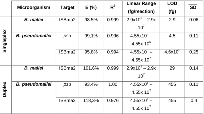
The duplex qPCR was firstly optimized, evaluated and compared with singlepex qPCR, using purified DNA from strains B. mallei NCTC 12938T and B. pseudomallei NCTC 12939T. Four hundred nM of each four primers proved to be the best concentration in the duplex reaction, while 200 nM were the appropriated concentration of the two probes targeting both ISBma2 and psu gene. The optimal annealing temperature, that gave detection of the target at the lowest quantification cycle (Cq) value, was 58.1 ºC. The limit of detection of the duplex qPCR was 29 fg and 455 fg for, respectively, B mallei and B. pseudomallei.
The coefficient variance percentages for the repeatability and reproducibility of the duplex qPCR were, respectively, 1.337% and 2.288%, a low variance that indicates high repeatability and reproducibility. The assay was also specific for B. mallei and B.
pseudomallei since it didn’t detect DNA from 13 other bacteria, including Mycobacterium tuberculosis, Pseudomonas aeroginosa and Burkolderia thailandesis. This methodology
applied to the prepared spiked samples was capable to detect both agents in pulmonary macerates until the less concentrated dilution (10-6) with corresponding Cq values between 15.93 (10-1) and 25.95 (10-6) for B. mallei and between 23.44 (10-1 – psu target) and 38.18
III (10-6 – psu target) for B. pseudomallei. For non–enriched swabs, both agents were also detected until the highest dilution 10-6, with Cq values ranging from 20.33 (10-1) to 39.25 (10
-6) for B. mallei and from 28.98 (10-1 – psu target) to 38.37 (10-6 – psu target) for B.
pseudomallei. Enriched swabs (incubation of swabs in BHIB 48h at 37ºC) but a slightly
improvement in the detection of both microorganisms. The alternative approach by performing the qPCR in B. pseudomallei isolated colonies showed an increase of sensitivity of the method resulting in Cq values as low as 27.75 for the psu target. The “gold standard” culture media method performed in parallel with the qPCR detection, presented some discrepancies mainly for B. mallei swabs that showed no growth, probably due to the absence of a specific culture media for this agent proving to be less sensitive than the qPCR.
Keywords: Burkholderia mallei; Burkholderia pseudomallei; quantitative polymerase chain reaction; Internal Amplification Control; Spiked Samples
IV RESUMO EM PORTUGUÊS
Mormo e melioidose são patologias causadas pelas bactérias Gram-negativas
Burkholderia mallei e B. pseudomallei, respectivamente. Sendo os equídeos o principal alvo
hospedeiro de mormo, cavalos, mulas e burros para exportação necessitam de procedimentos standard europeus de despistagem do agente através de ensaios de fixação do complemento pela detecção de anticorpos específicos. Erradicado de Portugal em 1952 e da Europa Ocidental, o mormo é ainda reportado em alguns locais da Ásia, África, Médio Oriente e América do Sul.
Nunca declarada em território português, a melioidose trata-se duma doença endémica em países como Tailândia e norte da Austrália, com expansão em países do continente asiático como as Filipinas, India, Indonésia, Laos, Singapura, Camboja e Vietname, alertando-se também para sua existência em zonas de África e América do Sul.
O potencial zoonótico de B. mallei é descrito na literatura, estando identificados como principais grupos de risco investigadores científicos, cujo alvo de estudo implica a manipulação e multiplicação do microorganismo, profissionais de medicina veterinária e funcionários de matadouros. Afectando os animais, o homem e ambiente, a capacidade zoonótica de B.pseudomallei não se encontra estabelecida. Contudo, estão declarados inúmeros factores de risco que contribuem para a transmissão da doença no hospedeiro humano, nomeadamente: diabetes, alcoolismo, doenças crónicas renais, hepáticas e pulmonares e terapias imunossupressoras.
As manifestações clínicas de ambas as patologias culminam, geralmente, em vastas complicações a nível pulmonar, podendo levar à morte. As vias de transmissão das duas doenças são principalmente cutânea, através de lesões expostas, inalação e, ocasionalmente, ingestão.
Não existem vacinas ou tratamentos 100% eficientes contra ambas as doenças. Contudo, algumas terapias com base em combinações de diversos antibióticos têm sido estabelecidas mas a sua eficácia depende do progresso de cada patologia e, portanto, qualquer caso de mormo e/ou melioidose deve ser tratado com o máximo de brevidade possível.
Devido à sua rápida disseminação, capacidade de infecção por inoculação e formação de aerossóis, alto factor de contágio e largo espectro de resistência antimicrobiana, B. mallei e
B. pseudomallei foram classificados como agentes de classe B pelo Centre of Disease
Control (CDC) e de risco 3 pelo Parlamento Europeu, segundo a Directiva 2000/54/CE. Numa reunião organizada em conjunto pela Organização Mundial da Saúde (WHO) e a Organização Mundial da Saúde Animal (OIE), especialistas alertaram para o risco eminente em países cujos mecanismos de preparação e prevenção para determinados agentes se encontram inactivos, tornando-os mais susceptíveis à libertação deliberada do agente.
V Tendo em conta as declarações acima descritas, protocolos foram estabelecidos para a detecção e diferenciação de B. mallei e B. pseudomallei, utilizando a metodologia biomolecular através da técnica quantitativa em tempo real de reacção de polimerização em cadeia (qPCR) em matrizes clínicas e ambientais.
Deste modo, e uma vez que não existem amostras clínicas e ambientais de mormo e/ou melioidose em Portugal, três matrizes foram seleccionadas para serem inoculadas com diluições decimais seriadas de B. mallei NCTC 10245 e B. pseudomallei NCTC 10276, de modo a obterem-se amostras experimentalmente infectadas ou spiked samples. A escolha das matrizes teve em consideração as amostras comumente recolhidas quando há suspeita de alguma destas infecções: zaragatoas, pois os exsudados ou feridas purulentas são normalmente colhidos com estas ferramentas; macerados pulmonares, visto que ambas as doenças proliferam a nível pulmonar e, no caso específico de melioidose, solos, uma vez que este é o reservatório natural de B. pseudomallei.
À excepção dos macerados pulmonares, a identificação de ambos microorganismos nas
spiked samples foi avaliada em dois tempos diferentes: imediatamente após a infecção, e
48 horas após incubação das matrizes a 37 ºC, comparando a sensibilidade de detecção do método de cultura com a metodologia de qPCR desenvolvida. O isolamento dos agentes através de cultura bacteriana foi realizado utilizando o meio de cultura Agar Ashdown’s, específico de B. pseudomallei, e o meio de Agar Columbia com 5% de sangue carneiro para
B. mallei. Enquanto B. pseudomallei produz colónias rosas rugosas morfologicamente
distinguíveis, as colónias de B. mallei não detêm características particulares que permitam a sua diferenciação doutras bactérias.
O duplex qPCR desenvolvido consiste num sistema capaz de identificar e diferenciar os dois microorganismos num só tubo de reacção. Dois alvos foram escolhidos para a detecção e diferenciação de B. mallei e B. pseudomallei: o gene psu que codifica para uma putativa acetiltransferase, pertencente ao cluster de genes do sistema tipo III de secreção de B. pseudomallei e um gene que codifica uma transposase ISBma2, uma sequência de inserção presente em cerca de 48 cópias e 6 cópias em B. mallei e B. pseudomallei, respectivamente. Deste modo, a amplificação e detecção de sinal por parte das sondas de hidrolisação dos dois genes alvo corresponde à identificação positiva de B. pseudomallei enquanto, a amplificação e detecção apenas do gene que codifica a transposase ISBma2 diz respeito a uma amostra positiva para B.mallei.
Junto desta plataforma de diagnóstico, foi também construído um controlo interno de amplificação (IAC – Internal Amplification Control), pNZYmyx, clonando o fragmento de 125 pares de base do gene diplóide m000.5 L/R da estirpe Laussane do mixoma vírus no vector pNZY28. A finalidade deste controlo consiste em aferir se a reacção de PCR detém
VI qualquer factor que resulte na inibição da reacção, afectando a amplificação dos genes alvo.
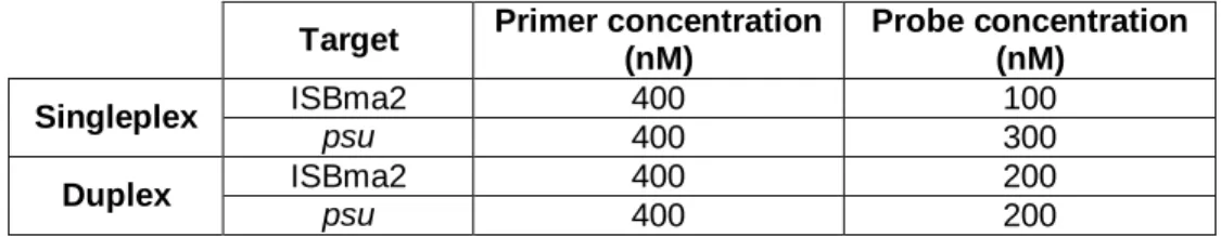
A adaptação deste sistema de qPCR necessitou de optimização dos oligonucleotídeos necessários à reacção (iniciadores ou primers e sondas) bem como o ajuste da sua temperatura de hibridação (annealing) utilizando as estirpes de referência B. mallei NCTC 12938T e B. pseudomallei NCTC 12939T. Esta optimização de reacção foi executada com os dois alvos em separado (singleplex) e em conjunto (duplex), seguida de testes de especificidade, sensibilidade, repetibilidade e reprodutibilidade. Em singleplex, a concentração final óptima para cada alvo provou ser 400 nM enquanto que a concentração óptima das sondas de hidrolisação foram 100nM e 300 nM para ISBma2 e psu, respectivamente. As concentrações finais óptimas em duplex de primers e sonda para ambos os alvos foram, respectivamente, 400 nM e 200nM e a temperatura de annealing que demonstrou o Cq (Quantification Cycle) mais baixo foi de 58.1 ºC. A especificidade do sistema foi provada testando o qPCR com 17 microorganismos, incluindo Mycobacterium
tuberculosis, Pseudomonas aeroginosa e a espécie geneticamente próxima, Burkholderia thailandensis, na qual o sinal de fluorescência foi somente detectado em B. mallei e B. pseudomallei. O sistema duplex qPCR provou ser capaz de detectar 29 fg e 455 fg de DNA
de B. mallei e B. pseudomallei, respectivamente. O coeficiente de variação calculado para avaliar a repetibilidade e reprodutibilidade obteve valores máximos de 1.337% e de 2.288 %, respectivamente comprovando este sistema ser altamente repetível e reproduzível.
A técnica de qPCR estabelecida foi capaz de identificar e distinguir os dois microorganismos em todas as matrizes inoculadas. No que diz respeito aos macerados pulmonares, o qPCR foi capaz de identificar correctamente os dois microorganismos até à diluição menos concentrada (10-6) detendo valores de Cq entre 15.93 (10-1) e 25.95 (10-6) em B. mallei e entre 23.44 (10-1 – alvo psu) e 38.18 (10-6 - alvo psu) para B. pseudomallei. Foi também possível identificar ambos agentes até à diluição menos concentradas para zaragatoas sem o passo de incubação, variando os valores de Cq entre 20.33 (10-1) e 39.25 (10-6) for B. mallei e entre 28.98 (10-1 - alvo psu) e 38.37 (10-6 - alvo psu) para B.
pseudomallei. A adição prévia do passo de incubação para as zaragotas demonstrou uma
variação ligeira indicando com valores de Cq inferiores comparativamente às zaragatoas não incubadas. A detecção de B. pseudomallei em solos sem incubação prévia foi igualmente possível até à diluição menos concentrada. Porém, a análise de colónias isoladas provou ser altamente sensível, detectando todas as amostras com valores de Cq inferiores a 30.
No entanto, o isolamento por cultura bacteriana (gold standard) provou ser um método de diagnóstico menos sensível comparando com o sistema de qPCR. A sensibilidade obtida por meio de cultura e qPCR para B. pseudomallei foi, respectivamente, 80% e 97%
VII indicando uma baixa percentagem de falsos negativos para as duas metodologias, contudo, o método de qPCR é mais sensível mostrando ser capaz de identificar amostras consideradas negativas pelo método de cultura. Comparativamente, o método de qPCR para B.mallei mostrou ser 100% sensível ao identificar o microorganismo em todas as amostras enquanto que a sensibilidade do método de cultura para a isolação deste agente é significativamente menor, 17%, possivelmente devido à falta de um meio de cultura específico para o isolamento deste microorganismo.
Desta forma, a identificação de B. mallei e B. pseudomallei por qPCR consiste num teste de diagnóstico sensível, específico, repetível e reprodutível capaz de identificar e diferenciar os dois agentes em amostras previamente inoculadas.
Palavras-chave: Burkholderia mallei; Burkholderia pseudomallei; duplex qPCR; Spiked Samples; Bioterrorismo
VIII TABLE OF CONTENTS
AKNOWLEDGEMENTS ...I ABSTRACT ... II RESUMO EM PORTUGUÊS ... IV TABLE OF CONTENTS ... VIII LIST OF FIGURES ...X LIST OF APPENDIX FIGURES ... XI LIST OF TABLES ... XII LIST OF APPENDIX TABLES ... XIII ABBREVIATIONS ... XIV
CHAPTER 1 - INTRODUCTION ... 1
1.0. Genus Burkholderia ... 1
1.1. Burkholderia mallei ... 1
1.1.1. Glanders ... 2
a) Epidemiology and Clinical Manifestations ... 2
b) Diagnosis ... 3
1.2. Burkholderia pseudomallei ... 3
1.2.1. Melioidosis ... 4
a) Epidemiology and Clinical Manifestations ... 4
b) Diagnosis ... 4
1.3. B. mallei and B. pseudomallei in biological warfare... 6
1.4. Real time Polymerase Chain Reaction (qPCR) ... 6
1.5. Internal Amplification Control (IAC) ... 7
1.6. qPCR as a diagnostic tool for glanders and melioidosis ... 8
1.7. Aims ... 9
CHAPTER 2 – MATERIAL AND METHODS ... 10
2.0. Bacterial strains ... 10 2.0.1. Bacterial growth ... 10 2.0.2. Spiked Samples... 11 a) Swabs ... 11 b) Pulmonary macerates... 11 c) Soils ... 12
2.1. Inactivation of the bacteria, DNA purification and quantification ... 12
2.2. qPCR system for the detection of B. mallei and B. pseudomallei ... 12
2.2.1. In silico analysis... 13
2.2.2. qPCR optimization ... 13
IX
b) Annealing Optimization ... 14
2.2.3. Estimation of the limit of detection (LOD), specificity, repeatability and reproducibility ... 14
2.3. Construction of an Internal Amplification Control (IAC) system for PCR reactions ... 15
2.3.1. Conventional PCR amplification ... 15
2.3.2. Cloning of the 125 bp fragment of m000.5L/R gene in pNZY28 vector – plasmid pNZYmyx construction ... 16
2.2.3. qPCR using pNZYmyx as IAC ... 16
2.3. Data analysis... 16
CHAPTER 3 – RESULTS AND DISCUSSION ... 17
3.0. Growth of B. mallei and B. pseudomallei strains in culture media ... 17
3.1. Spiked samples bacteriological culture ... 18
a) Swabs ... 18
b) Pulmonary macerates... 18
c) Soils ... 19
3.2. Optimization and evaluation of singleplex and duplex qPCR ... 20
3.2.1 Specificity of primers and probes ... 20
3.2.2 Concentration of primers/probes and annealing temperature ... 21
3.2.3 Comparison of Singleplex vs Duplex qPCR performance... 22
3.2.4 Efficiency, analytical specificity and limit of detection of qPCR ... 22
3.2.5. Repeatability and Reproducibility ... 25
3.3. qPCR analysis of the spiked samples ... 25
a) Swabs ... 25
b) Pulmonary macerates... 25
c) Soils ... 25
3.4. Sensitivity of the qPCR versus sensitivity of the culture method and significance of difference ... 26
3.5. pNZYmyx as an IAC ... 27
CONCLUSION ... 28
BIBLIOGRAPHY ... 30
X LIST OF FIGURES
Figure 1. Global distribution of B. pseudomallei.. ... 5
Figure 2. Colonies of B. pseudomallei and B. mallei in selective media. ... 17
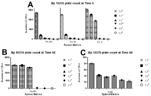
Figure 3. B. pseudomallei (Bp) 10276 spiked samples plate count.. ... 20
XI LIST OF APPENDIX FIGURES
Figure A 1. Sample processing with subsequent qPCR analysis. ...ii
Figure A 2. Decontamination strategies.. ... iii
Figure A 3. Optimization of the annealing temperature of duplex qPCR.. ...iv
XII LIST OF TABLES
Table 1. Key phenotypical features of B. mallei and B. pseudomallei. ... 5
Table 2. Resume table of PCR developments towards the identification of B. mallei and B. pseudomallei ... 8
Table 3. B. mallei and B. pseudomallei strains used. ... 10
Table 4. Oligonucleotides for the qPCR duplex Burkholderia system. ... 13
Table 5. Primers and probes for pNZYmyx. ... 15
Table 6. Primer and probe optimal final reaction concentrations for singleplex and duplex performances. ... 22
Table 7. Cq values of singleplex reaction against multiplex reaction. ... 22
Table 8. Efficiency and Limit of Detection (LOD) of singleplex and duplex systems, for B.mallei NCTC 12938T and B.pseudomallei NCTC 12939T... 23
XIII LIST OF APPENDIX TABLES
Table A 1. OD600 values andcorrespondentCFU/mL mean for each serial dilution of B.mallei
10245 and B.pseudomallei 10276 ... iii Table A 2. Repeatability and Reproducibility of the singleplex and duplex assay. ... v Table A 3. qPCR results for the spiked swabs and pulmonary macerates with B.mallei NCTC 10245 and B.pseudomallei NCTC 10276 ... v Table A 4. qPCR results for the spiked soils with B.pseudomallei 10276 ...vi
XIV ABBREVIATIONS µL - microliter µm – micrometer µM – micromolar A – Adenine
ATCC – American Type Culture Collection BHIB – Brain Heart Infusion Broth
bp – base pairs
BSL-2 – Biosafety Laboratory Level - 2 BSL-3 – Biosafety Laboratory Level – 3 C – Cytosine
CA/S – Columbia Agar with 5% Sheep Blood CFT – Complement Fixation Test
CFU - Colony Forming Units Cq – Quantification Cycle dsDNA – Double-Stranded DNA
ELISA – Enzyme-Linked Immunosorbent Assay EPF - End-Point Fluorescence
fg - femtograms G – Guanine H - Hours
IAC – Internal Amplification Control IPTG – Isopropyl-1-thio-β-D-galactoside IS – Insertion Sequence
LB – Luria Broth mL - mililiter mM - milimolar
NCTC – National Collection of Type Cultures nm - nanometer
nM – nanomolar
NTC – Non-Template Control
OIE - Organisation Mondiale de la Santé Animale/ World Organization of Animal Health PCR – Polymerase Chain Reaction
PPE – Personal Protective Equipment
qPCR – Real-Time Polymerase Chain Reaction/ quantitative Polymerase Chain Reaction rDNA – ribossomal Desoxyribonucleic Acid
XV T – Thymine
R – Adenine or Guanine TSA – Tryptic Soy Agar TSB – Tryptic Soy Broth
TTSS - Type III Secretion System WGS – Whole-Genome Sequencing
WRAIR - Walter Reed Army Institute of Research X-Gal - 5-bromo-4-chloro-3-indolyl-β-D-galactoside
1 CHAPTER 1 - INTRODUCTION
1.0. Genus Burkholderia
The β-Proteobacterial Burkholderia genus is composed by more than 40 species with wide
versatile ecological features. These bacilli shape Gram-negative bacteria ranging from 1–5 μm in length and 0.5–1.0 μm in width were previously classified in the heterogeneous Pseudomonas genus. The Burkholderia genus was proposed by Yabuushi et al. in 1992 on the basis of 16S ribosomal DNA (rDNA) sequence, DNA–DNA homology values, cellular lipid and fatty acid composition, and phenotypic characteristics 1,2. This genus includes plant pathogens, microbial biodegradation of pollutants, opportunistic human pathogens (B. cepacia complex interacts with cystic fibrosis patients) and primary pathogens B. mallei and B. pseudomallei, the etiological agents of glanders and melioidosis, respectively, with the ability to infect both humans and animals 3.
Despite glanders being recognized for centuries, multilocus sequence typing (MLST) and whole genome sequencing (WGS) have shown that B. mallei is a clone of B. pseudomallei, with a considerably smaller genome. The genome reduction resulted in the evolution of B.
pseudomallei to the mammalian-adapted pathogen B. mallei, unable to survive in the
environment outside its host 4.
1.1. Burkholderia mallei
Burkholderia mallei is the causative agent of glanders, a fatal disease with zoonotic
capa-bility 5. Described as “malis” by Hippocrates in 450 B.C., glanders adopted various names through history e.g. malleus given by Aristotle meaning “depicting a malignant disease”, equinia and droes. The skin form is often described as “farcy”, a designation recognized by the World Organization of Animal Health (OIE) 7,8.
It was first isolated by Friedrich Loeffler and Wilhelm Schütz in 1882 from the infected liver and spleen of a horse and since then, the pathogen has been classified as Loefflerella mallei,
Pfeifferella mallei, Malleomyces mallei, Actinobacillus mallei, Corynebacterium mallei, Myco-bacterium mallei, Pseudomonas mallei and Bacillus mallei 9,10.
The high infectiousness, zoonotic capability, aerosol transmission, absence of vaccines and antibiotic resistance characteristics qualifies this agent as a potential biological weapon 7,9,11. In fact, the use of B. mallei as a biological warfare agent during the American Civil War, World Wars I and II and the Russian invasion of Afghanistan has been reported 9.
2
B. mallei is non-motile, nonsporulating, facultative intracellular and obligate mammalian
pathogen. Outside the host, it represents susceptibility to heat, sunlight and common disinfect-ants. Even so, it can remain viable in water for up to 100 days and at room temperature 5,10.
Blood agar and other nutrient culture media are used for the growth of B. mallei since the microorganism has no specific culture media. Colonies become visualized after 48 hours of incubation at 37ºC (See key phenotypical features and growth medium conditions in Table 1). The 5.8 Mb genome of NCTC 12938T strain with G+C content of 68.5 %, contains numerous insertion sequence elements (IS) that mediated extensive deletions and rearrangements. Mutations in pseudogenes linked to flagellum biosynthesis and flagellum motor likely account for
B. mallei being non-motile and non-flagellated, unlike other closed related species as, for
example, B. pseudomallei and B. thailendensis 12.
1.1.1. Glanders
a) Epidemiology and Clinical Manifestations
With quarantine and veterinary control, glanders has been eradicated from most parts of Western Europe and North America since 1939 6. However, sporadic cases still occur in Asia, Africa, the Middle East, and South America. The most recent case of glanders in animals belongs to a dromedary in Baharin in 2011 13.
Glanders is transmitted by direct invasion of abraded or lacerated skin, inhalation with deep lung deposition and by bacterial invasion of the nasal, oral, and conjunctival mucous membranes 7,11. Solipeds (e.g. mules, donkeys, horses) are the natural reservoir of B. mallei 9,11. Carnivores can acquire glanders by eating infectious meat while small ruminants will obtain the disease if the contact with the infected horses is persistent 5.
Equine glanders generally takes an acute form in donkeys with high fever and respiratory signs (swollen nostrils, dyspnoea, and pneumonia) and death occurs within a few days. In horses, glanders generally takes a more chronic course with a variety of signs and symptoms dependent on the route of infection including mucopurulent nasal discharge, lung lesions and nodules in-volving the liver and spleen and horses may survive for several years 5,10. In the skin form, ”farcy”, lymphatics nodular abscesses are develop, fostering towards suppurative ulcers. It is also stated that vertical transmission from mare to foal and venereal transmission from stallions to mares is possible 10. Most human cases during the 20th century were occupational infections among laboratory scientists, horse handlers, butchers and veterinarians whose occupation exposes them to infection 6,14. Human-to-human transmission is rare but it may occur during occupational exposure in medical practice or at autopsies 7.
3 The mortality rate of human glanders can reach 95% within 3 weeks in untreated acute course. However, survival is possible if the infected person is treated early and aggressively with multiple systemic antibiotic therapies 5.
b) Diagnosis
OIE divides diagnostic techniques for glanders in two groups: Identification of the Agent and Detection of Immune Response.
Serological tests provide information regarding the prevalence of the disease in individuals and communities, contribution to eradication policies. Glanders has been eradicated in several countries due to the international implementation of complement fixation test (CFT) in horses, mules and camels. Immunoblot and Enzyme-Linked Immunosorbent Assay (ELISA) assays have been developed over time but difficulties have been reported in distinguishing B. mallei from the close relative species, B. pseudomallei 15.
Biochemical tests can be performed (Table 1.) but confirmation of the agent by Polymerase Chain Reaction (PCR) is recommended. Numerous PCR strategies have been developed for the identification of B. mallei in clinical samples, namely real-time PCR (qPCR) in which the fluorescence is measured alongside with the amplicon production, giving faster results and avoiding the electrophoresis analysis (See 1.4.).
1.2. Burkholderia pseudomallei
In 1912, Whitmore and Krishnaswami described a newly recognized septicemic disease in morphine addicts in Rangoon, Burma. They isolated a bacillus that was similar to B. mallei but motile. Whitmore noted the clinical similarity to glanders, and Stanton and Fletcher subsequently proposed the name melioidosis, derived from the Greek melis (distemper of asses) 4.
Burkholderia pseudomallei is a Gram-negative soil saprophyte, and its natural reservoir is water
and wet soils in rice paddy fields in endemic areas. The bacterium is motile, aerobic, and non-spore-forming. Ashdown’s selective medium is commonly used to culture the organism and colonies can take different characteristic intra and inter strains being the most common the pink rough texture (Table 1.) 16. B. pseudomallei is a facultative intracellular pathogen that invades and replicates inside polymorphonuclear leukocytes, macrophages, and some epithelial cell lines. Atkins et al. (2004) publicized the complete genome of Bp strain K96243, revealing two circular chromosomes with a total genome length of 7.25 Mb and G+C content of 68.06% 17.
4 1.2.1. Melioidosis
a) Epidemiology and Clinical Manifestations
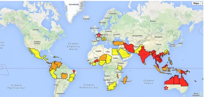
Melioidosis is endemic in several parts of Southeast Asia being northern Australia and Thailand the main endemic foci. Melioidosis is also being increasingly reported from many countries across south and east Asia as well as parts of South America, Papua New Guinea and the Caribbean (Figure 1.) 6,18. Sporadic cases were declared during and after World War II, in soldiers fighting in Vietnam, during the war of independence with France and the later conflict involving the USA. Incidence after post-natural disasters has been described 18,19.
Melioidosis is the third most frequent cause of death from infectious diseases in northeast Thailand and is the most common cause of community-acquired bacteraemic pneumonia in parts of northern Australia with mortality rates between 40% and 10% 4,18.
The commonest routes of infection are inoculation, inhalation and ingestion. There is no evidence to support direct human-to-human transmission via the respiratory route and its zoonotic power is still unclear 18. The clinical spectrum of disease ranges from localized cutaneous infection with no systemic manifestations to overwhelming sepsis and death. The incubation period ranges from 1 to 21 days for acute presentations.
Disease can be remitting and relapsing over months or years has been described and misdiagnosed as tuberculosis 4,6.
With rapid diagnosis, appropriate antibiotics, and state-of-the-art management of sepsis, death from melioidosis in those without identified risk factors such as diabetes, hazardous alcohol use, chronical lung and renal diseases and immunosuppressive therapy, is uncommon 4.
b) Diagnosis
Isolation of B. pseudomallei by culture methods is currently the “gold standard” diagnostic. For environmental sampling of B.pseudomallei is suggested a collection of 10 grams of soil with 30 cm in depth followed by the enrichment in 10 mL of Ashdown’s broth and incubation at 37 - 42 ºC for 48 hours. Isolated colonies are then obtained by plating the supernatant for another 48 hours 20. This process can take up to 7 days for culture, even for clinical samples. Serologic testing with indirect hemagglutination or various ELISA without culture confirmation is considered inadequate to confirm a diagnosis due to the background seropositivity rates in those living in endemic locations. Alternatively, flagged blood cultures or bacterial colonies on culture plates can now rapidly and accurately be identified using qPCR targeting the Bp (TTSS) gene cluster 4.
5 Figure 1. Global distribution of B. pseudomallei. Red background represents countries where B.
pseudomallei has been isolated from soil or water samples and melioidosis clinical reports are documented. Countries with orange background are those where only clinical melioidosis has been reported whilst yellow background countries can’t distinguish the isolated organism from other
Burkholderia spp. and no acquire melioidosis are reported (adapted from 20).
Table 1. Key phenotypical features of B. mallei and B. pseudomallei.
Feature B. mallei B. pseudomallei
Gram-stain morphology Gram-negative coccobacilli Bipolar Gram-negative bacilli Growth on medium Growth on blood agar within
24 to 48 hours
Growth on blood and Ashdown’s agar within 24 to 48 hours
Morphology of colonies Smooth texture with clear or yellow color
Blood Agar: White or yellow, smooth;
Ashdown’s Agar: Generally, rough and pink colonies
Motility Non-Motile Motile
Cytochrome oxidase activity Variable Positive
Catalase activity Positive Positive
Nitrate reduction to gas Negative Positive
Sugar utilization Non-fermenter Non-fermenter
Indole production Negative Negative
6 1.3. B. mallei and B. pseudomallei in biological warfare
Awareness of use of microorganisms for hostile proposes has been increasing since World War I catastrophes, by characterization of an infectious agent and preparation of national laboratories for rapid diagnosis tests and hospitals for potential therapeutics.
Three current organizations are updated with the bio warfare problematic worldwide: World Health Organization (WHO), an agency of the United Nations focused on international public health; World Organization for Animal Health (OIE), intergovernmental organisation responsible for improving animal health worldwide; Centre of Disease Control and Prevention (CDC), a United States of America agency to protect public health and safety through the control and prevention of disease, injury, and disability.
By July 2015, OIE together with the WHO hosted the first Global Conference on Biological Threat Reduction reuniting a variety of experts in the fields of public health, animal health, ecosystem health, and security sectors in order to highlight the framework for global preparedness against biological threats, its difficulties and possible solutions as well as sustainable investments in health system. Countries where certain diseases are eradicated or never been declared represent a special risk for the deliberate release of the agent once surveillance and control mechanisms are no longer, or have never been, active 21. Glanders, has been eradicated from Portugal since 1952 whilst melioidosis, has never been reported 22.
Due to their fast dissemination, ability to infect via inoculation, aerosols and ingestion, high contagiousness between humans, animals as well as the environment and resistance to a wide variety of antibiotics, B. mallei and B. pseudomallei are classified as Class B agents by the CDC and as level 3 risk agents by the European Parliament 11,23. The use of these microorganisms for terrorism attacks is not only acknowledgeable but recorded in past civil and world wars, as previously stated. The potential of these microorganisms for biological warfare leads to the need of strategic protocols to detect both agents in a rapid, effective and distinctive way.
1.4. Real time Polymerase Chain Reaction (qPCR)
Given the great impact of PCR as diagnostic technology, improvements have been made in the last few years by developing a new PCR platform called Real-Time PCR (qPCR), capable of monitoring by fluorescence detection, the accumulation of PCR products during cycling steps, eliminating the need of post-PCR procedures e.g. gel electrophoresis.
The detection of fluorescence in qPCR can be produced by two different chemistries: 1) DNA-binding dyes e.g. SYBR® Green, and 2) Fluorescence Resonance Energy Transfer (FRET) probes e.g. TaqMan®. By binding to non-specifically double-stranded DNA (dsDNA), SYBR®
7 Green allows the performance of a melting-curve at the end of the qPCR run to analyse the specificity of the reaction. SYBR® Green is incapable of performing multiplex reactions where qPCR is optimized to detect more than one target in one tube, whereas this is possible with hydrolysis probes, often referred to Taqman® probes. Hydrolysis probes consist in fluorescence labelled sequence-specific oligonucleotide with FRET chemistry. The probe contains a fluorescence reporter at the 5’ end and a quencher at the 3’ end of the oligonucleotide. During combined annealing/extension step of the amplification reaction the probe hybridizes the target the 5’→3’ exonuclease activity of the DNA polymerase, cleaves the reporter resulting in a fluorescence signal from the freed quencher. Hydrolysis probes assays include many advantages: 1) the use of another sequence-specific oligonucleotide offers a higher specificity; 2) the signal-to-noise ratio and 3) the ability to perform multiplex reactions 24,25.
The qPCR technology permits two forms of data analysis: qualitative and quantitative. Qualitative analysis indicates the presence or absence of the targeting genes, an approach widely use in pathogen diagnostics 26. The quantitative manner of qPCR allows the user to quantify the results and it can be done in two different ways: Relative quantification, most applied in gene expression case studies, measures the changes in the steady-state levels of a gene of interest relative to a housekeeping gene, using published mathematical equations like the ΔΔCq model and the Pfaffl model 27. Absolute quantification requires an independent
standard curve construction with the known diluted concentrations of the targeting genes in every analysis. Unknown samples are quantified by using the linear equation of the standard curve analysis 25.
1.5. Internal Amplification Control (IAC)
The Minimum Information for Publication of Quantitative Real-time PCR Experiments (MIQE) Guidelines suggests a Non-Template Control (NTC) in every qPCR assay. This NTC can be nuclease-free water and guarantees the reliability of the performance by detecting any source contamination in the sample run. However, it doesn’t grant if there is any inhibition factor as malfunction of thermal cycler, incorrect PCR mixture, poor DNA polymerase activity, or presence of inhibitory substances in the sample matrix 26.
A strategy commonly used to certify if the reaction was successful is the implementation of an IAC. An IAC is a non-target DNA sequence co-amplified simultaneously with the target sequence. This strategy can be approached in two ways: 1) In a competitive manner, where the set of primers hybridize with both target sequence and IAC, compromising the efficiency and detection limit of the performance, or 2) Non-competitive manner, in which the target and IAC
8 are amplified using a different primer set for each. Here, the IAC pair of primers targets a synthetic DNA (e.g., plasmid DNA) or a gene present in any microorganism and in higher copy number than the principal target gene (e.g., encoding rRNA) compromising the amplification of this target if the organism isn’t present. By using a non-competitive method, concentrations of primers and probe (if applied) of the IAC must be limited in order to limit the competition between the target for nucleotides and DNA polymerase. The practical advantage of this method is its extensive use of the conceived IAC in different qPCR assays 28.
1.6. qPCR as a diagnostic tool for glanders and melioidosis
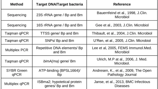
Real-time PCR tests have been extensively developed in clinical microbiology laboratories for routine diagnosis of infectious diseases, particularly bacterial diseases 29. For the identification of glanders and melioidosis etiologic agents, a numerous approaches have been developed through years (Table 2.).
Table 2. Resume table of PCR developments towards the identification of B. mallei and B.
pseudomallei
Method Target DNA/Target bacteria Reference
Sequencing 23S rRNA gene / Bp and Bm Bauernfeind et al., 1998, J.Clin. Microbiol
Sequencing 16S rRNA gene / Bp and Bm Gee et al., 2003, J.Clin. Microbiol Taqman qPCR TTSS gene/ Bp and Bm Thibault, et al., 2004, J.Clin. Microbiol Taqman qPCR SNPs/ Bp and Bm U’Ren, et al., 2005, J.Clin. Microbiol
Multiplex PCR Repetitive DNA elements/ Bp and Bm
Lee et al, 2005, FEMS Immunol.Med. Microbiol
Taqman qPCR bimA(ma) gene/ Bm Ulrich, M.P at al., 2006, J. Med. Microbiol.
SYBR Green qPCR
ATP-binding (BPSL1664)/ Bp
Andresen, K., et al., 2009, The Open Pathology Journal
Multiplex qPCR ISBma2; hypotetical protein genes/ Bp and Bm
Janse, et al., 2013, BMC Infectious Diseases
Bm – B. mallei; Bp – Burkholderia pseudomallei; Adapted from Botelho, A. in Accreditation of a PCR system for detection and differentiation of Burkholderia mallei and Burkholderia pseudomallei, IB-BIOALERTNET Conference, July, 2015, Madrid
9 1.7. Aims
In the frame of the project IB-BIOALERTNET (2013-2015) it was proposed to develop a qPCR that could detect and distinguish B. mallei and B. pseudomallei in different possible infected matrices, and to standardize and accredited the procedures in order to establish a net of prepared laboratories of bioterrorism alert in case of an emergency and deliberated realise of these agents.
Therefore, to attain these aims the following experimental strategy was implemented:
Preparation of spiked samples of pulmonary macerates, sterile swabs with B. mallei and the above mentioned and soils with B. pseudomallei reference strains;
Development of a qPCR system for the detection and differentiation of B. mallei and B.
pseudomallei, following an adaptation of Janse et al. 2013 work;
qPCR analysis with the purified DNA extracted from the spiked samples;
Development of a non-competitive Internal Amplification Control for the qPCR assay;
Establishment of standard operational protocols of biological alert, to be applied in case of suspicion of B. mallei and B. pseudomallei release.
10 CHAPTER 2 – MATERIAL AND METHODS
2.0. Bacterial strains
All procedures regarding the handling of B. mallei and B. pseudomallei strains were performed in a Biosafety Laboratory Level – 3 (BSL-3) facilities in a Class II biosafety cabinet (BSC II), using the required personal protective equipment (PPE) and following the protocols and Biosafety Manuals accessible on the Rede Laboratorial Portuguesa de Biossegurança- LABPTBIONET (http://www.labptbionet.ibmc.up.pt/) such as the Laboratory Biosafety Manual published by WHO (2013) and CDC Biosafety in Microbiological and Biomedical Laboratories (2009).
Two strains of B. mallei and two strains of B. pseudomallei, acquired to NCTC, United Kingdom (Table 3.) and received lyophilized, were reconstituted in Brain Heart Infusion Broth (BHIB) media and inoculated in Tryptone Soy Agar (TSA) plates. After incubation at 37ºC for 48 hours to 5 days, single colonies were inoculated in TSB (Tryptone Soy Broth) aliquots with 30% glycerol, stored at -20 ºC and defrosted when needed. Type strains NCTC 12939T and NCTC 12938T were selected for the optimization of the qPCR (See 2.1.), while the remaining two strains were used for the spiked sample process (See 2.0.2.).
2.0.1. Bacterial growth
For B. pseudomallei culture, Ashdown’s Agar (AA) and Broth (AB) (Appendix I – Ashdown’s Agar composition) were used. Brain Heart Infusion Agar (BHIA) supplement with 100 U/mL of Penicillin and 1:1000000 Crystal Violet (BHIA+Pen+CV)30 and Columbia Agar with 5% Sheep Blood (CA/S) were used for B. mallei culture.
Table 3. B. mallei and B. pseudomallei strains used. Strains (NCTC
reference)
Other References Characteristics
B. mallei
NCTC 12938T ATCC 23344 Clinical Isolate, Human, China 1944 31 NCTC 10245 ATCC 10399;
CHINA 5
Clinical Isolate, Horse, 1972 32
B. pseudomallei
NCTC 12939T ATCC 23343; WRAIR 286
Clinical Isolate, Human, USA
NCTC 10276 PRINCE Clinical Isolate, Human, UK, 1962 33
Standard Operation Procedures (SOPs) were elaborated according to ISO17025 legislation and audits have been squealed for accreditation of methods described in this work.
11 2.0.2. Spiked Samples
B. pseudomallei NCTC 10276 and B. mallei NCTC 10245 strains were chosen to spike three
different matrices: pulmonary macerates, sterile swabs and soils. Soils were only tested with B.
pseudomallei NCTC 10276 as B. mallei doesn’t persist in the environment.
For this procedure, a glycerol stock of each bacterial strain was defrosted and cultured on agar plates followed by incubation at 37ºC for 48h or until visualization of colonies. Single colonies were suspended in BHIB and incubated at 37ºC until the absorbance at 600 nm reached 0.5 (GeneQuant Pro Spectrophotometer, Pharmacia), the equivalent to approximately 1x10⁹ colony forming unites (CFU) per mL 34. These bacterial suspensions were serial tenfold diluted up to six orders of magnitude to spike the chosen matrices. Optical densities at 600 nm were measured and 10 µL of each dilution were plated in duplicate in the selective medium (See 2.0.1.).
To evaluate of the performance of the “gold standard” culture method and the molecular qPCR technique and enable comparison between them, the same spiked sample was tested in parallel by each method: plating in specific culture media and inactivation at 99ºC for 60 minutes, for subsequent DNA purification and qPCR analysis, out of the BSL3 facilities(Resume in Figure A 1. - Appendixes). For swabs and soils, a previous incubation procedure of the sample at 37ºC for 48 hours in culture media, was evaluated in comparison with no incubation.
a) Swabs
A single sterile swab was immersed in each dilution of each strain for 120 seconds and transferred into a 15 mL disposable tube (Sarstedt™, Germany) supplemented with 2 mL of BHIB. After a brief vortex, 10 µL were spread on the surface of selective medium and one millilitre was immediately inactivated for DNA extraction and purification, naming it Swab Time 0 (S T=0 H). The remaining inoculated broth was subjected to the incubation period (S T=48 H) prior to its plating and inactivation for DNA extraction and purification (See 2.1.).
b) Pulmonary macerates
Approximately 10 grams of a swine pulmonary tissue (Internal Code: 11251 8-5) were placed into a flask tube and mixed in a Stomacher (Stomacher 400, Colworth) with 8.5 % of sodium chloride saline solution, resulting in the pulmonary macerate.
In a BSC II in a Biosafety Laboratory Level – 2 (BSL-2), 25 mg of macerate were weight in a 2 mL screw cap micro tubes (Sarstedt™, Germany) for the spiked process.
In the BSL3 facilities, 800 µL/g of either B.mallei or B.pseudomallei culture dilution were inoculated in the respective micro tube. All micro tubes were briefly vortexed for homogenization and 10 µL were spread in the respective selective culture medium (See 2.0.1.). One hundred
Standard Operation Procedures (SOPs) were elaborated according to ISO17025 legislation and audits have been squealed for accreditation of methods described in this work.
12 and eighty µL of Digestion Buffer from PureLink® Genomic DNA Mini Kit (Invitrogen™) were added to 1 mL of the spiked pulmonary macerates before inactivation (See 2.1.).
c) Soils
Soil samples were collected in INIAV, Pólo Benfica (38º.44’55.84N, 9º.11’59.88’’O) territory near an orange tree irrigated hours before. To homogenise the collected sample, soil was spread on a bench in a cone shape starting from the centre to the periphery. Five grams of soil were collected from the top and distributed into 50 mL disposable tubes.
For the spiked process, 800 µL/g of each dilution of B. pseudomallei were added to the soil matrix. All tubes were vigorously shaken and 5 mL of AB were added. Ten µL were spread on AA and 1 mL of the supernatant was inactivated, defining these samples Soil culture at Time 0 (Sc T=0). The remaining spiked soils followed an incubation period of 48 hours at 37 ºC. One millilitre was withdraw from the incubated sample (Sc T=48) and 10 µL were plated in AA and incubated at 37ºC for 48 h. Typical B. pseudomallei colonies from Sc T=48, were selected and transferred to 1 mL of BHIB and inactivated for DNA extraction and purification naming these Soil culture Isolates (Sc I) (See 2.1.).
2.1. Inactivation of the bacteria, DNA purification and quantification
The samples collected from the spiked process were inactivated in a Labnet AccuBlock™ Digital Dry Bath (Citomed, Portugal) at 99º C for 60 minutes. Efficiency of inactivation was performed by plating 10 µL of the inactivated product into the selected bacteria medium (See 2.0.1.). Incubation was set at 37 ºC and plates were observed 6 days after, confirming the absence of any CFU, allowing the samples to be transferred to BSL-2 facilities for DNA purification.
DNA purification was performed using PureLink® Genomic DNA mini Kit (Invitrogen™) following the manufacturer’s instructions, according to the type of matrix, and Nanodrop 2000 UV-Vis Spectrophotometer (Thermo Fisher Scientific™) was used for DNA quantification.
2.2. qPCR system for the detection of B. mallei and B. pseudomallei
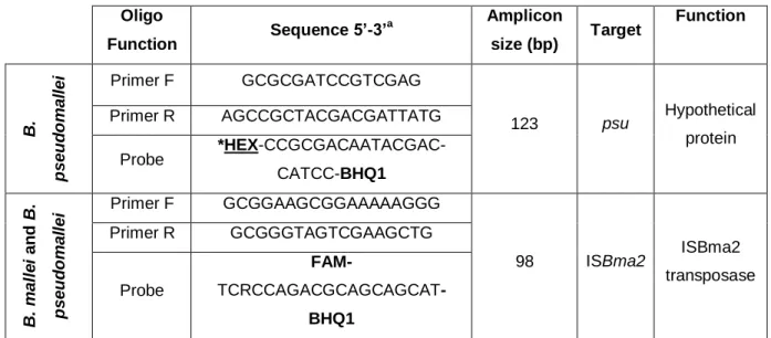
Development of the qPCR system was based on Janse et al. (2013) novel approach using the primers and probes depicted in Table 4. and synthetized by NZYTech (Lumiar, Portugal). Probe targeting psu gene with fluorophore CF590 was changed to HEX, a fluorophore calibrated for used thermocycler.
Standard Operation Procedures (SOPs) were elaborated according to ISO17025 legislation and audits have been squealed for accreditation of methods described in this work.
13 2.2.1. In silico analysis
The specificity of primers, probes and target sequences depicted in Table 4. and Table 5. was tested and confirmed using BLASTn software (http://blast.ncbi.nlm.nih.gov/Blast.cgi). All oligonucleotides were analysed in Thermo Scientific webtool, Multiple Primer Analyzer, (http://www.thermoscientificbio.com/webtools/multipleprimer/) to check for self-dimer and cross-dimer reactions.
Table 4. Oligonucleotides for the qPCR duplex Burkholderia system.
a - Based on Janse et al, 2013 ; *-Modifications made from Janse et al work; Primer F – Primer Forward; Primer R –
Primer Reverse
2.2.2. qPCR optimization
B.mallei NCTC 12938T and B. pseudomallei NCTC 12939T purified DNA was used as DNA templates for the qPCR optimization. qPCR reactions were carried out in a Bio-Rad CFX96™ Thermal Cycler (Bio-Rad Laboratories Srl, Redmond, USA) using Bio-Rad CFX Manager, version 3.0 software for data analysis. Cycling conditions were adapted from Janse et al. (2013): Enzyme activation at 95 ºC for 5 minutes and 44 cycles of 5 seconds at 95 ºC and 35 seconds at 60ºC. In a reaction volume of 20 µL, 3 µL of DNA template were added in a concentration of 10 ng/µL. For Non-Template Controls (NTC), ultra-pure water was as template in every experimental set, to rule out any source of contamination.
The qPCR system was optimized by testing different final concentrations of primers and probes in singleplex and duplex reactions. Annealing temperature was optimized for the duplex reaction. Oligo Function Sequence 5’-3’ a Amplicon size (bp) Target Function B. p s e u d o m a ll e i Primer F GCGCGATCCGTCGAG 123 psu Hypothetical protein Primer R AGCCGCTACGACGATTATG Probe *HEX-CCGCGACAATACGAC-CATCC-BHQ1 B. m a ll e i a n d B . p s e u d o m a ll e i Primer F GCGGAAGCGGAAAAAGGG 98 ISBma2 ISBma2 transposase Primer R GCGGGTAGTCGAAGCTG Probe FAM- TCRCCAGACGCAGCAGCAT-BHQ1
Standard Operation Procedures (SOPs) were elaborated according to ISO17025 legislation and audits have been squealed for accreditation of methods described in this work.
14
a) Primers and probe concentration optimization
SsoFast™ EvaGreen® Supermix (Bio-Rad Laboratories Srl, Redmond, USA) was used to test final primer concentrations from 100 nM to 400 nM with 100 nM iterations of each set of primers, separately. Cycling conditions were as described above, adding a final step of 65ºC to 95ºC with increments of 0.5ºC in 5 seconds each for the melting curve analysis in order to exclude primer combinations that produce any inefficient or primer-dimer products.
Final primer concentrations ranging 100 nM to 400 nM and final probe concentrations ranging 100 nM to 300 nM with 100 nM iterations were tested in a duplex reaction using polymerase NZY qPCR Master Mix 2x, NZYTech (Lumiar, Portugal) and the cycling conditions above mentioned.
The combination that exhibits the earliest quantitative cycle (Cq) and the highest end-point fluorescence (EPF) values while minimizing non-specific amplification was chosen as the optimal primer concentrations.
b) Annealing Optimization
Given the optimized concentrations of primers and probes for the duplex reaction, annealing temperature was tested by subjecting qPCR reactions to a gradient of annealing temperatures ranging from 62.9, 62.5, 61.6, 60.0 to 58.1 ºC. The temperature at which the Cq value and EPF gave the highest values, was chosen as the optimal temperature.
c) Evaluation of Cq variance between singleplex and duplex assays
DNA templates in a concentration, respectively, of 2.9 ng/µL and 4.5 ng/µL of per reaction, were used in duplicates to evaluate the optimized singleplex and duplex assays in the same run to determine significate differences between the Cq values of each platform.
2.2.3. Estimation of the limit of detection (LOD), specificity, repeatability and reproducibility
DNA extracted from B. mallei NCTC 12938T and B. pseudomallei NCTC 12939T cultures were used to determine the linearity and sensibility of singleplex and duplex reactions. Ten-fold dilutions ranging 10-0 to 10-10 from 10 ng/µL of DNA template were prepared and standard curves were constructed, with the qPCR results from two replicates per dilution, by plotting the Cq values to the logarithm of the DNA concentration per reaction (fg/reaction). Slopes were used to obtain the qPCR efficiency percentage by the following equation: 101/slope1x 100. Estimation of the limit of detection (LOD), sensitivity, was determined by the lowest concentration of template per reaction that produced positive results in both replicates.
Standard Operation Procedures (SOPs) were elaborated according to ISO17025 legislation and audits have been squealed for accreditation of methods described in this work.
15 For specificity, purified DNA from B. mallei, B. pseudomallei and other bacteria were used as DNA templates in the optimized qPCR duplex system.
For the intra-assay (repeatability) and inter-assay (reproducibility) variability of singleplex and duplex assay, 3 dilutions of each B. mallei NCTC 12938T and B. pseudomallei NCTC 12939T were tested. Each dilution was tested in duplicate and in two independent runs performed in different days by different operators. The mean Cqs values, standard deviation and percent coefficient of variation were calculated independently for each DNA dilution.
2.3. Construction of an Internal Amplification Control (IAC) system for PCR reactions DNA of myxoma virus Lausanne strain, kindly provided by Doctor Margarida Duarte, (INIAV, IP), was used to construct an IAC by PCR amplification of a 125 bp fragment of the m000.5L/R gene and cloning into pNZY28 vector.
2.3.1. Conventional PCR amplification
Conventional PCR was performed using primers described in Duarte et al (2014)35 (Table 5.). For the master mix reaction, High Fidelity PCR Master Mix (Roche Diagnostics, Indianapolis, IN, USA) was used in the total reaction of 25 µL using 2 µL of DNA template at a concentration of 10ng/µL and a final concentration of 1 µM for each primer. Amplifications were performed with forty cycles of denaturation at 95ºC for 30 seconds, annealing at 50ºC for 45 seconds and extension at 72ºC for 30 seconds, followed by a final step of extension at 72ºC for 5 minutes. Reactions were processed in a MJ Mini™ Personal Thermocycler (Bio-Rad Laboratories Srl, Redmond, USA) and PCR products were subjected to agarose gel electrophoresis on 2% low melting SeaPlaque® GTG® Agarose (FMC® Bioproducts, USA) stained with 2 mg/mL ethidium bromide (UltraPure™ Ethidium Bromide, Invitrogen™) in parallel with 100 bp DNA ladder (Promega).
Table 5. Primers and probes for pNZYmyx.
Primer F – Primer Forward; Primer R – Primer Reverse
Microorganism Oligo Function Sequence 5’-3’ Amplicon size (bp) Myxoma Virus Primer F CGACGTAGATTTATCGTATACC 125 Primer R GTCTGTCTATGTATTCTATCTCC Probe [FAM]TCGGTCTATCCTCGGGCAGAC ATAGA[TAMRA]
Standard Operation Procedures (SOPs) were elaborated according to ISO17025 legislation and audits have been squealed for accreditation of methods described in this work.
16 2.3.2. Cloning of the 125 bp fragment of m000.5L/R gene in pNZY28 vector – plasmid pNZYmyx construction
The 125 bp amplicon was excised from the gel with the help of a scalpel under a UV transilluminator (White/UV Transilluminator, UVP). Purification of the amplicon was done following the instructions of the NZY Gelpure kit (NZYTech, Lisbon, Portugal) and DNA quantification was measure in a Nanodrop 2000 UV-Vis Spectrophotometer (Thermo Scientific). Cloning procedure was done using the pNYZ28 vector (Figure A 4. - Appendixes) of the NZY-A PCR Cloning Kit, NZYTech (Lumiar, Portugal), using 1:3 ratio of vector:insert for the ligation reaction and transformation into E.Coli NZYStar Competent Cells (Genotype: endA1 hsdR17(rk-,
mk+) supE44 thi -1 recA1 gyrA96 relA1 lac[F´ proA+B+ lacIqZΔM15 :Tn10(TcR)].
Transformed cells were plated in LB agar plates supplemented with antibiotics (100 µg/mL ampicillin and 15 µg/mL tetracycline) and lactose analogues (100 µg/mL X-Gal and 0.5 mM of IPTG) and incubated overnight at 37 ºC. White colonies with recombinant plasmid (pNZYmyx) were selected, plasmid DNA extracted and, to confirm the effectiveness of the cloning pro-cedure, a conventional PCR targeting the inserted fragment of the plasmid pNZYmyx was performed, using the above primers. The cells confirmed to have the recombinant plasmid (recombinants) were transferred to 10 mL cryotubes with 2 mL LB broth with the antibiotics above mentioned. Ten percent of glycerol (v/v) was added and the cryotubes were stored at -80 ºC and defrosted whenever needed.
2.2.3. qPCR using pNZYmyx as IAC
The qPCR for the pNZYmyx recombinant plasmid was performed following the NZY qPCR Master Mix 2x (Lumiar, Portugal) recommended conditions: enzyme activation step at 95 ºC for 2 minutes, 95 ºC for 10 seconds and annealing with fluorescence measurement at 60 ºC for 20 seconds. Primers and probe concentrations were 0.4 µM and 0.1 µM, respectively, in a final reaction volume of 20 µL with 2 µL of DNA template at a concentration of 10ng/µL.
2.3. Data analysis
Statistical values and graphics were done in GraphPad Prism 5.03 and Microsoft Excel 2007 Office Tool.
Sensitivity and McNemar tests were computed using the clinical research calculators of the online VassarStats software (http://vassarstats.net) and GraphPad online software, QuickCalcs (http://graphpad.com/quickcalcs/).
Standard Operation Procedures (SOPs) were elaborated according to ISO17025 legislation and audits have been squealed for accreditation of methods described in this work.
17 CHAPTER 3 – RESULTS AND DISCUSSION
3.0. Growth of B. mallei and B. pseudomallei strains in culture media
Colonies of B. pseudomallei NCTC 10276 showed different morphologic aspects inter and intra-species. The most common colony features was its pink colour, irregular form and mucoid texture (Figure 2. - C).
Brain Heart Infusion Agar (BHIA) supplement with 100 U/mL of Penicillin and 1:1000000 Crystal Violet (BHIA+Pen+CV) displayed difficulties for the growth of B. mallei NCTC 10245. When colonies were present, they assumed a darkish grey colour with smooth texture, forming agglomerates in the periphery of the Petri dish. This fact created an obstacle for the determination of colony forming units (CFU). Therefore, Columbia Agar with 5% Sheep Blood (CA/S) was selected for the isolation of B. mallei, where colonies presented two distinctive forms: pin-point clear colonies (Figure 2. - A) or opaque yellow with variable dimension (Figure 2. - B).
A
B
C
Figure 2. Colonies of B. pseudomallei and B. mallei in selective media. Characteristic
colonies of B. mallei 10245 are shown in (A) and (B), while B. pseudomallei 10276 most common colony morphology is shown in (C). Translucid pin-point colonies of B. mallei are indicated by arrows in plate (A) and irregular shape with yellow color in CA/S plate (B), inoculated with 10 µL of B. mallei NCTC 10245. Plate (C) represents colonies of B. pseudomallei in Ashdown’s media, from a 10-2 dilution spiked soil sample.
18 To evaluate the number of CFU/mL to be inoculated in each spiked matrices and further evaluate the sensitivity of both culture and PCR methods, the absorbance at 600 nm (OD600)
was measured for each ten-fold serial dilution of B. mallei and B. pseudomallei cultures that were, in parallel, plated in agar plates with the respective selective bacterial media and incubated at 37ºC for 48 hours. The number of CFU/mL was calculated by counting the number of CFU in each dilution plate and multiplying by the correspondent inoculum factor.
B. mallei platted dilutions presented growth until the third ten-fold dilution, with an OD600 of
0.006 corresponding to 250 CFU/mL (Table A 1. - Appendixes). For B. pseudomallei fourth ten-fold dilution was established as the limit for CFU visualization (650 CFU/mL), corresponding to an OD600 of 0, a value associated with absence of any bacteria in culture. Human manipulation
errors or spectrophotometer inaccuracy might explain these incongruences.
3.1. Spiked samples bacteriological culture
Spiked sample process was performed by inoculating each 10-1 to 10-6 dilution from bacterial cultures corresponding, respectively, to 2.5x10⁴, 4.4x10³, 2.5x10² and 0 CFU/mL for B.mallei 10245, and 5.9x10⁴, 3.7x10⁴, 2.7x10⁴, 6.5x10² and 0 CFU/mL for B.pseudomallei (Table A 1. - Appendixes). Ten µL of each swab and soil spiked sample, was plated into selective culture media (See 2.0.1.) without previous enrichment incubation period and after 48 hours incubation at 37ºC in selective media. No previous enrichment was performed for pulmonary macerates (See 2.0.2. and Figure A 1. - Appendixes) due to the natural presence, in this type of clinical sample, of other bacteria that could easily over grow, competing with Burkholderia and unable its detection.
a) Swabs
For B. pseudomallei it was possible to detect 4.0x10² CFU/mL for S T=0 and 1.35x10⁵ CFU/mL for S T=48 (Figure 3. - A and B). Swabs spiked with B. mallei showed no growth in any dilution either with (S T=48) or without incubation period (S T=0) (results not shown). The absence of specific and sensitive culture media for isolation of B.mallei might have had influence in these results. The plating of 10 µL of suspension instead of the direct smear of the swab in the culture media could also have had influence since the inoculum might have been too less.
b) Pulmonary macerates
For B. pseudomallei, the forth ten-fold dilution represents the limit of CFU visualization corresponding to 2.0x10² CFU/mL (Figure 3. – A - Lungs). For B.mallei spiked pulmonary macerates lowest dilution bacterial growth detection was the second ten-fold dilution corresponding to 3.3x10³ CFU/mL (Figure 4.). These results show that the isolation of B.