2019
Genetic study of Toxoplasma gondii strains isolated from humans and animals
“Documento Definitivo”
Doutoramento em Biologia
Especialidade de Microbiologia
Anabela Barreiro Gomes Vilares Bendada Esteves
Tese orientada por:
Doutor João Paulo dos Santos Gomes
Professor Doutor Rogério Paulo de Andrade Tenreiro
2019
UNIVERSIDADE DE LISBOA FACULDADE DE CIÊNCIAS
Genetic study of Toxoplasma gondii strains isolated from humans and animals Doutoramento em Biologia
Especialidade de Microbiologia Anabela Barreiro Gomes Vilares Bendada Esteves
Tese orientada por:
Doutor João Paulo dos Santos Gomes
Professor Doutor Rogério Paulo de Andrade Tenreiro
Júri: Presidente:
Professor Doutor Rui Manuel dos Santos Malhó, Professor Catedrático, Faculdade de Ciências da Universidade de Lisboa.
Vogais:
Doutor Agostinho Luís da Silva Cruz, Professor Coordenador com Agregação, Escola Superior de Saúde do Instituto Politécnico do Porto;
Doutor João Paulo dos Santos Gomes, Investigador Auxiliar com Habilitação, Instituto Nacional de Saúde Doutor Ricardo Jorge (orientador);
Doutora Mónica Alexandra de Sousa Oleastro, Investigadora Auxiliar, Instituto Nacional de Saúde Doutor Ricardo Jorge;
Doutor Luís Manuel Madeira de Carvalho, Professor Associado com Agregação, Faculdade de Medicina Veterinária da Universidade de Lisboa;
Doutor Ricardo Pedro Moreira Dias, Investigador Auxiliar Convidado, Faculdade de Ciências da Universidade de Lisboa.
Fundação da Ciência e Tecnologia com a bolsa de doutoramento (SFRH/BD/75154/2010)
E aprendes que realmente podes suportar...
que realmente és forte, e que podes ir muito
mais longe depois de pensar que não se pode mais.
VII
AGRADECIMENTOS
À Maria João Gargaté (INSA), obrigada por seres como és, que sem ti este trabalho não teria sido possível, de todo, obrigada por teres sempre acreditado que eu era capaz, pelo apoio incondicional, pela amizade e por todo o teu esforço e empenho no meu futuro, nunca terei palavras ou ações que te possam compensar.
Às minhas companheiras de laboratório (INSA) (Assunção António, Cláudia Júlio, Idalina Ferreira, Susana Martins e Tânia Reis) que fizeram parte de todo este processo, que me ajudaram, trabalharam, encorajaram, me ouviram nos dias mais terríveis, foram indiscutivelmente umas queridas, um muito obrigado a todas para sempre. No entanto, tenho que agradecer em especial à Idalina Ferreira por tudo o que me ensinaste, à Cláudia Júlio por teres sido uma das mentoras deste processo e me encorajares, à Susana Martins por seres a boa disposição deste laboratório e por fim, pelo companheirismo, não apenas neste processo, mas também nos longos 18 anos de convivência.
Ao meu orientador Doutor João Paulo Gomes (INSA), pela oportunidade que me deu, pelo conhecimento, experiência, paciência e disponibilidade com que sempre me presenteaste, foi sem dúvida uma agradável surpresa.
Doutor Vítor Borges (INSA) por seres uma pessoa tão especial, sempre com um sorriso, amabilidade e saber único. Obrigado por todo o conhecimento que me transmitiste neste último trabalho (capítulo VI), sem ti teria sido tudo muito, mas mesmo muito mais difícil.
Ao meu orientador Professor Doutor Rogério Paulo de Andrade Tenreiro (BioISI) que permitiu que este trabalho fosse possível, que sempre se disponibilizou para me ouvir e ajudar e por todo o seu conhecimento e simpatia.
VIII
nomeadamente aos que fizeram de alguma forma parte deste trabalho, e em especial aos do sector da UTI pela paciência e debate na melhoria dos resultados deste trabalho, do capítulo IV ao VI.
Ao Conselho Diretivo do Instituto Nacional de Saúde Doutor Ricardo Jorge (INSA), que me permitiu a possibilidade de realizar este trabalho.
À Fundação de Ciência e Tecnologia (FCT), pela concessão de diferentes financiamentos.
À vous Marie-Laure Dardé (CHU Limoges) pour être une inspiration, pour toute la votre connaissance, amitié et encouragement et à vous Daniel Ajzenberg (CHU Limoges) pour la votre capacité d’innovation et connaissance. Je ne vous oublierai jamais!
Doutora Helga Waap (Laboratório de Parasitologia - INIAV) pelo teu empenho, pelas noites e dias que trabalhámos em conjunto no projeto que originou o Capitulo IV desta tese. Doutor Jacinto Gomes e equipa (Laboratório de Parasitologia - INIAV) por terem possibilitado a nossa parceria.
Doutora Ana Patrícia Lopes (UTAD) pela amizade, simpatia e parceria neste trabalho que resultou tão bem (Capítulo III).
Dra. Luísa Costa Gomes (CML), responsável pelo departamento de controlo sanitário da Câmara Municipal de Lisboa, e sua equipa, Dra. Filomena Oliveira, Dra. Ana Machado, Dr. Leonel Fernandes, Dr. Augusto Batista, Dr. Vasco Ribeiro (CML) pela valiosa colaboração neste estudo (Capítulo IV).
A todos os veterinários que tornaram possível este estudo. São tantos que não posso vos nomear com receio de me esquecer de algum (Capítulo III e IV).
IX
e pela amizade com que sempre me presenteou. Lembrei-me várias vezes das suas sábias palavras.
Ao Dr Correia Neves, Dr. Jorge Canena, Dra Margarida Canas, Dr Manuel Liberato e Dr António Marques um obrigada muito especial nestes tempos difíceis, que sem o vosso saber e disponibilidade eu não me encontraria neste mundo, quanto mais a defender este trabalho.
À minha família, a ti mãe, por seres a minha referência, o meu ídolo, o meu herói, por acreditares que eu venceria desde os tempos de gestação, LUTADORAS!!!! A ti pai pelo teu humor, por me passares a tua perseverança e princípios únicos de vida que fazem de mim o que sou hoje. A vocês, mana e manos por me ajudarem a crescer desta forma tão maravilhosa. The last but not the least, a ti Tiago, Dinis e Duarte por me fazerem rir, chorar, encorajar e amar incondicionalmente em todo este processo, que se revelou um caminho árduo, com muitos obstáculos para TODOS.
XI
ABSTRACT:
Toxoplasma gondii is responsible for toxoplasmosis, a zoonosis spread worldwide. This PhD thesis aimed to provide a more comprehensive overview of the genetic characteristics and virulence of T. gondii strains isolated from humans and animals in Portugal. We enrolled T. gondii strains collected from intermediary (farm, urban and sylvatic animals) and the definitive (cat) hosts, as well as from human toxoplasmosis. In general, we applied Sag2 and microsatellites classical genotyping, next-generation sequencing and inoculation in mice.
As major results, we highlight: i) the majority of isolated strains were avirulent in mice bioassay, where virulent strains belonged almost exclusively to Sag2 type I; ii) type II strains were the most identified type in strains from human toxoplasmosis as well as from all animal species, followed by type I strains; iii) this is the first report that shows type II T. gondii strains isolated from cattle, boars, foxes and hares in north of Portugal and recombinant strains isolated both from infected cats and pigeons; iv) the development of a rapid multi-loci-based NGS scheme, which allowed evaluating multi loci polymorphism and genomic mosaicism simultaneously. This schema is now implemented as the surveillance tool in the National Reference Laboratory of Parasitic and Fungal Infections (URSZ-INSA) to perform molecular surveillance of T. gondii; v) with this extended typing scheme, we identified a high rate (63 %) of recombinant strains isolated from humans, which had been previously identified as type I and II, as well as new putative genetic markers of virulence.
Overall, this PhD shed some light on the genetic and virulence diversity of T. gondii strains circulating in Portugal and revealed a surprising scenario of rampant genomic exchange in this parasite. These data highlight the need for further extended genomic studies to better understand the genotype/phenotype associations in this important human pathogen and thus contribute to better infection control measures.
XIII
RESUMO:
Toxoplasma gondii é o parasita responsável pela toxoplasmose e pode infetar a grande maioria dos animais vertebrados, incluindo o Homem. Este parasita tem uma distribuição mundial e apresenta um elevado interesse médico e veterinário. A infeção no Homem está maioritariamente associada ao consumo de carnes pouco cozinhadas ou cruas e à contaminação ambiental, sendo esta infeção na sua maioria assintomática. No entanto, pode ser grave ou mesmo levar à morte quando associada ao hospedeiro imunodeprimido ou à infeção primária materna por transmissão fetal, podendo ainda causar aborto ou doenças congénitas graves (ex. corioretinite, cegueira, linfoadenopatia, etc). A progressão e a gravidade da infeção têm sido associadas a diferentes fatores, incluindo características genéticas do parasita. Diferentes estudos têm sido desenvolvidos na genotipagem de T. gondii, os quais têm revelado que a maioria das estirpes isoladas na Europa e nos Estados Unidos da América pertencem ao tipo II e raramente se verificam isolados do tipo I, recombinantes ou atípicos. As estirpes recombinantes e atípicas apresentam normalmente uma maior gravidade na infeção, podendo levar à morte do hospedeiro imucomprometido ou mesmo do imunocompetente. No entanto, em Portugal, o conhecimento da distribuição, do tipo e da virulência dos genótipos circulantes é extremamente diminuto quer em humanos quer em animais. Para colmatar esta falta de conhecimento no nosso país, esta tese de doutoramento teve como objetivo geral a caracterização genética de estirpes de T. gondii de origem humana e animal, isoladas em Portugal. Neste sentido, delinearam-se diferentes estudos com objetivos específicos, correspondendo a diferentes capítulos desta tese e estando alguns deles publicados em jornais científicos.
No primeiro estudo (capítulo III) pretendeu-se caracterizar geneticamente estirpes de T. gondii isoladas de 209 produtos animais provenientes de dois grupos diferentes (silvestres e domésticos) da região Norte de Portugal (Grupo I – 12 bovinos, 34 ovinos, 5 caprinos e 16 porcos, Grupo II – 6 cães errantes, 59 raposas, 49 javalis e 28 lebres). Destes animais, serologicamente positivos para IgG anti T. gondii, foram colhidas diferentes amostras, nomeadamente coração, cérebro, diafragma e língua. Em todas as amostras foi realizada a técnica de PCR clássica do gene B1 onde 89 (43 %) animais revelaram resultados positivos. No que se refere à genotipagem de T. gondii realizaram-se PCRs das extremidades 5’ e 3’ do gene Sag2 onde 29 % (60/209) dos animais revelaram resultados positivos. Destas 60 estirpes, 77 % (46/60) pertenciam ao tipo II, 10 % (6/60) ao tipo I e em 13 % (8/60) das amostras não foi possível diferenciar o tipo I do tipo II. Neste estudo foi demonstrado pela primeira vez no
XIV
norte de Portugal a existência de estirpes do tipo II em vacas, javalis, raposas e lebres e foi também confirmada a predominância de estirpes do tipo II em porcos nesta mesma região. Foi também a primeira vez em que se procedeu à caracterização genética de T. gondii em cabras, javalis, raposas e lebres em Portugal. Demonstrou-se assim um maior leque de possíveis fontes de infeção para o Homem e para outros animais, bem como a importância da contaminação ambiental do ciclo silvático e sinantrópico de T. gondii.
Estes resultados vieram demonstrar a necessidade de extender os estudos de caracterização genética a outros hospedeiros, nomeadamente ao hospedeiro definitivo (gato), e a outras regiões do país com maior densidade populacional (capítulo IV). No segundo estudo (capítulo IV) pretendeu-se proceder à caracterização genética e fenotípica de estirpes de T. gondii isoladas de 41 cérebros de pombos e de 164 cérebros de gatos com anticorpos IgG anti-T. gondii provenientes da região de Lisboa. Os gatos e os pombos representam excelentes modelos de contaminação ambiental, e o gato é o hospedeiro definitivo de T. gondii sendo o único animal com capacidade de excretar oocistos (forma de resistência) nas zonas urbanas, logo mais populosas. Os cérebros de gatos e pombos foram colhidos e inoculados em ratinhos de laboratório de forma a estudar a virulência das estirpes, onde três isolados apresentaram fenótipo de virulência, sendo um proveniente de gato e dois de pombo. De forma a determinar o genótipo das estirpes de T. gondii realizaram-se duas técnicas distintas, nomeadamente a PCR clássica das extremidades 5’ e 3’ do gene Sag2 e a análise por electroforese capilar dos produtos da PCR multiplex de 5 microssatélites (B17, B18, TgM-A, W35 e Tub2). Com base no gene Sag2, a genotipagem foi alcançada em 70.7 % (29/41) dos isolados de pombos e em 50 % (82/164) dos isolados de gatos. Dos 29 isolados de pombos, 26 foram identificados como tipo II, dois como tipo III e um como tipo I, enquanto que dos 82 isolados de gatos, 72 foram identificados como tipo II e em 10 não se conseguiu fazer a distinção entre tipo I e tipo II. Com o intuito de aumentar o conhecimento na diferenciação de T. gondii, realizou-se a análise por electroforese capilar dos produtos da PCR multiplex de 5 microssatélites onde se identificaram duas estirpes (uma proveniente de gato e uma de pombo) recombinantes que tinham sido anteriormente identificadas como tipo II pela técnica de PCR clássica das extremidades do gene Sag2. Neste estudo, foi possível, pela primeira vez, identificar os genótipos de T. gondii circulantes no hospedeiro definitivo em Portugal e também a existência de estirpes recombinantes a circular neste país. Estes resultados vêm reforçar a importância destes animais no ciclo sinantrópico bem como demonstram a
XV
importância da contaminação ambiental, neste caso na região de Lisboa, zona mais populosa de Portugal.
Ao estudar estes modelos de contaminação ambiental e possíveis fontes de infeção, quer animal quer humana, foram-se estudar também geneticamente e fenotipicamente (análise de virulência no ratinho) estirpes circulantes em Portugal isoladas de produtos humanos (capítulo V). O estudo da virulência e da diversidade genética de estirpes de T. gondii isoladas de produtos humanos nunca tinha sido realizado em Portugal anteriormente. Para alcançar este objetivo realizaram-se as mesmas técnicas utilizadas no capítulo anterior. Analisaram-se 48 estirpes (provenientes de casos de toxoplasmose congénita e adquirida) colhidas durante as duas últimas décadas, pertencentes à coleção do Laboratório Nacional de Referência de Infecções Parasitárias e Fúngicas do Departamento de Doenças Infecciosas do Instituto Nacional de Saúde Doutor Ricardo Jorge (URSZ-INSA). Sete das 48 estirpes (14.6 %) analisadas foram virulentas no ratinho de laboratório, em que três foram identificadas como tipo I, três recombinantes e uma do tipo II. A genotipagem pela PCR clássica das extremidades 5’ e 3’ do gene Sag2 não permite a identificação de estirpes recombinantes, no entanto permitiu a identificação de 35 (73 %) estirpes do tipo II e 13 (27 %) estirpes do tipo I. Contudo, com a análise por electroforese capilar dos produtos de PCR multiplex dos 5 microssatélites foi possível a identificação de 10 estirpes (21 %) recombinantes, anteriormente identificadas como type II ou como tipo I pela sequenciação das extremidades do gene Sag2. Este estudo constitui a primeira evidência de estirpes recombinantes a circular em Portugal provenientes da infeção congénita em humanos.
A identificação destas estirpes recombinantes provenientes da infeção humana gerou o interesse em efetuar a caracterização genética mais profunda das estirpes de T. gondii, a qual constituiu o objetivo do capítulo VI. Desenhou-se assim uma estratégia de caracterização genética, baseada em sequenciação de nova geração (NGS), tendo como alvos vários loci, os quais estão distribuídos pelos vários cromossomas e pensa-se estarem associados a funções de adaptação e virulência do parasita. A opção por esta abordagem multi-loci prende-se com o facto da metodologia de sequenciação total do genoma ter ainda um custo e dificuldade técnica muito elevados para T. gondii, uma vez que o genoma deste parasita é constituído por 14 cromossomas com um total de cerca de 65 Mb. A estratégia implementada permitiu, não só descriminar geneticamente a maioria das 68 estirpes estudadas [52 estirpes isoladas de produtos humanos em Portugal (48 isolados estudados no capítulo V, mais três novos isolados e uma estirpe de referência RH mantida em laboratório por vários anos no URSZ-INSA e
XVI
16 estirpes de referência mundial (GT1, ME49, VEG, ARI, CAST, COUG, CtCo5, FOU, GAB2-2007-GAL-DOM2, MAS, P89, RUB, TgCATBr5, TgCATBr9, TgCatPRC2 e VAND)], como também revelou um marcado cenário de mosaicismo genómico. De facto, cerca de dois terços (n = 43) das estirpes foram classificadas como recombinantes, sendo que esta recombinação parece estar ligada diretamente às linhagens das tipologias I, II e III (“archetypes”). As estirpes anteriormente classificadas pela sequenciação das extremidades do gene Sag2 como pertencendo ao tipo I revelaram um mosaicismo genómico superior (92 %) em relação aos outros tipos, nomeadamente às estirpes do tipo II, das quais 45 % revelaram recombinação. Contrariamente ao observado para o tipo I, a maioria das estirpes do tipo II, independentemente da recombinação existente, não apresentou virulência no ratinho de laboratório. De realçar ainda que, no âmbito da virulência, para além do Sag2, novos loci estudados (nomeadamente os alelos tipo I e tipo I-like dos loci Sag1, B17, PK1 e Sag3 e os alelos tipo III e III-like do TgM-A) revelaram-se candidatos muito promissores para uma rápida identificação de virulência no ratinho de laboratório. Esta abordagem baseada em NGS, a qual foi, no decurso deste estudo, já implementada no URSZ-INSA para efeitos de vigilância, permitiu também identificar variações no tamanho dos microssatélites, o que diminui drasticamente o trabalho do anterior método por deteção através de PCR clássico e electroforese capilar.
Em conclusão, o conjunto dos resultados desta tese contribui para um maior e melhor conhecimento da dinâmica da população de T. gondii que circula em Portugal, nomeadamente em termos da sua diversidade genética e identificação de recombinação entre estirpes. Em última análise, esta tese de doutoramento, em particular a metodologia de NGS recentemente implementada, a qual revelou um elevado poder discriminatório e de caracterização genética, poderá ser utilizada como uma ferramenta útil para estudos de epidemiologia molecular, os quais são fundamentais para o controlo desta importante infeção em saúde pública.
XVII TABLE OF CONTENTS: AGRADECIMENTOS ... VII ABSTRACT ... XI KEY WORDS ... XI RESUMO ... XIII TABLE OF CONTENTS ... XVII LIST OF TABLES ...XX LIST OF FIGURES ... XXI LIST OF ABBREVIATIONS ... XXII NOTES OF THE AUTHOR: THESIS ORGANIZATION ... XXV
PART I ... 1
CHAPTERI–INTRODUCTION ... 3
1.1 General Introduction ... 5
1.2 History ... 5
1.3 Toxoplasma gondii infection ... 7
1.3.1 Clinical features ... 7
1.4 Biology and Life Cycle ... 9
1.5 Molecular epidemiology ... 10
1.5.1 Genotypes ... 10
1.5.2 Genotyping Methods ... 12
1.5.2.1 Multilocus Enzyme Electrophoresis (MLEE) ... 12
1.5.2.2 RAPD-PCR ... 13
1.5.2.3 PCR - RFLP ... 13
1.5.2.4 Microsatellite analysis ... 14
1.5.2.5 Sequencing ... 14
1.5.2.6 Serotyping ... 14
1.5.3 Genotypes and virulence ... 15
1.5.4 Geographical distribution of T. gondii genotypes ... 15
1.6 Toxoplasma gondii in Portugal ... 16
1.6.1 T. gondii epidemiology in Portugal ... 16
1.6.2 Genotypes circulating in Portugal ... 20
XVIII
2.1 AIMS AND OUTLINES ... 23
PART II ... 25
CHAPTER III – Genotyping characterization of Toxoplasma gondii in animals from the North of Portugal ... 27
3.1 Abstract ... 29
3.2 Introduction ... 30
3.3 Methods ... 30
3.3.1 Animals and samples ... 30
3.3.2 DNA extraction and identification of T. gondii ... 31
3.3.3 Genotyping ... 31
3.4 Results ... 32
3.5 Discussion ... 35
CHAPTER IV – Isolation and molecular characterization of Toxoplasma gondii isolated from pigeons and stray cats in Lisbon, Portugal ... 39
4.1 Abstract ... 41
4.2 Introduction ... 42
4.3 Methods ... 43
4.3.1 Samples ... 43
4.3.2 Sample preparation ... 43
4.3.3 In vivo assay - Inoculation in mice ... 44
4.3.4 DNA extraction and identification of T. gondii ... 44
4.3.5 Genotype analysis of T. gondii ... 45
4.4 Results ... 45
4.4.1 Identification of T. gondii ... 45
4.4.2 In vivo assay ... 46
4.4.3 Genotype analysis ... 46
4.5 Discussion ... 48
CHAPTER V – Molecular and virulence characterization of Toxoplasma gondii strains isolated from humans in Portugal ... 53
5.1 Abstract ... 55
5.2 Introduction ... 56
5.3 Methods ... 57
XIX
5.3.2 T. gondii DNA extraction ... 58
5.3.3 Genotype analysis of T. gondii ... 59
5.4 Results ... 59
5.4.1 Inoculation in mice ... 60
5.4.2 Genotype analysis ... 60
5.5 Discussion ... 62
CHAPTER VI – ... Towards a rapid sequencing-based molecular surveillance and mosaicism investigation of Toxoplasma gondii ... 65
6.1 Abstract ... 67
6.2 Introduction ... 68
6.3 Methods ... 70
6.3.1 Strains ... 70
6.3.2 PCR, Sanger sequencing and Next-generation Sequencing ... 71
6.3.3 Bioinformatics analyses ... 71
6.3.4 Allele nomenclature ... 72
6.3.5 Strain nomenclature ... 73
6.3.6 Intra-host heterogeneity targeting tandem repeats of microsatellites ... 74
6.4 Results ... 75
6.4.1 Analysis of the discriminatory power by locus and genotyping strategy ... 75
6.4.2 Interlocus recombination ... 80
6.4.3 Atypical and atypical-like profiles and insights on microsatellites-driven intra-host diversification ... 83
6.4.4 Insights on the association between loci/strain classification and mouse virulence 84 6.5 Discussion ... 85
PART III ... 89
CHAPTER VII– Final discussion, conclusions and future directions ... 91
7.1 Final discussion and conclusions ... 93
7.2 Future Directions ... 99
REFERENCES ... 101
XX
LIST OF TABLES:
Table 1.1 History of the identification of Toxoplasma gondii ... 6
Table 1.2 Seroprevalence of T. gondii infection in humans from Portugal ... 17
Table 1.3 Seroprevalence of T. gondii in animals from Portugal ... 19
Table 3.1 Sag2 genotype of T. gondii strains from Group I ... 33
Table 3.2 Sag2 genotype of T. gondii strains from animals of Group II ... 34
Table 3.3 Toxoplasma gondii strains detected in samples from Group II ... 34
Table 4.1 Typeable Toxoplasma gondii isolates from pigeons ... 47
Table 4.2 Typeable Toxoplasma gondii isolates from cats ... 47
Table 5.1 Virulence and genotyping analysis of Toxoplasma gondii strains ... 61
Table 6.1 T. gondii loci analysed in the present study ... 75
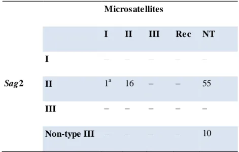
Table 6.2 Identification of interlocus recombination: use of two traditional loci panels (Sag2 classical + Microsatellites) versus combination of all loci ... 82
Supplementary Table S1 ... 121
Supplementary Table S2 ... 123
Supplementary Table S3 ... 124
Supplementary Table S4 ... 125
Supplementary Table S5A ... 126
Supplementary Table S5B ... 127
Supplementary Table s S6 ... 128
XXI
LIST OF FIGURES:
Figure 1.1 Toxoplasma gondii tissues cysts with multiple bradyzoites enclosed in mouse
brain cells ... 9
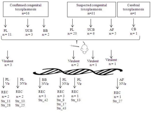
Figure 1.2 Toxoplasma gondii life cycle ... 10 Figure 1.3 Population genetic structure of T. gondii ... 13 Figure 5.1 Flowchart of the study. The figure summarizes the mice inoculation results as
well as the genotyping data concerning exclusively the recombinant strains ... 59
Figure 6.1 Minimum spanning tree (MST) of the 68 T. gondii isolates based on Sag2 5’
and 3’ ends nucleotide diversity colored according to classical genotyping classification (A) and mouse virulence (B) ... 77
Figure 6.2 Discriminatory power provided by each locus or combination of loci ... 77 Figure 6.3 Minimum spanning tree (MST) based on the multi-loci-based combined allelic
profiles colored according to classical Sag2 archetypes (A), Sag2 sequence alelles (B) and mouse virulence (C) ... 79
Figure 6.4 T. gondii genetic mosaicism ... 81 Supplementary Figure S1 ... 137
XXII
LIST OF ABBREVIATIONS: 3R – Reduce, Reuse, Recycle ABI – Applied Biosystems
AIDS – Acquired Immunodeficiency Syndrome AF – amniotic Fluid BB – Baby Blood bp – Base pair CB – Cerebral Biopsy CI – Confidence Intervals cM – Centimorgan cm - centimetres
CML – Câmara Municipal de Lisboa DMEM - Minimal Essential Medium DNA – Desoxyribonucleic acid
ELISA – Enzyme-Linked Immunosorbent Assay ENA – European Nucleotide Archive
Gra – Dense granule protein
Hsd: ICR (CD-1®) – Harlan Sprague Dawley, Inc., obtained a breeding stock from Charles
River Breeding Laboratories.
HIV – Human Immunodeficiency Virus HG – Haplogroup
Hp – Haplotype ID – Index Diversity IgG – Immunoglobulins G IntraRec – Intra Recombinant LD – Lethal Dosis
MAT – Modified Agglutination Test Mb – Mega base pair
ml – millilitres
MLST – Multilocus Sequence Typing MST – Minimum Spanning Tree NGS – New Generation Sequencing NIH – National Institute of Health PBS – Phosphate Buffered Saline PCR – Polymerase Chain Reaction
XXIII
p.i. – post inoculation PL – Placenta PT – Portuguese
RAPD - Random Amplified Polymorphic DNA RFLP – Restriction Fragment Length Polymorphisms RNA – Ribonucleic Acid
Rop – Roptry
Sag – Surface Antigen
SNP – Single Nucleotide Polymorphism TgMA – Myosine A
Tub2 – Beta Tubulina
UCB – Umbilical Cord Blood UI – International Units UK – United Kingdom
URSZ-INSA – Laboratório Nacional de Referência de Infecções Parasitárias e Fúngicas do
Instituto Nacional de Saúde Doutor Ricardo Jorge
USA – United States of America
UTAD – Universidade de Trás os Montes e Alto Douro WGS – Whole Genome Sequencing
XXV
NOTES OF THE AUTHOR: THESIS ORGANIZATION
This PhD thesis is organized in three parts. Its contents are essentially based on four manuscripts (listed below) that are presented as individual chapters (III to VI) in PART II, where three of them have already been published (the last one was submitted for publication at the time this thesis was submitted) in peer reviewed international journals.
The chapter arrangement does not perfectly reflect the chronological order of the manuscripts' publication, not only because some studies were developed simultaneously, but also because their publication time depended on the review process and journal requirements. In addition, despite of the main goals defined in the beginning of this PhD project, specific goals were raised on the course of the results obtained throughout the entire project, which influenced the publication priorities. As such, for the sake of clarity, the order of the chapters was decided to provide a better understanding of the approaches that were followe d to achieve the proposed goals instead of reflecting the chronological order of their release to the scientific community. The three parts are the following:
PART I - Presents the literature overview (CHAPTER I) and the major objectives of
this work (CHAPTER II). It aims to provide the reader with an overview of the state of art underlying the subject of this thesis. CHAPTER I addresses a general introduction about Toxoplasma gondii, clinical features, biology, life cycle of the parasite, history, general geographical mapping, virulence, an approach of some widely used molecular methods to detect and differentiate T. gondii, and the epidemiology that concerns serology and genotyping of T. gondii in Portugal. CHAPTER II comprises the questions that lead to the objectives of this work.
PART II – This part describes the experimental approaches that were undertaken to
achieve the proposed objectives. It contains several chapters, where most of them correspond to data that were already published in peer-reviewed scientific journals. CHAPTER III – Genotyping characterization of Toxoplasma gondii in animals from the North of Portugal. Contains a published study that was enriched with additional information. Lopes A.P.,
Vilares A., Neto F., Rodrigues A., Martins T., Ferreira I., Gargaté M.J., Rodrigues M.,
Cardoso L. 2015. Genotyping characterization of Toxoplasma gondii in Cattle, Sheep, Goats and Swine from the North of Portugal. Iranian Journal of Parasitology. 10(3):465-72.
CHAPTER IV – Contains a published study: Vilares A., Gargaté M.J., Ferreira I., Martins
S., Júlio C., Waap H., Ângelo H., Gomes J.P. 2014. Isolation and molecular characterization of Toxoplasma gondii isolated from pigeons and stray cats in Lisbon, Portugal. Veterinary
XXVI
Parasitology. 205(3-4):506-11. doi: 10.1016/j.vetpar.2014.08.006. CHAPTER V – Contains the following published study: Vilares A., Gargaté M.J., Ferreira I., Martins S., Gomes J.P. 2017. Molecular and virulence characterisation of Toxoplasma gondii strains isolated from humans in Portugal. Parasitology Research. 116(3):979-985. doi: 10.1007/s00436-017-5374-5. CHAPTER VI – Contains the following study that was recently submitted for publication:
Vilares A., Borges V., Sampaio D., Ferreira I., Martins S., Vieira L., Gargaté M.J., Gomes
J.P. 2019. Towards a rapid sequencing-based molecular surveillance and mosaicism investigation of Toxoplasma gondii. In review at Parasitology Research.
PART III – This part is constituted by CHAPTER VII that provides a general
discussion of the results obtained throughout the previous chapters and conclusions. Of note, in order to avoid redundancies, this section solely summarizes and discusses the major findings of this work because a detailed discussion of specific results was already provided in each chapter. It also presents the future perspectives enrolling specific research lines that may be addressed on the course of the obtained results. Considering the different layouts required by the different journals where the manuscripts were published, including tables, figures and references, all chapters were formatted in a unique style, with all references being cited by the name of the first author and year of the publication and listed in a single section - "References" - according alphabetical order.
1
3
CHAPTER I
5
1.1 General introduction
T. gondii belongs to the Apicomplexa phylum and it is the causal agent of toxoplasmosis. It is a wide spread protozoan parasite of humans and warm-blooded animals, including mammals and birds. In humans, this intracellular parasite is commonly acquired by the oral ingestion of tissue cysts containing bradyzoites, however it can also be transmitted by the ingestion of oocysts containing sporozoites that are the product of a sexual cycle shed by cats, the definitive host (Halonen and Weiss, 2013) and which can contaminate water and food. Classically, consumption of undercooked meat has been described to be the major risk factor for acquisition of toxoplasmosis (Tenter et al., 2000). T. gondii is the only known species in the genus Toxoplasma and is considered one of the most successful eukaryotic pathogen in the world in terms of the number of host species and percentage of animals infected worldwide (Tenter et al., 2000; Grigg and Suzuki, 2003; Su et al., 2010). Serological studies suggest that chronic infection rates in humans can vary from less than 10 % to more than 70 % depending on geographic region and various risk factors (Joynson, 2003).
1.2 History
Toxoplasma gondii was simultaneously described in tissues of different animals by Nicolle and Manceaux, and Splendore (Table 1.1). The genus was named by Nicolle and Manceaux as Toxoplasma for its bow-like shape (from Greek: toxo = bow or arc; plasma = creature). Over the years, different authors described T. gondii-like organisms, however it is believed that these cases were likely associated with infection by Leishmania spp. (Weiss and Dubey, 2009).
Janku (1923) and Torres (1927) described similar cases in infants who died with convulsions at two days of age. Although Janku only referred to the observed organisms as Sporozoa and Torres as Encephalitozoon chagasi, it was Levaditi (1928) who suggested that both cases were due to T. gondii. Later, T. gondii still be mistakenly named as E. brumpti when identified in the spinal fluid of a 17 year old boy from Corsica who died of meningitis.
The first viable organism was isolated by Albert Sabin in 1937 from laboratory mice during a routine virus testing experiment (Sabin and Olitsky, 1937), and later from humans by inoculating infected tissues into mice (Wolf et al., 1940). Sabin speculated that the parasite was an important zoonosis and demonstrated that this human strain was neither biologically
6
nor immunologically different from isolates from other animals. It was since then that the parasite was progressively recognized as the agent of a widespread zoonosis (Halonen and Weiss, 2013).
In 1939 Sabin isolated and inoculated T. gondii RH (Table 1.1) in mice and on the thirteenth day after inoculation, one of the mice exhibited a distended abdomen and a large amount of peritoneal exudate in which Toxoplasma organisms (Sabin, 1938) . Reinoculation of the tissues from the mice in healthy mice showed that Toxoplasma produced a fatal toxoplasmosis. This laboratory strain, it has been passed in mice in many laboratories. After these prolonged passages its pathogenicity for mice has been stabilized (Dubey et al., 1977) and it has lost the capacity to produce oocysts in cats (Frenkel et al., 1976).
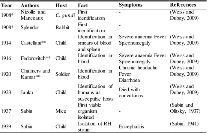
Table 1.1 History of the identification of Toxoplasma gondii.
Year Authors Host Fact Symptoms References
1908* Nicolle and Manceaux C. gundi First identification - (Weiss and Dubey, 2009)
1908* Splendor Rabbit First
identification - 1914 Castellani** Child Identification in smears of blood and spleen
Severe anaemia Fever Spleenomegaly
(Weiss and Dubey, 2009)
1916 Fedorovitch** Child Identification in blood
Severe anaemia Fever Spleenomegaly (Weiss and Dubey, 2009) 1920 Chalmers and Kamar** Soldier Identification in blood Chronic headache Fever Diarrhoea (Weiss and Dubey, 2009) 1923 Janku Child Identification of humans as susceptible hosts Died with convulsions (Weiss and Dubey, 2009) 1937 Sabin Mice First viable organism isolated - (Sabin and Olitsky, 1937)
1939 Sabin Child Isolation of RH
strain Encephalitis
(Sabin, 1941) *T. gondii was identified for the first time simultaneously by these authors in different regions; **Authors could miss associated Leishm ania spp to T. gondii.
7
Other forms of Toxoplasma including tissue cysts were recognized to exist by several researchers in 1951, but it was not until the 1960s and 1970s that the parasite was identified as a coccidian and Apicomplexa phylum. In 1972, the cat was identified as the definitive host harbouring the sexual parasitic cycle and spreading oocysts through faeces (Jewell et al., 1972).
The importance of maternal and congenital transmission has long been recognized since 1939; when a neonate from New York developed toxoplasmosis (Jones et al., 2001), and the growing role of Toxoplasma infection in immunocompromised patients was acknowledged in the mid-1970s (Luft and Remington, 1992). A more complete appreciation of the symptoms associated with acute acquired toxoplasmosis was achieved with the report of outbreaks of acute toxoplasmosis in adults in the USA (Teutsch et al., 1979) and Canada (Bowie et al., 1997).
1.3 Toxoplasma gondii infection 1.3.1 Clinical features
In animal husbandry toxoplasmosis is the major cause of reproductive failure by leading to early embryonic and fetal death with abortions and resorption with fetal mummification (Dubey and Kirkbride, 1989; Dubey, 2009, 2010a). The severity of infection is associated with the stage of gestation at which the livestock becomes infected. The earlier infection in gestation, the more severe will be the consequences (Dubey, 2009). However, the majority of T. gondii infections in most host species are subclinical or asymptomatic, and chronic (Dubey, 2010a) and the parasite can remain dormant in the tissues until reactivation or until the host is eaten by a predator. Also, the associations of chronic infection with changes in behaviour, memory, and neurologic disorders (Berdoy et al., 1995, 2000; Webster, 2001; Torrey and Yolken, 2007; Berenreiterová et al., 2011; Dass et al., 2011; House et al., 2011; Pedersen et al., 2011; Torrey et al., 2012; Webster et al., 2013) are fascinating, as illustrated for example, by the cases of the preys that lost the natural fear of predators (Haroon et al., 2012). Consequently, the healthy aspect and the fear loss of preys could increase the perpetuation of T. gondii cycle in human’s and in other hosts.
In humans, toxoplasmosis was implicated, about two decades ago, as the third most common cause of food borne infection in the USA (Mead et al., 1999). It is recognized as a category B priority pathogen by the National Institutes of Health, Bethesda, USA due to its
8
risk of transmission through contamination of food or water and importance as an opportunistic pathogen. Hence, studies performed in T gondii are often used as model to related but less tractable pathogens such as Cryptosporidium, another category B Biodefense Agent that causes severe diarrheal disease, and Plasmodium, the causative agent of malaria (Kim and Weiss, 2004).
Most human T. gondii infections are often asymptomatic, however, in several of its hosts it is associated with congenital infection and abortion (Gilbert et al., 1999; McLeod et al., 2006; Oz, 2014). Congenital toxoplasmosis can manifest with severe complications, such as miscarriage, fetal developmental retardation, encephalitis, neurological and mental illnesses, visual and auditory inflammatory disorders, cardiovascular abnormalities, and pains (Gilbert et al., 1999; McLeod et al., 2006; Oz, 2014). In addition, T. gondii can also cause encephalitis or systemic infections in the immunocompromised, particularly in individuals with HIV/AIDS. Toxoplasma infection was also implicated in etiologies of neurodevelopmental and neurocognitive disorders like-schizophrenia (Torrey and Yolken, 2007). Severe cases of toxoplasmosis have been reported in immunocompetent patients in association with atypical T. gondii genotypes (Ajzenberg et al., 2004; Demar et al., 2007; Elbez-Rubinstein et al., 2009; Vaudaux et al., 2010; Wendte et al., 2011; Pomares et al., 2011; Sobanski et al., 2013). Although current available drugs, like pyrimethamine and sulphadiazine, can control the proliferative form of the parasite and treat Toxoplasma infections, they are poorly tolerated, they have severe side effects such as allergic reactions, and they are ineffective against chronic Toxoplasma infections. In addition, resistance to some of these drugs has been noted (Aspinall et al., 2002b; Baatz et al., 2006; Blader and Saeij, 2009). Pathogenesis is typically associated with the ability of these parasites to replicate and proliferate within host cells. However, pathogenicity can also vary with the host and the morphological stage ingested (Dubey, 2010a; Dubey et al., 2012).
A study performed in the USA estimated that the annual cost of illnesses caused by Toxoplasma is about $3 billion (Hoffmann et al., 2012). A study in ewes (n=1613) from Uruguay estimated annual losses due to toxoplasmosis during gestation of 1.4 to 3.9 %, accounting to approximately $ 1.4 to 4.7 million (Freyre et al., 1997). The economic losses due to lamb mortality and missed lactation are estimated at 10 million Euros per year in Italy (Masala et al., 2003). In UK, the impact of toxoplasmosis in sheep industry is between £ 12 million and £ 24 million each year (http://www.apd.rdg.ac.uk/AgEcon/livestockdisease/index.htm). However, clinical and detailed economic information is lacking in Europe and particularly in
9
Portugal. Overall, toxoplasmosis results in increased production costs, diminished marketability of meat, less replacement animals, retardation of genetic progress and constitutes a major source of human infection (Leighty, 1990; Freyre et al., 1997).
1.4 Biology and Life Cycle
T. gondii is a tissue cyst-forming coccidia with a heteroxenous life cycle in which an asexual reproduction in intermediate hosts is linked to a sexual reproduction in definitive hosts (Tenter and Johnson, 1997).
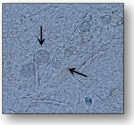
T. gondii has three morphological stages: tachyzoite (2 x 6 μm), bradyzoite (1-3 x 5-8.5 μm) and sporozoite (2 x 6-8 μm) (Sabin and Olitsky, 1937; Jones et al., 1972; Cowper et al., 2012). Bradyzoites and sporozoites are the infective forms typically acquired by ingestion. Bradyzoites are enclosed in tissue cysts in chronically infected hosts (Figure 1.1).
Figure 1.1. T. gondii tissue cysts with multiple bradyzoite enclosed in mouse brain cells. Note : T his figure belongs to the URSZ-INSA acquis.
Eight sporozoites are present in each completed sporulated oocyst (11 x 13 μm), millions of which are shed into the environment, unsporulated, by the definitive hosts (felids) of the parasite, over 4 days after the first infection. It is an obligatory intracellular parasite of the nucleated cells of its hosts. They possess a nucleus, a mitochondrion, a Golgi complex, ribosome, an endoplasmic reticulum, and an apicoplast (Roos, 1999). The oocyst is the only form of life of the parasite that can persist outside the host for a long period of time (Yilmaz and Hopkins, 1972; Frenkel and Dubey, 1973; Dubey, 1998c, 2010a).
After ingestion, tissue cysts or oocysts invade the host cells and differentiate into tachyzoites which divide rapidly within the host cells and together with the host immune response are responsible for the clinical manifestations of infection. Tachyzoites infect
10
nucleated host cells and utilize monocytes, macrophages, and dendritic cells as “Trojan Horses” to escape the host immune defence (Elsheikha and Khan, 2010), to bypass the blood– brain barrier (Bierly et al., 2008) and the placenta barricade, and to spread and cause systemic disease.
Tachyzoites differentiate into latent bradyzoites, which can be induced by exposure of the organism to stress conditions such as an immune response. Tissue cysts can persist indefinitely for the life of the host (Figure 1.2). If an individual becomes immunocompromised these tissue cysts serve as a reservoir from which disseminated or local infections can develop. Tissue cysts have a predilection for neural and muscle tissue as well as the eye in humans, with most cases of reactivation disease presenting as encephalitis or chorioretinitis (Kim and Weiss, 2008).
Figure 1.2. Toxoplasm a gondii life cycle. Note : Adapted from Nature Reviews
1.5 Molecular epidemiology 1.5.1 Genotypes
The genome size of T. gondii is between 60 - 70 Mb. In 2001 only 11 chromosomes had been identified (Ajioka et al., 2001) but now it is known that T. gondii genome consists of
11
14 chromosomes (Khan et al., 2005a). In the early days, isolates of T. gondii were grouped according to virulence in mice. Later on, the first phylogenetic studies of T. gondii strains indicated that their genetic complexity was much smaller than expected (Sibley and Boothroyd, 1992a; Dardé et al., 1992) so, for a long time, T. gondii was considered to be clonal with three genetic types (Types I, II, III) (Dardé et al., 1992; Howe and Sibley, 1995; Ajzenberg et al., 2004) and very small differences between clonal lineages. In fact, comparative sequence analysis of individual genes indicated low allelic diversity within the clonal lineages (about 1 % divergence). In addition, limited genetic diversity between and within clonal lineages indicated that they have evolved quite recently from a common ancestor, 10,000 years ago at the most (Su, 2003). Also, recent advances in the knowledge of the virulence associated with some genotypes have been achieved (Saeij et al., 2005) with the development of new genotyping tools and the multiplication of field studies (Mercier et al., 2011). Also, recent studies on T. gondii in different animal populations worldwide started to reveal the extensive diversity of the parasite (Dubey et al., 2002; Lehmann et al., 2006; Pena et al., 2008; Khan et al., 2011a).
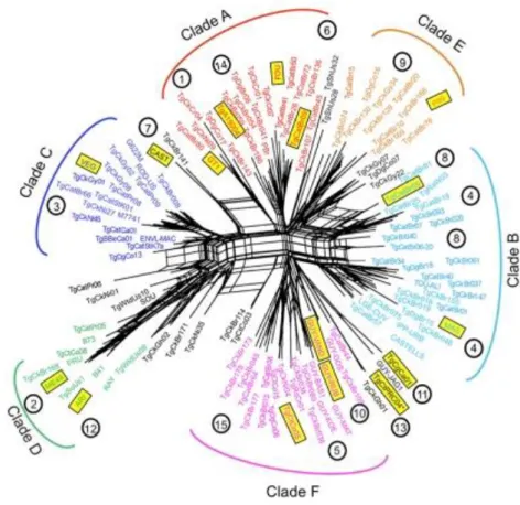
A fourth clonal lineage, designated haplotype 12, is largely confined to North America, where it is more common in wild animals (Khan et al., 2011a). Other studies subdivided T. gondii in I, II, III and 4 to 14 haplotypes based on only five loci (Khan et al., 2011b; a). Also, Su and colleagues classified T. gondii strains into 15 different haplotypes, defining six major clades (Figure 1.3) (Su et al., 2012). However, Minot and colleagues showed that the observed extent of Toxoplasma genetic diversity does not fit the scenario of the proposed haplotypes. Instead, most strains appear to have been originated through recent recombination events and it is believed that some of them led to particularly fit genotypes that geographically spread or swept (Minot et al., 2012). The majority of genotypes were identified either by polymerase chain reaction followed by restriction fragment length polymorphism (PCR-RFLP) or by microsatellite analysis using different genetic markers and variable number of loci. In America, 85.7 % of parasites were typed by using three to nine protein-coding genetic markers by PCR-RFLP, while the majority of T. gondii genotyping in Europe (67.4 %, n = 126) and Oceania (100 %, n = 2) was performed by microsatellite analysis. All variants from Asia and most from Africa (70.4 %) were identified with one coding-gene marker (Rico-Torres et al., 2016).
12
Currently, there is no gold-standard for genotype designation. As long as different methods and genes are used to type a variety of isolates, and each method has its own classification scheme, genotype designation remains a confusing issue.
Figure 1.3. Population genetic structure of T. gondii. Note : adapted from Su et al 2012. Neighbor-net analysis was conducted
using 11 multilocus RFLP markers plus 1 marker for the apicoplast and four intron sequences from the 138 representative strains representing unique haplotypes. Neighbor-net analysis showed various routes for gene flow between different populations (interconnecting lines between representative strains). Six major clades (A through F) are indicated on the basis of ST RUCT URE analysis. Strains in black lettering do not correspond to major clades. Haplogroups are shown in circled numbers. Representative strains for each haplogroup are indicated by yellow boxes.
1.5.2 Genotyping Methods
Numerous studies of the T. gondii population structure were based on genotyping using a single marker, mostly Sag2 (Howe et al., 1997; Fuentes et al., 2001; Sabaj et al., 2010) and particularly, due to its polymorphisms and sensitivity, Gra6 (Fazaeli et al., 2000; Messaritakis et al., 2008). However, genotyping with a single marker does not allow identification of nonclonal strains. To determine more precisely the presence of polymorphisms in the population, application of multilocus PCR and microsatellite analysis of multiple markers is necessary (Ajzenberg et al., 2005; Su et al., 2006). The paragraphs below describe the most used techniques in T. gondii genotyping.
13 1.5.2.1 Multilocus Enzyme Electrophoresis (MLEE)
MLEE differentiates strains by assessment of differences in migration of metabolic enzymes by electrophoresis. These migration differences are determined by the sequences of amino acids (molecular weight and enzyme charge) and could be associated to allelic variation at the corresponding gene locus. When a group of enzymes are analyzed all together, the different migration produces a unique finger print of each strain representing a multilocus genotype (Dardé et al., 1988, 1992).
The first study on strain diversity in T. gondii based on MLEE analysis was developed by Dardé et al. (Dardé et al., 1988). Six polymorphic enzymatic systems exhibited 12 zymodemes with the majority of stocks clustering into three main zymodemes Z1, Z2 and Z3 (Dardé et al., 1988, 1992; Ajzenberg et al., 2002a). The disadvantages of MLEE are the low
resolution in
T. gondii genetic studies, the requirement of large numbers of parasites (approximately 7x10⁶ tachyzoites) and the purity of the samples (Dardé et al., 1992).
1.5.2.2 RAPD-PCR
RAPD-PCR is based on the amplification of genomic DNA using arbitrary primers to identify unknown DNA polymorphisms. T. gondii have been classified into virulent and avirulent strains based on the murine virulence by RAPD-PCR using arbitrary primers (Guo et al., 1997), however nowadays the study of virulence strains require a much more complex interpretation. The results of this technique are difficult to reproduce and it requires a high DNA purity (Guo and Johnson, 1995; Ferreira et al., 2004).
1.5.2.3 PCR- RFLP
The PCR-RFLP is based on the ability of restriction endonucleases to recognize single nucleotide polymorphisms (SNPs), digest PCR products and then display distinct DNA banding patterns on agarose gels by electrophoresis (Sibley et al., 1992; Howe and Sibley, 1995). How and Sibley identified for the first time T. gondii genotypes with PCR-RFLP using 6 markers. Since then, several different sets of multilocus PCR-RFLP markers have been developed to genotype T. gondii isolates (Howe and Sibley, 1995). As the conventional multilocus PCR-RFLP requires a large amount of DNA, multiplex nested PCR-RFLP was developed to overcome this problem. However, the possibility of contamination is high and to avoid this problem multiple negative controls must be used in all PCRs (Sibley et al., 1992; Cristina et al., 1995; Howe and Sibley, 1995; Khan et al., 2005b; de Melo Ferreira et al.,
14
2006; Dubey et al., 2007a; Pena et al., 2008; Soares et al., 2011; Su et al., 2012; Bacci et al., 2015).
1.5.2.4 Microsatellite analysis
Microsatellite sequences are tandem short DNA motif repeats that are widespread in eukaryotic genomes and the sequences usually change due to insertion or deletion of repeat units. The numbers of repeat units differ in a population, thus producing multiple alleles at a microsatellite locus. The tandem repeats in T. gondii are composed of 2 to 6 nucleotides and occur 2–20 times (Blackston et al., 2001; Ajzenberg et al., 2002a, 2010; Li et al., 2014). This length polymorphism of microsatellite regions can be assessed with fluorescent primers after electrophoresis on an automatic sequencer. A total of 5 markers have been used (Tub2, W35, TgM-A, B18, B17) to differentiate types I, II, III, recombinants and atypical genotypes (Ajzenberg et al., 2002b). More recently, Ajzenberg and colleagues developed a method for T. gondii genotyping in a single multiplex PCR assay using 15 microsatellite markers (Tub2, W35, TgM-A, B18, B17, M33, IV.1, XI.1, M48, M102, N60, N82, AA, N61, and N83) (Ajzenberg et al., 2010). Although microsatellite multiplex has been considered an accurate assay to differentiate T. gondii strains and to identify mixed infections (Ajzenberg et al., 2009; Mercier et al., 2010) the presence of small amounts of DNA (usually from clinical samples or wild strains with low rate of taquizoites or cysts) may impair the results (Ajzenberg et al., 2010).
1.5.2.5 Sequencing
MLST was first proposed in 1998 as a typing approach enabling the unambiguous characterization of bacterial isolates in a standardized, reproducible, and portable manner using the human pathogen Neisseria meningitides as the model organism (Maiden, 2006). The typical MLST for T. gondii is based on DNA sequence polymorphisms of seven housekeeping genes, including the single nucleotide polymorphisms (SNPs), deletion and insertion of nucleotides (Su et al., 2012). However different virulence loci have been used by different authors and also different sequencing approaches, such as Sanger (classic) or NGS (Khan et al., 2011a, 2014; Cheng et al., 2015; Lorenzi et al., 2016). Once more, although this approach is the most discriminatory method it is hardly applicable to clinical samples, as a large quantity of genomic DNA is required for the successful amplification of several loci.
15 1.5.2.6 Serotyping
Serotyping is a typing method based on a serological test using strain-specific peptides. For T. gondii, different authors have used specific peptides derived from dense granule antigens, like GRA5, GRA6 and GRA7 (Sousa et al., 2008; Maksimov et al., 2013). Serotyping was shown to be capable of distinguishing type II from non-type II infections (Kong et al., 2003; Xiao et al., 2009) and may be used to determine which strains are associated with symptomatic or asymptomatic infections (Sousa et al., 2008). As serotyping is fast, inexpensive, relatively noninvasive, and there is no need to isolate parasites, this technique could have the potential to become the method of choice for typing T. gondii in humans and animals. However, some limitations arise, such as cross-reactivity issues, the low sensitivity of the selected peptides in detecting recombinant strains (Sousa et al., 2009a) and the potential no viability of the serum (dead animals). In particular, immunosuppressed patients may not produce sufficient specific antibodies to reach the detection threshold (titer of 1:64) and infection with the rare genotypes may induce highly different humoral responses that may not be detectable using the custom polymorphic polypeptides(Kong et al., 2003).
1.5.3 Genotypes and virulence
In terms of mouse virulence, Type I isolates are considered as the most virulent, and can lead to death of mice less than 10 days after the first inoculation. In contrast, strains of Type II and Type III are avirulent (although type II isolates can cause low virulence in mice) and usually cause chronic infection and shaped tissue-cysts. Cystogenic T. gondii strains can be grouped according to their virulence, however, all non-cystogenic isolates present high virulence in mice (de Melo Ferreira et al., 2006).
In Humans, type I and recombinant strains isolated from immunocompetent individuals have been associated with some severe forms of toxoplasmosis or atypical ocular toxoplasmosis (Howe et al., 1997; Fuentes et al., 2001; Grigg et al., 2001a). Also, in southern Brazil and French Guiana, atypical strains have been associated with severe disseminated disease (Dardé et al., 1998; Blaizot et al., 2019) and with debilitating ocular toxoplasmosis and death in healthy adults (Carme et al., 2002; Jones et al., 2006a; b; Demar et al., 2012; Silveira et al., 2015).
16
1.5.4 Geographical distribution of T. gondii genotypes
Several studies have been conducted in diverse countries aiming at analyzing T. gondii genotypes of isolates circulating in diverse hosts and countries. Most of them performed a multilocus genotyping by PCR and RFLP markers (Sundar et al., 2008; Sun et al., 2013; Wang et al., 2015), and less frequently by microsatellite markers (Ajzenberg et al., 2010), gene sequencing or NGS (Lorenzi et al., 2016). Studies are most often carried out in birds, as sentinels of the environmental contamination (Lehmann et al., 2006), whereas genotypes infecting wild species or other domestic animals are less frequently studied. Although epidemiological studies presenting genotypes infecting humans may be biased due to travel or to consumption of imported food, they usually reflect circulating genotypes (Ajzenberg et al., 2015).
In Europe and North America, T. gondii exists as four distinct clonal lineages that show marked virulence differences in laboratory mice (Howe and Sibley, 1995; Khan et al., 2011a). It is accepted that type II is the predominant type in cases of congenital toxoplasmosis in Europe and North America (Howe and Sibley, 1995; Howe et al., 1997; Ajzenberg et al., 2002b). A fourth clonal lineage, referred to as type 12, has recently been described in North America where it is commonly found in wildlife (Khan et al., 2011a). All four clonal lineages show evidence of overly abundant, highly similar multilocus genotypes and high levels of linkage disequilibrium (i.e., infrequent recombination). While it has been established that type II is predominant in Europe and North America (Dardé et al., 1992; Howe and Sibley, 1995; Howe et al., 1997), there are significant regional differences. Type I and some recombinant strains were isolated from immunocompetent individuals suffering from severe or atypical ocular toxoplasmosis in United States (Grigg et al., 2001a).
One French study has shown that of the 86 isolates (85 %) from cases of suspected and confirmed congenital toxoplasmosis and 88 isolates from immunosuppressed patients (61.4 %) were type II (Ajzenberg et al., 2002b, 2009). In other studies with immunosuppressed patients, also from France, it was shown that type II isolates were also predominant, while types I and III were rarely isolated (Howe et al., 1997; Honoré et al., 2000). Research in congenital toxoplasmosis from Spain and Egypt showed the presence of type I (Fuentes et al., 2001; Eldeek et al., 2017), while genotyping of isolates from Crete, Cyprus showed the predominance of type III (Messaritakis et al., 2008), however it must be noted that these studies have been conducted using only one or four markers.
17
Furthermore, strains of atypical genotypes were isolated from immunocompetent patients in French Guiana and Brazil (Carme et al., 2002; Demar et al., 2012; Silveira et al., 2015).
1.6 Toxoplasma gondii in Portugal
1.6.1 T. gondii epidemiology in Portugal
In Portugal, there is a lack of human data regarding T. gondii as few studies are available, and most of them are serological estimates of T. gondii seroprevalence in humans (Table 1.2) and in several animal hosts (Table 1.3).
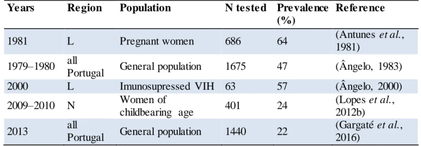
There were a limited serologic studies performed in humans, in Portugal (Ângelo, 1983, 2003; Antunes, 1984; Sevivas, 2011; Lopes et al., 2012b; Gargaté et al., 2016), however, only two were performed in the general population. The first serological survey reported 47 % of seropositivity, which is considered a high value when compared with the one obtained in 2013 (Ângelo, 1983; Gargaté et al., 2016), where the overall seroprevalence was 22 % (Gargaté et al., 2016). Of note, despite this apparent strong decreasing trend of the T. gondii seroprevalence in Portugal during the last decades, the risk of congenital toxoplasmosis is higher, since these data reveal that the majority of potential pregnant women are thus susceptible to primary infection (Table 1.2).
Table 1.2 Seroprevalence of T. gondii infection in humans from Portugal
Years Region Population N tested Prevalence
(%)
Reference
1981 L Pregnant women 686 64 (Antunes et al.,
1981) 1979–1980 all
Portugal General population 1675 47 (Ângelo, 1983)
2000 L Imunosupressed VIH 63 57 (Ângelo, 2000)
2009–2010 N Women of
childbearing age 401 24
(Lopes et al., 2012b)
2013 all
Portugal General population 1440 22
(Gargaté et al., 2016)
L: Lisbon; N: North
T. gondii antibodies were also detected in cats (Lopes et al., 2008; Duarte et al., 2010; Esteves et al., 2014), sheep (Sousa et al., 2009b; Lopes et al., 2012a), cattle (Lopes et al., 2012a), dogs (Lopes et al., 2011b), wild boar (Lopes et al., 2011a; Coelho et al., 2014), goats
18
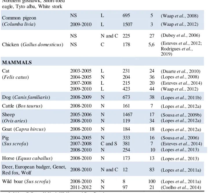
(Lopes et al., 2012a), pigs (Sousa et al., 2006; Lopes et al., 2012a; Esteves et al., 2014), horses (Lopes et al., 2013), chickens (Dubey et al., 2006; Rodrigues et al., 2019), birds (Waap et al., 2008, 2012; Lopes et al., 2011a), genets, wolf, red foxes and badgers (Lopes et al., 2011a) (Table 1.3). Waap and colaborators studied a large number of pegeons describing a low prevalence rate, however, when considered at flock level, seropositive animals were identified in nearly one third of feeding sites sampled, with seroprevalence rates ranging between 5 % and 62.5 % (Waap et al., 2008, 2012). In cats, the serological prevalence varied from 20 to 44 %; the highest being from stray cats from Lisbon (Table 1.3). The stray cats were captured from different areas of Lisbon and revealed the high serological titres, 18000 to 162000 (Waap et al., 2012). These results may be due to the T. gondii type strains circulating or a very high environmental contamination leading to repeated infections in cats (Lopes et al., 2014, 2017). In meat animal consumption, T. gondii prevalence varies between 7 % in cattle and pigs and 100 % in wild boars (Table 1.3). The three surveys performed in pigs demonstrated prevalences between 7 % and 16 %, which are not different from the heterogeneous values that have been reported in Europe (Dubey, 2009; Santoro et al., 2017). Of note, most authors have used in-house ELISAs and thus these results are difficult to be compared due to the need for standardization of cut-off values, antigens and specific methodology procedures. In addition to meat from domestic pigs, meat from wild boars may also be an important source of T. gondii infection in humans.
Lopes and collaborators (2011) found 100 % of T. gondii prevalence in wild boars, however another study performed also in north of Portugal achieved the prevalence of 21 % when increased the number of wild boars studied (Table 1.3) (Lopes et al., 2011a; Coelho et al., 2014). In Portugal there is some tradition of wild boar meat consumption. This meat is widely used to cook uncontrolled homemade sausages that prepared without any heat processing and, therefore, are consumed raw, increasing the likelihood of T. gondii as a foodborne pathogen for humans (Coelho et al., 2014). Additionally, infected viscera and carcasses left by hunters may pose a threat to scavenging susceptible animal.
It is believed that in Europe up to 63 % of human toxoplasmosis is attributed to the consumption of undercooked or cured meat products, however all the animals, like cats, wild animals or birds, could represent the zoonotic transmission but also the environmental contamination (Cook et al., 2000; Berger-Schoch et al., 2011; Belluco et al., 2018).