RESUMO.- [Ocorrência e caracterização de isolados de Campylobacter spp. em cães, gatos e crianças.] Com o objetivo de melhorar o entendimento das infecções por Campylobacter spp. em cães, gatos e crianças no Brasil, fo-ram avaliadas 160 amostras fecais de crianças e 120 swabs retais de pets (103 cães e 17 gatos). Do total das amostras das crianças, 6,87% foram positivas para Campylobacter
spp. e em cães e gatos a positividade foi de 18,3%. Das 33 amostras positivas para Campylobacter spp., 57,6% foram identificadas como C. jejuni e 33,4% foram identificadas como C. coli. Mais de 50% das amostras isoladas de pets foram resistentes a ceftiofur, sulphazotrim, norfloxacina e tetraciclina. Em crianças, a maioria das amostras foi resis-tente a amoxilina, cefazolina, ceftiofur, eritromicina e nor -floxacina. De 19 isolados de C. jejuni, 11 isolados de crian-ças e cinco (5) de cães tinham dois (2) dos quatro (4) genes de virulência flaA, pldA, cadF or ciaB. Associação positiva entre a presença de Campylobacter spp. e diarreia em cães e gatos foi observada em animais desverminados e com he-mograma sugestivo de infecção bacteriana. Também houve associação positiva entre a presença dos genes de virulên-cia e a ocorrênvirulên-cia de diarreia, e entre o uso de antibióticos e a positividade para Campylobacter spp. em suabes fecais de pets. Os dados desse trabalho indicam que cepas virulentas
Occurrence and characterization of
Campylobacter
spp.
isolates in dogs, cats and children
1Cecilia G. Rodrigues2, Roberta T. Melo2, Belchiolina B. Fonseca2*, Pedro A. Martins2, Fernando A. Ferreira3, Maria B.J. Araújo4 and Daise A. Rossi2
ABSTRACT.- Rodrigues C.G., Melo R.T., Fonseca B.B., Martins P.A., Ferreira F.A., Araújo M.B.J. & Rossi D.A. 2015. Occurrence and characterization of Campylobacter spp. isolatesin dogs, cats and children. Pesquisa Veterinária Brasileira 35(4):365-370. Laboratório de Biotecnologia Animal Aplicada, Faculdade de Medicina Veterinária, Universidade Federal de Uberlândia, Rua Ceará s/n, Bloco 2D, Sala 43, Bairro Umuarama, Uberlândia, MG 38402-018, Brazil. E-mail: [email protected]
To improve the understanding of implications of Campylobacter spp. infections in pets and children of different environments were analysed 160 faecal samples from children and 120 from pets (103 dogs and 17 cats). Campylobacter spp. were detected in 6.87% of the children and in 18.3% of the dogs and cats. From 33 stool samples positive for Campylo-bacter spp., 57.6% were identified as C. jejuni, and 33.4% were identified as C. coli. More than 50% of the isolates in pets were resistant to ceftiofur, sulphazotrim, norfloxacin and tetracycline. In humans, most of the isolates were resistant to amoxicillin, cefazolin, ceftio -fur, erythromycin and norfloxacin. From 19 isolates of C. jejuni, 11 isolates from children and 5 from dogs contained two to four of the virulence genes flaA, pldA, cadF or ciaB. We found an association between the presence of virulence genes and diarrhoea. Furthermore, an association was observed between the presence of Campylobacter spp. and diarrhoea in dewormed pets with blood picture suggestive of bacterial infection, and the therapeutic use of antibiotics was associated with more positive detection of Campylobacter spp. in the faeces of pets. Our data indicate that virulent strains of Campylobacter spp. can be risk factor to diarrhoea in animals, and that high resistance to antimicrobial agents is common in pets.
INDEX TERMS: Campylobacter spp., dogs, cats, children, diarrhoea, infection, epidemiology, virulence genes.
1 Received on February 15, 2014.
Accepted for publication on December 5, 2014.
2 Laboratório de Biotecnologia Animal Aplicada, Faculdade de Medicina Veterinária, Universidade Federal de Uberlândia, Rua Ceará s/n, Bloco 2D, Sala 43, Bairro Umuarama, Uberlândia, MG 38402-018, Brazil. *Corres-ponding author: [email protected]
3 Hospital Veterinário da Faculdade de Medicina Veterinária, Universi-dade Federal de Uberlândia, Rua Ceará s/n, Bloco 2D, Sala 43, Bairro Umu-arama, Uberlândia, MG 38402-018.
de Campylobacter spp. são fatores de risco para diarreia em cães e a resistência antimicrobiana é comum em isolados de cães.
TERMOS DE INDEXAÇÃO: Campylobacter spp., cães, gatos, crian-ças, diarreia, infecção, epidemiologia, genes de virulência.
INTRODUCTION
In humans, Campylobacter sp. is the most common bacte-rial cause of foodborne enteritis (EFSA 2009, EFSA 2010). In addition to ingestion of contaminated food and water, di-rect contact with animals may be a possible source of Cam-pylobacter jejuni infection (Damborg et al. 2004, Houf et al. 2008). In dogs, Campylobacter spp. are frequently isolated from animals with symptoms of enteritis and asymptomatic animals (Baker et al. 1999, Moser et al. 2001, Chaban et al. 2010). Although symptoms of enteritis may be associated with campylobacteriosis, few veterinarians understand the importance of this bacterium in clinical practice. Antimi-crobial resistance is an important factor for Campylobacter spp. in different regions of the world in humans and pets.
Dogs and cats are increasingly present in family life, especially with children. In many cases, close contact with animals occurs regularly, and Campylobacter spp. can be transmitted to humans by contact (Lengerh et al. 2013).
Knowledge of the incidence, association of clinical symptoms of virulent strains of Campylobacter spp., anti-microbial resistance, and epidemiology of Campylobacter spp. infection in pets and children enables measures to prevent human infections and guide the treatment and care provided for animals.
This study aimed to identify dogs, cats and children po-sitive for Campylobacter spp. through faecal samples, to es-tablish associations between the presence of the microor-ganism and gastrointestinal disorders, and to evaluate the presence of virulence genes and antimicrobial resistance in isolated strains.
MATERIALS AND METHODS
We analysed 103 samples from dogs, 17 samples from cats of different ages, and 160 samples from up to 5-year-old children. After authorization, the samples were collected from patients in the Department of Internal Medicine of Small Animal Veterina-ry Hospital (HVet-UFU) and sectors of the Emergency and Clinic Department of the Clinics Hospital (HC-UFU), both at the Federal University of Uberlândia. Samples were collected from July 2010 to March 2011. Dogs and cats with clinical symptoms of diarrhea or other diseases (dermatitis, ear infections, ocular infections and accidents) and children with symptoms of diarrhea or anal itching were chosen randomly. Type of diarrhea (as colour, appe-arance of faeces, time course) and antibiotic usage in the animals and children were recorded. Blood picture of dogs and cats with diarrhoea showed changes suggestive of bacterial infection. The experiment was approved by the Ethics Committee on Animal Use and the Ethics Committee on Humans from the Federal University of Uberlândia, number 089/2010.
The collection of animal faeces was performed directly from the rectum of animals with sterile swabs. Faeces samples were collected from children and stored in sterile jars by health pro-fessionals. The samples were immediately transported to the laboratory and analysed. We collected information regarding the
presence of pets in the residences of children positive for Cam-pylobacter spp. We also verified whether the animals treated at HV-UFU that were positive for Campylobacter spp. lived in the res-idences of children.
In the laboratory, Bolton broth (Oxoid®) with an antibiotic
supplement (Selective Supplement Oxoid®) was added, and the stool samples were homogenised. The samples were supplemen-ted with 5% horse blood hemolysate and subjecsupplemen-ted to pre-en-richment in a microaerophilic atmosphere (Probac of Brazil®) with anaerobic jars at 37°C for 24 hours. After pre-enrichment, the material was seeded in selective Campylobacter Blood-Free Selective Agar Base modified medium (mCCDA) (Oxoid®), added
to the supplement antibiotic (Oxoid®) and 5% horse blood hemo-lysate and incubated at 37ºC for 48 hours in anaerobic jars under microaerobic conditions (Probac of Brazil®). Suspected colonies
were confirmed by the modified Gram stain by observing the mor -phology of spiral and curved rods or “gull wings”.
The phenotypic identification of differentiated species was performed as recommended by Silva et al. (2007) using the ox -idase test, catalase production, hippurate hydrolysis, nitrate re-duction, H2S production, growth at 25°C and 42°C and the DNAse test. All the tests were performed in duplicate, and C. jejuni (ATCC 33291) was used as the positive control.
In parallel with the phenotypic identification, typical colonies were subjected to molecular identification using a multiplex PCR technique. The DNA was extracted from colonies by thermal ex -traction and amplified using the primer described by Harmon et al. (1997). The reaction solution (Invitrogen®) was prepared and
the amplification cycle (Eppendorf®) was performed according to Harmon et al. (1997). As a positive control, we used the standard strains of Campylobacter jejuni ATCC 33291 and Campylobacter coli ATCC 43478. The amplified products were separated by elec -trophoresis, stained with SYBR® solution Safe DNA gel stain (In-vitrogen®), supplemented with a 100 bp molecular weight marker (Invitrogen®) and visualised in UV light in a transluminator (L PIX EX Loccus Biotechnology).
The primer pairs and PCR assay described for Hänel et al. (2004) and Zheng et al. (2006) were used for the analysis of vir-ulence factors associated with adhesion and cell invasion using multiplex PCR analysis. The final volume of the amplification re -action (50µL) comprised 20ng of bacterial DNA and the following reagents: 10mM Tris-HCL, 50mM KCl, 200 mM of each deoxy -nucleotide triphosphate (dNTP), 2.6mM MgCl2, 10 picomoles of primer for flaA and pldA, 40 picomoles for gene cadF and ciaB and 1.25 U Taq DNA polymerase. The determination of each gene was performed individually. The positive control C. jejuni NCTC 11351 and a negative control were used in all of the amplification reac -tions. Amplification was performed in a thermocycler (Eppendorf
®) according to the following cycles: 1 cycle starting at 95°C for 10 min, 35 cycles consisting of 3 (three) steps including denaturation at 95°C for 1 min, annealing at 45°C for 1 min and extension at 72°C for 2 min and a final cycle with extension at 72°C for 10 min. Separation and visualisation of the amplified products
were performed using the same technique as described for multiplex PCR to identify the species.
To determine the epidemiological association measures, we used chi-square tests and odds ratios (OR) (p≤0.05) using the statistical program Graphpad Prism 5.
RESULTS AND DISCUSSION
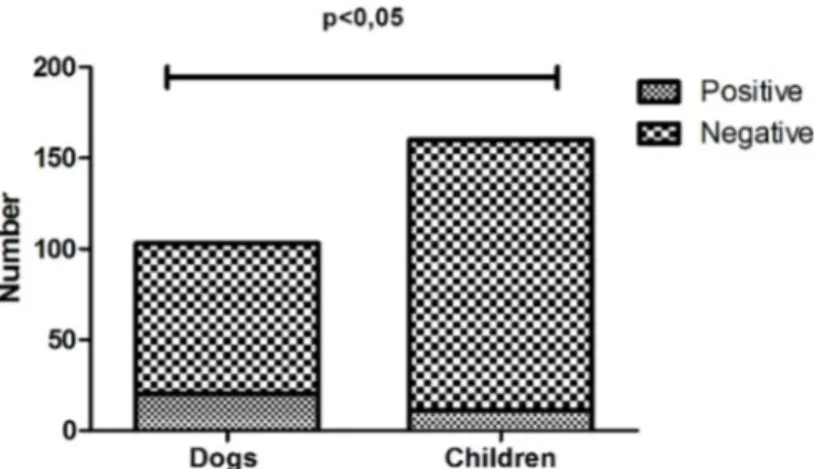
In the children, 6.87% (11/160) of the faecal samples tes-ted were positive for Campylobacter spp. The 120 stool samples analysed from pets included 103 samples from dogs and 17 from cats. Overall, 18.3% (22/120) of the pet samples tested were positive for Campylobacter spp. inclu-ding 19.41% (20/103) of dog samples and 11.7% (2/17) of cat samples.
The rate of Campylobacter spp. isolated from children was slightly lower compared with the results of other au-thors. In Rio de Janeiro, Mangia et al. (1993) found that 9.9% (15/153) of stool samples from children under 5 years of age with and without diarrhoea were positive for Campylobacter spp.
Only two of the children positive for Campylobacter spp. were reported to have had contact with animals, and the corresponding animals negative for Campylobacter spp. Among all the positive pets that lived with children, only 5 cases provided corresponding stool samples from chil-dren, and all were negative for Campylobacter spp. faeces. Wolf et al. (2001), Damborg et al. (2004) and Houf et al. (2008) studied possible transmission of Campylobacter from household pets to humans via constant direct physi-cal contact. In our study it was not possible to evaluate the importance of dogs as reservoirs for the infection of chil-dren, because few samples were obtained from children living with pets positive for Campylobacter.
Our study included only a small number of cats, but the number of dogs allowed to verify the high occurrence of bacteria in the dogs (19.41%). In Nigeria, Salihu et al. (2010) noted the importance of healthy dogs and cats as reservoirs of Campylobacter (27.7% of dogs and 18.3% of cats were positive for Campylobacter spp.). In an impover-ished community in Buenos Aires, Lopez et al. (2003) re-ported a prevalence of Campylobacter sp. of 16.96% and 20% in the faeces of dogs and cats, respectively. Chaban et al. (2010) found that 58% of the healthy dogs and 97% of the diarrhoeic dogs shed detectable levels of Campylobacter spp. using qPCR (quantitative PCR) in a region of Canada. These findings show the importance of pets as reservoirs of Campylobacter spp.
The presence of Campylobacter spp. was higher in dogs compared with children (Fig.1). As studies comparing the occurrence of Campylobacter spp. in children and animals are not common, we had difficulty to interpret this result. Despite the importance of our study to show the incidence and virulence genes of the strains of Campylobacter spp. in dogs, cats and children, the majority of children and dogs of this study did not live with each other and samples were from different locations, different hygiene habits and social classes.
The results of the occurrence of Campylobacter spp. in faeces of children, and dogs are shown in Table 1. The genotypic and phenotypic identification for Campylobacter jejuni and Campylobacter coli using multiplex PCR demon
-strated homology of 100%. There was no significant differ -ence between the percentage of isolation of C. jejuni (57.6% (19/33)) and C. coli (33.3% (11/33)). Among the 20 isol-ated from dogs, 15 were identified as C. jejuni, 4 were iden-tified as C. coli and 1 (one) was identified as Campylobacter sp. (Table 1). Although C. upsaliensis has been reported to be the most prevalent species in dogs in different parts of the world (Steinhauserova et al. 2000, Tsai et al. 2007, Sa-lihu et al. 2010, Parsons et al. 2011), this was not verified in our study. The prevalence of the strain may vary from region to region.
High antimicrobial resistance was observed in Campy-lobacter spp. isolates. The antimicrobial resistances ob-served in C. jejuni and C. coli from animals and humans are shown in Table 2. Campylobacter coli showed high resist-ance to the β-lactams amoxicillin and ceftiofur, and C. je-juni showed high resistance to ceftiofur in the isolates from Fig.1. Occurrence of Campylobacter spp. in faeces of children and dogs seen at the Veterinary Hospital and Clinics Hospital of the Federal University of Uberlândia.
Table 1. Species identification of Campylobacter spp. isolated from feces of dogs, cats and children by multiplex PCR
C. jejuni C. coli Campylobacter sp.
N % N % N %
Dogs 15 79.0 4 36.4 1 33.4
Cats 0 0.0 2 18.1 0 0.0
Children 4 21.0 5 45.4 2 66.6 N = Number.
Table 2. Antimicrobial resistance of Campylobacter jejuni
and C. coli isolated from pets and children
Campylobacter jejuni Campylobacter coli
Children Petsa Children Pets
Nalidixic acid 25.0% 33.3% 40.0% 33.0%
Amoxicillin 75.0% 26.7% 60.0% 50.0%
Cefazolin 75.0% 40.0% 60.0% 67.0% Ceftiofur 75.0% 53.3% 40.0% 50.0%
Enrofloxacin 0.0% 40.0% 40.0% 50.0%
Erythromycin 75.0% 46.7% 60.0% 67.0% Gentamicin 0.0% 33.3% 20.0% 50.0% Lincomycin 0.0% 60.0% 60.0% 67.0%
Neomucin 0.0% 40.0% 0.0% 17.0%
Norfloxacin 50.0% 46.7% 40.0% 67.0%
pets (Table 2). Modolo et al. (2003) also observed isolated highly resistant to β-lactams. More than 50% of the isolates of C. coli showed resistance to enrofloxacin and norfloxa -cin. Kuana et al. (2008) also found high rates of resistance to quinolones, particularly enrofloxacin, in Campylobacter spp. Rossi et al. (2008) reported that the extensive use of fluoroquinolones in dogs and cats in Italy resulted in a high rate of resistance to nalidixic acid and ciprofloxacin in Campylobacter spp. In our study, more than 50% of the isol-atesof C. coli and C. jejuni were resistant to erythromycin and tetracycline. Our results showing resistance to these antibiotics are highly relevant because these antibiotics are the drugs of choice for the treatment of infections (Kuana et al. 2008).
The isolates in our study showed high levels of resist-ance. Resistance was observed mainly for drugs class com-monly used in the clinical treatment of gastroenteritis in animals, such as sulphazotrim. Resistance to antibiotics is concerning and shows the necessity of proper antimicro-bial use in the clinic to minimize the selection pressure for these microorganisms.
Of the isolates from children identified as C. jejuni, 75% were resistant to the antibiotics amoxicillin, cefazolin, erythromycin and ceftiofur. Higher resistance to amoxicil -lin, cefazo-lin, erythromycin and lincomycin (60%) was ob-served in isolates identified as C. coli. Isolated from pets and children showed more resistance to β-lactam, macrolide and quinolone antimicrobials. Macrolides and fluoroquino -lones are the antibiotics of choice for treatment in children (Engberg et al. 2001, Moore et al. 2005).
Due to the self-limiting nature of campylobacteri-osis, antimicrobials are generally not recommended for treatment in humans except in severe cases in which fluoroquinolones and macrolides are recommended (Yates 2005). However, resistance to norfloxacin (fluoroquino -lone) was confirmed in this study. Studies conducted in the United States, Poland and other EU countries have also con-firmed this trend (Gupta et al. 2004, Rozynek et al. 2008, NARMS 2007, EFSA 2010).
The index of association OR showed that children posit -ive for Campylobacter spp. in faeces were 3.57 times more likely to have diarrhoea compared with the children that tested negative; however, the association was not statist-ically significant . In São Paulo, Brazil, Palma et al. (1997) found that the second most common agent isolated from cases of childhood diarrhoea was Campylobacter spp. (3/40, 7.5%). Our findings revealed asymptomatic children that were positive for Campylobacter spp., which is consist-ent with a previous study. Mendes et al. (1987) mconsist-entioned that people in developing countries did not always exhibit clinical symptoms when infected with Campylobacter spp. The recovery of Campylobacter organisms from children without diarrhoea is common in developing countries. Ac-quisition of the pathogen as a result of poor sanitation and contact with animals early in life may cause infections in healthy children (De Wit et al. 2001). Blaser (1997) found that healthy children and adults in developing countries were constantly exposed to Campylobacter spp. antigens in the environment. As a consequence, serum antibodies to
Campylobacter spp. develop early in life in children in de-veloping countries, and the levels of the antibodies tend to be higher compared with children in developed countries.
There was no association between antibiotic use and the occurrence of Campylobacter spp. in children. We were unable to effectively evaluate this finding, and little or no data concerning this topic exist in the literature.
There was an association between positive test results for Campylobacter spp. and clinical symptoms of diarrhoea in pets. The frequency of Campylobacter spp. infection was 7.38 times higher in animals with diarrhea (Table 3). An association between the presence of Campylobacter spp. in stools and diarrhoea in dogs was also observed by Burnens et al. (1992), who analysed the rates of Campylobacter spp. in animals with gastroenteritis and asymptomatic anim-als. Chaban et al. (2010) found that 58% of healthy dogs and 97% of diarrhoeic dogs shed detectable levels of Cam-pylobacter spp. using qPCR (quantitative PCR) in a region of Canada. Rossi et al. (2008) studied different species of Campylobacter strains isolated from dogs and cats and found a possible association between the isolation of these microorganisms in animals with clinical disease and anti-microbial resistance of the strain.
Table 3. Association between clinical symptoms of diarrhea and positivity for Campylobacter spp. animal feces pets
treated at Veterinary Hospital
Campylobacter spp. Diarrhea Asymptomatic Total
Positive 17 5 22
Negative 31 67 98
p<0.05; OR=7.38.
dogs and cats and the presence of autoimmune diseases (Yu et al. 2006) should be studied in future clinical pets.
A total of 17 of the 22 positive animals had diarrhoea, and 5 (five) were asymptomatic. Only 2 (two) of the 17 an -imals positive for Campylobacter spp. and diarrhoea were cats. Only the cats with diarrhoea tested positive for Cam-pylobacter spp., and these cats were less than 3 months old. Of the 15 dogs with positive test results and diarrhoea, 10 dogs were approximately 5 (five) months old, 4 (four) dogs were 2 (two) years old and 1 (one) dog 11 years old.
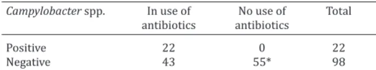
A positive association was observed between Campy-lobacter and concurrent use of antibiotics (Table 4). Pets treated with antimicrobials were 57.41 times more likely to test positive in faeces compared with untreated pets (Table 4). The previous use of antimicrobials may reduce the nat-ural microflora of the gut and favours the colonisation and multiplication of Campylobacter after infection. Antimicro-bial use in dogs and cats should be evaluated. In healthy hu-mans the disease is self-limiting and in most cases it does not need treatment. In dogs and cats very little is known about the pathogenesis and disease progression. Thus, it would be interesting knowledge of isolated by antimicro-bial resistance test.
ant and can be used as a reference in the study of the mech-anisms of pathogenicity of C. jejuni.
The flaA, cadF, pldA and CiaB genes encode proteins involved in adhesion and invasiveness of C. jejuni. The flaA gene is required for the adhesion and invasion of epi-thelial cells (Wassenaar et al. 1991), and the ciaB gene en-codes a protein involved in cell invasion (Rivera-Amill et al. 2001). The cadF gene encodes a protein that interacts with fibronectin in the extracellular matrix of the host cell participating in the colonisation (Monteville et al. 2003). The Plda gene is related to cellular invasion and encodes a protein involved in the synthesis of outer membrane phos-pholipase (Ziprin et al. 2001). These results show that the genes studied in this work help identify virulent strains of C. jejuni in animals and humans.
CONCLUSIONS
This study shows high antimicrobial resistance in strains of Campylobacyter jejuni and C. coli isolated from children and pets.
It also shows that the presence of Campylobacter spp. is associated with diarrhoea in animals, which may be asso-ciated with the presence of virulence genes. In addition, an-timicrobial therapy is associated with increased likelihood of positive Campylobacter spp. in the faeces of pets.
Conflict of interest statement.- None of the authors has any financial or personal relationships that could inappropriately influence or bias the
content of the paper.
Acknowledgements.- This study was financed by the “Fundação de Am -paro a Pesquisa de Minas Gerais” (FAPEMIG) (Minas Gerais Research
Sup-port Foundation) and “Conselho Nacional de Desenvolvimento Científico
e Tecnológico” (CNPq) (Federal Brazilian Research Support Foundation).
REFERENCES
Acke E., Whyte P., Jones B.R., McGill K., Collins J.D. & Fanning S. 2006. Pre-valence of thermophilic Campylobacter species in cats and dogs in two animal shelters in Ireland. Vet. Rec. 158(2):51-54.
Aquino M.H., Filgueiras A.L., Ferreira M.C., Oliveira S.S., Bastos M.C. & Ti-bana A. 2002. Antimicrobial resistance and plasmid profiles of Campy-lobacter jejuni and Campylobacter coli from human and animal sources. Lett. Appl. Microb. 34(2):149-153.
Baker J., Barton M.D. & Lanser J. 1999. Campylobacter species incats and dog in South Australia. Aust. Vet. J. 77(10):662-666.
Blaser M.J. 1997. Epidemiologic and clinical features of Campylobacter je-juni infections. J. Infect. Dis. 176(2):103-105.
Burnens A.P., Wick B.A. & Nicolet J. 1992. Comparison of Campylobacter
carriage rates in diarrheic and healthy pet animals. J. Vet. Med. 39:175-180.
Chaban B., Ngeleka M. & Hill J.E. 2010. Detection and quantification of 14
Campylobacter species in pet dogs reveals an increase in species rich-ness in feces of diarrheic animals. BMC Microbiol. 10:73.
Damborg P., Olsen K.E.P., Nielsen E.M. & Guardabassi L. 2004. Occurrence of Campylobacter jejuni in pets living with human patients infected with
C. jejuni. J. Clin. Microbiol. 42(3):1363-1364.
De Wit M.A.S., Koopmans M.P.G., Kortbeek L.M., Van Leeuwen N.J., Bartelds A.I.M. & Van Duynhoven Y.T.H.P. 2001. Gastroenteritis in sentinel general practices, the Netherlands. Emerg. Infect. Dis. 7:82-91.
EFSA - European Food Safet Authority 2009. The Community Summary Report on trends and sources of zoonoses and zoonotic agents in the European Union in 2007. EFSA Journal 223:17-18.
Table 4. Association between antibiotic use and presence of
Campylobacter spp. in animal feces pets treated at Veterinary Hospital
Campylobacter spp. In use of No use of Total
antibiotics antibiotics
Positive 22 0 22
Negative 43 55* 98
p<0.05; OR=57.41; * asymptomatic dogs.
Table 5. Association between the presence of virulence genes in Campylobacter jejuni and the occurrence of
diarrhea in children under 5 (five) years treated at Clinics
Hospital and pets treated at Veterinary Hospital
Virulence genes Diarrhea Asymptomatic Total
Presence of genes 12 0 12
No presence of genes 5 2 7
p<0.05; OR=11.36.
import-EFSA - European Food Safety Authority 2010. Analysis of the baseline survey on the prevalence of Campylobacter in broiler batches and of Campylobacter and Salmonella on broiler carcasses in the EU, 2008. EFSA Journal 8(3):1503.
Engberg J., Nachamkin I., Fussing V., McKhann G.M., Griffin J.W., Piffaretti
J.C., Nielsen E.M. & Gerner Smidt P. 2001. Absence of clonality of Campy-lobacter jejuni in serotypes other than HS:19 associated with Guil-lain-Barré syndrome and gastroenteritis. J. Infect. Dis. 184(2):215-220.
Gupta A., Nelson J.M., Barrett T.J., Tauxe R.V., Rossiter S.P., Friedman C.R.,
Joyce K.W., Smith K.E., Jones T.F., Hawkins M.A., Shif-Eraw B., Beebe J.L., Vugia D.J., Rabatsky-Her T., Benson J.A., Root T.P. & Ângulo F.J. 2004. Anti-microbial resistance among Campylobacter strains, United States, 1997-2001. Emerg. Infect. Dis. 10:1102-1109.
Hänel I., Muller J., Muller W. & Schulze E. 2004. Correlation between inva-sion of Caco-2 eukaryotic cells and colonization ability in the chick gut in Campylobacter jejuni. Vet. Microbiol. 101:75-82.
Harmon K.M., Ramsom G.M., Wesley I.V. 1997. Differentiation of Campy-lobacter jejuni and Campylobacter coli by polymerase chain reaction. Mol. Cell Probes 11:195-200.
Houf K., De Smet S., Baré J. & Daminet S. 2008. Dogs as carries of the emerging pathogen Arcobacter. Vet. Microbiol. 130(1/2):208-213. Lengerh A., Moges F., Unakal C. & Anagaw B. 2013. Prevalence, associated
risk factors and antimicrobial susceptibility pattern of Campylobacter
species among under five diarrheic children at Gondar University Hos -pital, Northwest Ethiopia. BMC Pediatr. 21(13):82.
Lopez C., Agostini A., Giacoboni G., Cornero F., Tellechea D. & Trinidad J.J. 2003. Campylobacteriosis in a low income community in Buenos Aires, Argentina. Revta Scient. Technol. 22(3):1013-1020.
Kuana S.L., Santos L.R., Rodrigues L.B., Borsoi A., Moraes H.L., Salle C.T.P. & Nascimento V.P. 2008. Antimicrobial resistance in Campylobacter spp
isolated from broiler flocks. Braz. J. Microbiol. 39:738-740.
Mangia A.H., Duarte R., Silva L.A., Bravo V.L. & Leal M.C. 1993. Aetiology of acute diarrhea in hospitalized children in Rio de Janeiro City, Brazil. J. Trop. Pediatr. 39(6):365-367.
Mendes E.N., Queiroz D.M.M., Cisalpino E.O., Peres J.N., Penna F.J. & Figuei-redo Filho P.P. 1987. Ocorrência de Campylobacter jejuni em crianças com e sem diarréia em Belo Horizonte. Revta Microbiol. 18:25-30. Modolo J.R., Giuffrida R. & Lopes C.A.M. 2003. Antimicrobial susceptibility
of 51 Campylobacter strains isolated from diarrheic and diarrhea-free dogs. Arqs Inst. Biológico, São Paulo, 70(3):283-286.
Monteville M.R., Yoon J.E. & Konkel M.E. 2003. Maximal adherence and
invasion of INT 407 cells by Campylobacter jejuni requires the CadF
outer membrane protein and microfilament reorganization. Microbiol.
149:153-165.
Moore J.E., Corcoran J.E., Dooley J.S.G., Fanning S., Lucey B., Matsuda M., McDowell D.A., Megraud F., Millar B.C., O’Mahony R., O’Riordan L., O’Rourke M., Rao J.R., Rooney P.J., Sails A. & Whyte P. 2005. Campylobac-ter. Vet. Res. 36(3):351-382.
Moser I., Rieksneuwohner B., Lentzsch P., Schwerk P. & Wieler L.H. 2001. Genomic heterogeneity and O antigenic diversity of Campylobacter up-saliensis and Campylobacter helveticus strains isolated from dogs and cats in Germany. J. Clin. Microbiol. 39:2548-2557.
NARMS 2007. Enteric Bacteria: executive report. National Antimicrobial
Resistance Monitoring System, Rockville. 99p. <http://www.fda.gov=A- nimalVeterinary=SafetyHealth=AntimicrobialResistance=NationalAnt-imicrbia ResistanceMonitoringSystem=ucm164662.htm> Accessed Jan. 20, 2010.
Palma D., Oliva C.A., Taddei J.A. & Fagundes-Neto U. 1997. Acute diarrhea: stoolwater loss in hospitalized infants and its correlation with etiologic agents and lactose content in the diet. Arq. Gastroenterol. 34:86-195. Parsons B.N., Williams N.J., Pinchbeck G.L., Christley R.M., Hart C.A.,
Gaskell R.M. & Dawson S. 2011. Prevalence and shedding patterns of
Campylobacter spp. in longitudinal studies of kennelled dogs. Vet. J. 190(2):249-254.
Rivera-Amill V., Kim B.J., Seshu J. & Konkel M.E. 2001. Secretion of the viru-lence associated Campylobacter invasion antigens from Campylobacter jejuni requires a stimulatory signal. J. Infect. Dis. 183:1607-1616. Rossi M., Hänninen M.L., Revez J., Hannula M. & Zanoni R.G. 2008.
Occur-rence and species level diagnostics of Campylobacter spp., enteric Heli-cobacter spp. and Anaerobiospirillum spp. in healthy and diarrheic dogs and cats. Vet. Microbiol. 129(3/4):304-314.
Rozynek E., Dzierzanowska-Fangrat K., Korsak D., Konieczny P., Wardak S., Szych J., Jarosz M. & Dzierzanowska D. 2008. Comparison of antimicro-bial resistance of Campylobacter jejuni and Campylobacter coli isolated from humans and chicken carcasses in Poland. J. Food Prot. 71:602-607. Salihu M.D., Magaji A.A., Abdulkadir J.U. & Kolawale A. 2010. Survey of
thermophilic Campylobacter species in cats and dogs in north-western Nigeria. Vet. Ital. 46(4):425-430.
Steinhauserova I., Fojtikova K. & Klimes J. 2000. The incidence and PCR detection of Campylobacter upsaliensis in dogs and cats. Lett. Appl. Mi-crobiol. 31:209-212.
Silva N., Junqueira V.C.A. & Silveira N.F.A. 2007. Campylobacter, p.201-211. In: Ibid. (Eds), Manual de Métodos de Análise Microbiológica de Alimen-tos. 3ª ed. Varela, São Paulo.
Tsai H.J., Huang H.C., Lin C.M., Lien Y.Y. & Chou C.H. 2007. Salmonellae and Campylobacters in household and stray dogs in northern Taiwan. Vet. Res. Commun. 31(8):931-939.
Wassenaar T.M., Bleumink-Pluym N.M. & Van der Zeijst B.A. 1991. Inac-tivation of Campylobacter jejuni flagellin genes by homologous recom -bination demonstrates that FlaA but not FlaB is required for invasion. EMBO J. 10:2055-2061.
Wolf T.F., Duim B., Geelen S.P., Rigter A., Thomson-Carter F., Fleer A. & Wa-genaar J.A. 2001. Neonatal sepsis by Campylobacter jejuni: genetically proven transmission from a household puppy. Clin. Infect. Dis. 32(5):97-99.
Yates J. 2005. Traveler’s diarrhea. Am. Fam. Phys. 71:2095-2100.
Yu R.K., Usuki S. & Ariga T. 2006. Ganglioside molecular mimicry and its pathological roles in Guillain-Barre syndrome and related diseases. Infect. Immun. 74:6517–6527.
Zheng J., Meng J.H., Zhao S.H., Singh R. & Song W.X. 2006. Adherence to and invasion of human intestinal epithelial cells by Campylobacter jejuni
and Campylobacter coli isolates from retail meat products. J. Food Prot. 69:768-774.