1. Unidade de Reumatologia do Hospital Pediátrico Carmona da Mota – Coimbra)
Goldbloom’s Syndrome – a case report
ACTA REUMATOL PORT. 2013;38:51-55
abstRaCt
The Goldbloom’s syndrome (GS) is a rare clinical con-dition of unknown aetiology, occurring exclusively in the pediatric population. It consists in an idiopathic periosteal hyperostosis with dysproteinemia, whose symptoms can mimic a neoplastic disease. We present a case report illustrating the diagnostic challenge of this condition. The exclusion of the common causes of bone pain, associated with generalized periostitis and in-creased gammaglobulins suggested the diagnosis of GS. The self-limited symptoms, the resolution of radiolo-gical findings in four months and the normalization of laboratory abnormalities within ten months, allowed to establish definitely the diagnosis of GS. GS must be considered when diffuse bone pain, prolonged fever and weight loss are present after exclusion of malignant disease with bone involvement.
Keywords: Goldbloom’s Syndrome; Difuse Periostitis;
Bone Pain; Dysproteinemia; Child.
IntRoduCtIon
The Goldbloom’s syndrome (GS) is an idiopathic periosteal hyperostosis associated with
dysproteine-mia1-5.This is a rare clinical condition affecting only the
pediatric population, without gender preference, with
limited number of cases reported in the literature1-5.
The clinical, laboratorial and radiological recovery is complete with only supportive care but may last
months to years1-5.
We describe the case of a two-year-old girl with fe-ver, bone pain and weight loss, whose investigation and follow-up led to the diagnosis of GS. This is the only case of GS described in the Portuguese literature. Taken this into consideration, the authors present a brief des-cription of some cases of GS published.
Case RepoRt
A two and a half year old girl was first admitted to a lo-cal community hospital with one month history of pain localized to the limbs’ diaphysis, refusal to walk, inter-mittent fever and weight loss (two kilograms). The pain was described as occurring all day long and over night, reaching the upper and lower limbs, making it impos-sible for her to go up and down stairs and even to hold a spoon. The progressive clinical deterioration led to the inability to walk. Additionally, she had a daily peak of fever (maximum 38 °C), anorexia, fatigue, cutaneous pallor, night sweats, as well as occasional headache, with photo and phonophobia. There were no respira-tory, cardiac, gastrointestinal or urinary symptoms.
The past medical history was unremarkable and the stature-weight growth was in the fifth percentile. About a month before the beginning of the symptoms, she had a febrile suppurative otitis, treated with amoxicillin with clavulanic acid. The family history was irrelevant. Complementary investigation showed: normochromic normocytic anaemia (haemoglobin 10,3 g/dl), normal peripheral blood smear, thrombocytosis (798,000/ /mcL); erythrocyte sedimentation rate (ESR) of 102 mm/1st hour and C-reactive protein (CRP) of 9,4 mg/dl. The values of lactate dehydrogenase, alkaline phosphatase, uric acid and serum ferritin were within normal limits. Blood cultures and Mantoux test (two UI) were negative and serological tests for cytomegalo-virus and toxoplasmosis did not show acute infection. The anti-nuclear antibodies, ENA screen and anti-DNA antibodies and fractions of the C3 and C4 complement were also negative.
On the third day of hospitalization (D3), tempera-ture reached values of 39°C associated with periods of either irritability or prostration. An abdominal ultra-sound, a chest X-ray and a dorsal-lumbar magnetic re-sonance (MRI) were performed and were considered normal. At D9, she was transferred to the Central Hos-pital. The physical examination revealed a “sick look”, cutaneous pallor and intense pain on palpation of the limbs’ diaphysis. The echocardiogram was normal. Ho-Sónia Santos1, Paula Estanqueiro1, Manuel Salgado1
ta b Le I. C a se s d es CR Ib ed In t h e In te R n a tI o n a L LI te R a tu R e, t o w h IC h w e a d d o u R C a se P ri o r C li n ic al A n al y ti ca l R ad io lo g ic al G en d er A g e in te rc u rr en t m an if es ta ti o n s ch an g es ch an g es E v o lu ti o n G o ld b lo o m e t al 1 ♂ 1 4 y ea rs S ca rl et f ev er -B o n e p ai n S L , IL , ja w -A n em ia N N P er io st ea l re ac ti o n , L ab o ra to ri al a n d -F ev er -In cr ea se d E S R u ln a, s h in b o n e, f em u r, ra d io lo g ic al -W ei g h t lo ss -In cr ea d ed d y sp ro te in em ia : m an d ib le , h u m er u s, n o rm al iz at io n 5 .5 h y p o al b u m in em ia , fi b u la , m et at ar su s y ea rs l at er α an d γ g lo b u li n s an d m et ac ar p u s ♀ 1 0 y ea rs T o n si ll it is -B o n e p ai n I L -A n em ia P er io st ea l re ac ti o n C li n ic al r es o lu ti o n -F ev er -In cr ea se d d y sp ro te in em ia : u ln a, f em u r, f ib u la in t w o w ee k s h y p o al b u m in em ia , α 2 a n d an d s h in b o n e γ g lo b u li n s C am er o n e t al 2 ♂ 1 2 y ea rs P h ar y n g it is -A n o re x ia -A n em ia N N P er io st ea l re ac ti o n C li n ic al a n d -F ev er - N o rm al E S R m an d ib le , ra d iu m , ra d io lo g ic al -W ei g h t lo ss -In cr ea se d C R P u ln a, s h in b o n e an d im p ro v em en t; b u t -B o n e p ai n h an d , w ri st , -In cr ea se d h y p o al b u m in em ia , fi b u la a n d p h al an x af te r tw o y ea rs k ep t an k le a n d j aw α 1 ,α 2 a n d γ g lo b u li n s th e d y sp ro te in em ia R o d rí g u ez e t al 3 ♀ 1 3 y ea rs U p p er -E x tr em it ie s p ai n -A n em ia N N P er io st ea l re ac ti o n C o m p le te R es p ir at o ry -W ei g h t lo ss - In cr ea se d E S R u ln as , ra d iu m , ti b ia , re so lu ti o n e le v en In fe ct io n - In cr ea se d h y p o al b u m in em ia , m et at ar su s an d m o n th s la te r γ g lo b u li n m et ac ar p u s G er sc o v ic h e t al 4 ♂ 3 y ea rs U p p er -F o re ar m p ai n -A n em ia P er io st ea l re ac ti o n o f C o m p le te R es p ir at o ry -F ev er - In cr ea se d E S R th e u ln a, r ad iu m a n d re so lu ti o n o n e In fe ct io n - In cr ea se d h y p o al b u m in em ia , fe m u r y ea r la te r α 1 , α 2 a n d γ g lo b u li n s K u w as h im a et a l 5 ♀ 4 m o n th s N S -F ev er -In cr ea se d E S R a n d C R P P er io st ea l re ac ti o n N S -P ai n a n d s w el li n g -In cr ea se d h y p o al b u m in em ia , h u m er u s, u ln a, k n ee , el b o w , w ri st γ g lo b u li n ra d iu m a n d p h al an x an d f in g er s (m ig ra to ry p ai n ) S an to s S e t al ♀ 2 .5 y ea rs F eb ri le -F ev er - A n em ia N N P er io st ea l re ac ti o n -C li n ic al a n d S u p p u ra ti v e -B o n e p ai n s S L a n d I L -In cr ea se d E S R a n d ra d iu m , u ln a, t ib ia , ra d io lo gi ca l re so lu ti o n O ti ti s - W ei g h t lo ss -In cr ea se d α 1 , α 2 a n d γ an d f ib u la in f o u r m o n th s -A n o re x ia g lo b u li n s -A n al y ti ca l re so lu ti o n in t en m o n th s L eg en d : N S N o t st at ed , S L – S u p er io r L im b s, I L – I n fe ri o r L im b s, E S R – E ry th ro cy te S ed im en ta ti o n R at e, C R P C -r ea ct iv e p ro te in ; N N n o rm o ch ro m ic n o rm o cy ti c
wever, X-rays of the long bones (Figures 1a and 2a) and bone scintigraphy (Figure 3) revealed signs of dif-fuse periostitis. Further laboratorial investigation an high total proteins value of 89.3 g/L [Reference value (RV) 54-75 g/L] and protein electrophoresis revealed a reduction in the percentage of albumin 37.1% (N 60-70%) and increased alpha fraction 1 [4.3% (RV 1.4-2.9%)], alpha 2 [17.2% (RV 7-11%)], beta [14.5% (RV 8-13%)] and gammaglobulin [26.9% (RV 9-16%)] by polyclonal hypergammaglobulinemia. The serological screening for syphilis was negative and the bone marrow examination was normal.
The diagnosis of GS was made, she was treated with ibuprofen (10 mg/kg/dose 3 id), being discharged from the hospital four days later. Four months later on fol-low-up she showed complete clinical and radiological remission, but laboratory abnormalities only normali-zed after ten months of disease duration.
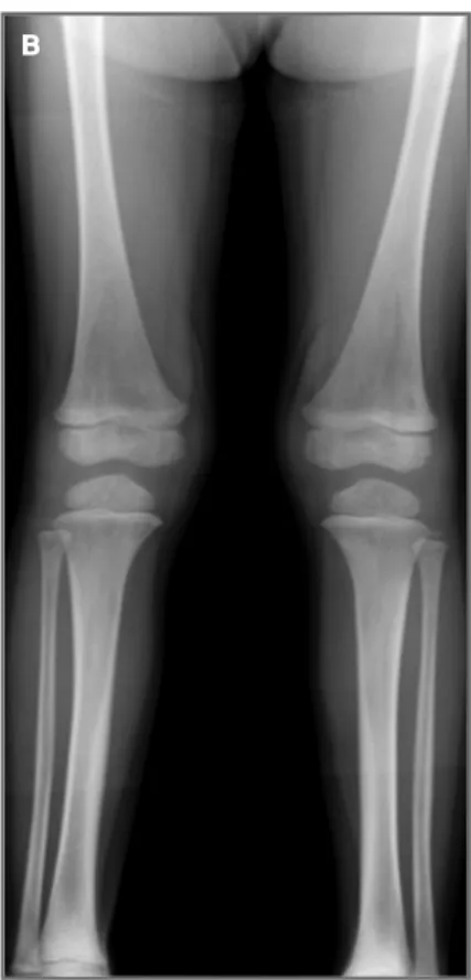
She maintained rheumatological follow-up until she was four years of age with clinical, laboratorial and ra-diological resolution (Figures 1b and 2b) and with a good/regular growth.
dIsCussIon
In 1966, Goldbloom et al. reported two cases of fever and bone pain in a ten and a fourteen year old child, with periostitis and dysproteinemia in both, which de-signated as idiopathic periosteal hyperostosis
associa-ted with dysproteinemia1. Later this entity was
recog-nized in other pediatric patients as Goldbloom’s
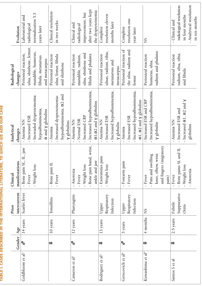
Syn-drome2-5. In Table I are described schematically the
ca-ses published since 1966, to which we add our case. More than 40 years after the description of the first two
cases of GS1, its aetiology remains undetermined2,5. The
virusal aetiology is the most probable, although there are cases reported with evidence of previous strepto-coccal infection (see Table I). In our patient, an infec-tious cause was considered since the symptoms were preceded by a suppurative otitis.
The bone pain often disproportionate to clinical
fin-dings is the most obvious sign1-5. Fever1,2,4,5and weight
loss1-3are common features in GS, and were also
pre-sent in our patient.
Radiologically, the signs of periostitis are typical, af-fecting preferencially long bones, in decreasing order of involvement: radio, femur, humerus, ulna and
ti-bia5. This was demonstrated in our case report
(Figu-re 1a and 2a). The involvement of short tubular bo-nes2,3,5or the mandible1,2is rare. Radiological changes are confined to the periosteum, although plasma cell
infiltration can occur at bone marrow level2.
The differential diagnosis of GS, due to the presen-ce of fever, bone pain and radiological findings of pe-riostitis must be done with lymphoproliferative
disea-se6-11, malignant bone disease6,8 and osteomyelitis4-6
with one or more focus. Therefore a thorough investi-gation should be performed and should reveal a nor-mal white blood cell count, peripheral blood smear
FIGuRe 1a. Signs of periostitis of the upper limb (marked with arrow) in November 2007
a
FIGuRe 1b. Radiological resolution in March 2008
age and absence of relevant personal history or vitamin A chronic ingestion. The pachydermoperiostosis (or Touraine-Solente-Golé Syndrome) is an autosomal do-minant disease characterized by periostitis in addition FIGuRe 2a. Signs of periostitis of the lower limb (marked with
arrow) in November 2007 a
FIGuRe 2b. Radiological resolution in March 2008
b
and bone marrow examination and a negative blood culture. Periostitis is a radiologic manifestation pre-sent in about 1.9% to 35% of acute lymphoblastic
leu-kemia (ALL)11. Thus, the association of periostitis,
bone pain and fever requires malignancy to be
exclu-ded, especially ALL7-11.
No specific laboratory test exists for GS but univer-sal findings include: increased acute phase reactants
(ESR and CRP)1-5, anaemia1-4(mainly normocytic
nor-mochromic), dysproteinemia with hypoalbuminemia, increased gamaglobulins and variable values of alpha
1, alpha 2 and beta globulins1-5. Such laboratorial
abnormalities were found in our patient. These fin-dings, together with the radiological abnormalities and after exclusion of other clinical entities, allowed to evo-ke the diagnosis of GS.
Other differential diagnoses to consider are: Caffey
disease12(child cortical hyperostosis), occurring in
in-fants with preferential involvement of the mandible
but without dysproteinemia4and chronic intoxication
by vitamin A, which may present with bone pain and
radiological abnormalities but without fever13. These
to digital clubbing and coarse facial features4,6, that didn’t exist in this case. Secondary hypertrophic os-teoarthropathy was also excluded since there weren’t signs of cardiac, respiratory or digestive involvement
which could suggest a chronic disease14.
The treatment of the GS is symptomatic and the prognosis is good, with clinical improvement in weeks to several months, although complete laboratorial and
radiological normalization may take years1,2,4,14. Our
patient, after four months on follow-up showed both clinical and radiological recovery. However laborato-ry abnormalities have normalized only after ten months.
The GS is a rare clinical entity and may be under-diagnosed. Therefore it is important to consider GS as a possible diagnosis when symptoms of diffuse bone pain, prolonged fever and weight loss are present, and malignancy with bone involvement or infectious di-seases, such as osteomyelitis, are ruled out.
CoRRespondenCe to
Sónia Alexandra Pinto dos Santos Unidade de Reumatologia
Hospital Pediátrico Carmona da Mota 3000 Coimbra, Portugal
E-mail: soniafmuc@gmail.com
ReFeRenCes
1. Goldbloom RB, Stein PB, Eisen A, McSheffrey JB, Brown BS, Wiglesworth FW. Idiopathic periosteal hyperostosis with dys-proteinemia. A new clinical entity. N Engl J Med 1966; 274: 873-878.
2. Cameron BJ, Laxer RM, Wilmot DM, Greenberg ML, Stein LD. Idiopathic periosteal hyperostosis with dysproteinemia (Gold-bloom’s syndrome): case report and review of the literature. Arthritis Rheum 1987; 30: 1307-1312.
3. Rodríguez JC, Horzella RR, Zolezzi PR. Hiperostosis perióstica idiopática transitória con disproteinemia (síndrome de Gold-bloom). Rev Child Pediatr 1989; 60: 36-39.
4. Gerscovich EO, Greenspan A, Lehman WB. Idiopathic perios-teal hyperostosis with dysproteinemia – Goldbloom’s syndro-me. Pediatr Radiol 1990; 20: 208-211.
5. Kuwashima S, Nishimura G, Harigaya A, Kuwashima M, Ya-mato M, Fujioka M. A young infant with Goldbloom syndro-me. Pediatr Int 1999; 41: 110-112.
6. Mantadakis E, Valsamidis A, Chatzimichael A. A case report and review of clinical and laboratory pointers of leukemia in children with bone pain. IJCRI 2010; 1: 1-6.
7. Guillerman RP, Voss SD, Parker BR. Leukemia and Lymphoma. Radiol Clin N Am 2011; 49: 767-797.
8. Raab CP, Gartner JC. Diagnosis of childhood cancer. Prim Care Clin Office Pract 2004; 36: 671-684.
9. Marwaha RK, Kulkarni KP, Bansal D, Trehan A. Acute lymp-hoblastic leukemia masquerading as juvenile rheumatoid arth-ritis: diagnostic pitfall and association with survival. Ann He-matol 2010; 89:249-254.
10. Gupta D, Singh S, Suri D, Ahluwali J, Das R, Varma N. Arthri-tic presentation of acute leukemia in children: experience from a tertiary care centre in North India. Rheumatol Int 2010; 30:767-770.
11. Tafaghodi F, Aghighi Y, Yazdi HR, Shakiba M, Adibi A. Predic-tive plain X-ray findings in distinguishing early stage acute lymphoblastic leukemia from juvenile idiopathic arthritis. Clin Rheumatol 2009; 28:1253-1258.
12. Kamoun- Goldrat A, le Merrer M. Infantile cortical hyperosto-sis (Caffey disease): a review. J Oral Maxillofac Surg 2008; 66:2145-2150.
13. Eledrisi MS. Vitamin A Toxicity [Internet]. The Emedicine Medscape website [updated 2012 January 3; cited 2012 Fe-bruary 28]. Available from: http://www.emedicine.com. 14. Dhawan R, Ahmed MM, Menard HA. Hypertrophic
osteoarth-ropathy [Internet]. The Emedicine Medscape website [updated 2011 August 23; cited 2012 February 28]. Available from: http://www.emedicine.com.


