r e v b r a s o r t o p . 2015;50(2):239–242
w w w . r b o . o r g . b r
Case
report
Gorham’s
disease:
clinical
case
夽
Pedro
Sá
a,∗,
Pedro
Marques
a,
Carolina
Oliveira
a,
André
Sá
Rodrigues
b,
Nelson
Amorim
b,
Rui
Pinto
baUnidadeLocaldeSaúdeAltoMinho,VianadoCastelo,Portugal
bCentroHospitalarSãoJoão,Porto,Portugal
a
r
t
i
c
l
e
i
n
f
o
Articlehistory: Received25April2014 Accepted28April2014 Availableonline28March2015
Keywords:
Essentialosteolysis/pathology Essentialosteolysis/diagnosis Rarediseases/etiology
a
b
s
t
r
a
c
t
Gorham’sdisease,alsoknownasidiopathicmassiveosteolysis,isararepathological condi-tioncharacterizedbyvascularproliferationthatresultsindestructionandreabsorptionof thebonematrix,ofunknownetiology.ItwasfirstdescribedbyJacksonin1838,butitwas GorhamandStout,in1955,whodefinedthisdiseaseasaspecificentity.Ithasvariable clin-icalpresentationandgenerallyhasprogressivebehavior.Controversycontinuesregarding thetreatmentandthereisnostandardtreatment.Thispathologicalconditiongenerally presentsafavorableprognosis.Here,acaseofGorham’sdiseasewithinvolvementofthe lefthipispresented,inamalepatientwithoutrelevantantecedents.
©2015SociedadeBrasileiradeOrtopediaeTraumatologia.PublishedbyElsevierEditora Ltda.Allrightsreserved.
Doenc¸a
de
Gorham:
caso
clínico
Palavras-chave:
Osteóliseessencial/patologia Osteóliseessencial/diagnóstico Doenc¸asraras/etiologia
r
e
s
u
m
o
Adoenc¸adeGorham,tambémconhecidaporosteólisemacic¸aidiopática,éumapatologia rara,caraterizadaporumaproliferac¸ãovascularqueresultanadestruic¸ãoereabsorc¸ãoda matrizóssea,deetiologiadesconhecida.Foidescritapelaprimeiravezem1838porJackson, masforamGorhameStout,em1955,quedefiniramadoenc¸acomoumaentidadeespecífica. Comumaapresentac¸ãoclinicavariável,geralmentetemumcomportamentoprogressivo.O tratamentopermanececontroverso,nãoháumtratamentopadrão.Essapatologiaapresenta geralmenteumprognósticofavorável.Éapresentadoumcasodedoenc¸adeGorhamcom envolvimentodoquadrilesquerdo,numdoentedosexomasculino,semantecedentesde relevo.
©2015SociedadeBrasileiradeOrtopediaeTraumatologia.PublicadoporElsevierEditora Ltda.Todososdireitosreservados.
夽
WorkdevelopedattheSãoJoãoHospitalCenter,Porto,Portugal. ∗ Correspondingauthor.
E-mail:[email protected](P.Sá).
http://dx.doi.org/10.1016/j.rboe.2015.03.004
240
rev bras ortop.2015;50(2):239–242Introduction
Gorham’sdiseaseconsistsofprogressiveidiopathicosteolysis ofaboneorbonesadjacentandsurroundingafocus,without respectingjointborders.
It can affect any part of the skeleton. The sites most affectedarethecranium,shoulderandpelvis.1
The clinical presentation depends on the site involved and many months or years may pass bybefore it is diag-nosed.Insomecases,thediseaseismanifestedacutely,with incapacitatingpain. Thefirst manifestationsare commonly spontaneousfractures.In other patients,Gorham’s disease presentsaninsidiouscourse,withprogressivemuscle weak-ness.
Thisdiseaseisofprogressivenatureinmostofthepatients andmay,incertaincases,beself-limiting.Thenaturalcourse ofthis pathologicalconditionisgenerallybenign. However, ithascomplicationscomprisingpericardialandpleural effu-sionsand theseare potentially fatalif the mediastinumis reached.
Radiological examinations, especially X-rays, computed tomography(CT)andmagneticresonanceimaging(MRI),in associationwithbonebiopsies,areessentialformakingthe differentialdiagnosisofthisrarepathologicalcondition.
ThemedicaltreatmentofGorham’sdiseaseincludesuse ofradiotherapy,antiosteoclasticdrugs(bisphosphonates)and interferon ␣-2b. The surgical options include resection of thelesion andfilling withautologousor heterologousgraft materialorreconstructionofthejointthrougharthroplastic resources.
Case
report
Thepatientwasa48-year-oldwhitemanwithahistoryofuse ofintravenousdrugs.Hewasseenatanexternalorthopedic consultationwithacomplaintofpaininhisleftthighthathad started5yearsearlier,withprogressivelossofrangeofmotion andthecapacitytowalk,andincreasingneedforexternal sup-portinordertowalk.Hedidnothaveanyprevioushistoryof trauma.
Attheobjectiveexamination,hepresentedpainupon pal-pationofthelefttrochantericregionandalsoduringpassive internaland externalrotation.Itwas observedthat during activemovements,hisflexionofthelefthipwaslimitedto 30◦,internalrotationto10–15◦andexternalrotationto15–20◦. HepresentedaHarrisHipScoreof36.65.
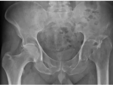
HeunderwentX-rayexaminationwiththepelvisbearing weight(Fig.1),fromwhichapparentbonenecrosisofthe left-sidefemoralheadandneckwasobserved.ACTscanontheleft hip(Fig.2AandB)showedthatcompletebonereabsorption oftheleft-sidefemoralheadandneckhadoccurred.MRIon thelefthip(Fig.3)alsoshowedcompletebonereabsorption oftheproximalfemur,alongwiththepresenceofavascular “mass”inthejointspaceandinvasionofthefossaandthe upperwalloftheleftacetabulumandwingoftheipsilateral ilium.Bonescintigraphywasalsoperformedandthisrevealed destructionoftheleftcoxofemoraljoint,withlatefixationof theradiopharmaceutical.
Fig.1–X-rayofthepelvis,withweightbearing(APview).
Abiopsywasperformed,directedbymeansofCTonthe lefthip.Theanatomopathologicalresultwascompatiblewith Gorham’s disease and revealed the presence of dispersed small-calibervessels.
Thepatientiscurrentlybeingmonitoredasan orthope-dicsoutpatientandtotalarthroplastyofthelefthiphasbeen proposed.
Discussion
Gorham’sdisease,alsoknownasGorham–Stoutsyndrome,is arareidiopathicdiseasethatischaracterizedbyextensiveloss ofbonematrix,whichisreplacedbyfibrotictissueand prolife-rativevascularandlymphaticcanals.Approximately200cases havebeenreportedintheliterature.2Itwasfirstdescribedby
Jacksonin1838,3,4inacaseofanadultmaleinwhom
com-pletehumeralbonereabsorptionwasobservedoveran11-year period.In1955,GorhamandStout5definedthispathological
conditionasaspecificentitythroughpublishingareviewof 24casesofthedisease.Theyreportedthat“Gorham’sdisease isgenerallyassociatedwithangiomatosisofbloodandlymph vessels.
Most of the cases occur inchildren or in adults under the age of40years.Approximately 60% ofthe casesoccur among male patients and there is no apparent genetic predisposition.6Theshoulder7,8andthepelvis9arethe
loca-tionsmostaffectedbythissyndrome,althoughitmayaffect anybone.10Whenitaffectstheribs,scapulaorthoracicspine,
it mayleadtotheappearanceofchylothorax,duetodirect invasion oflymphangiectasia into pleuralcavity or via the thoracic duct.11 In these cases, surgical drainage needs to
beperformed. If this isnotdone,there are highmorbidity andmortalityrates.11Nonetheless,thissyndromegenerally
presentsbenignevolution.
Gorham’s disease is ofprogressive nature, but insome casesitpresentsasaself-limitingpathologicalcondition.Its etiologyandpathophysiologyremainunknown.The patholog-icalprocessischaracterizedbyreplacementofnormalbone bynon-neoplasticvasculartissueofaggressivebehavior,10,12
rev bras ortop.2015;50(2):239–242
241
Fig.2–(A)CTscanofthepelvis(axialslice)and(B)CTscanofthepelvis(coronalslice).
stageofthelesion,theboneisreabsorbedand replacedby hypervascularand/orangiomatousfibrousconnectivetissue. Histologically,thin-walledvesselsareseentobepresent,and thesemaybecapillary,sinusoidalorcavernous.Atlaterstages, progressivebonedestructiontakesplace,withtheappearance offibrotictissueandmassiveosteolysis.13
Theexactnatureofthepathologicalprocessisunknown. Gorhamand Stout5stated thathyperemia,localalterations
ofpHandmechanicalforceswere responsibleforthe bone absorptionandrulesoutanyroleforosteoclasts.Devlinetal.13
suggestedthattheosteolysisthatispresentinGorham’s dis-easewasduetoincreasedactivitybyosteoclasts,mediatedby increasedlevelsofinterleukin-6(IL-6).Molleretal.14published
sixcasesofGorham’sdisease,inwhichthehistopathological resultsshowedthattherewasanincreasednumberof osteo-clasts.Hirayamaetal.15 suggestedthatthe increaseinthe
numberofosteoclastswasduetogreater sensitivityofthe precursorsforhumoralfactors,whichpromotesformationof osteoclastsandbonereabsorption.
Theclinicalmanifestationsarevariableanddependonthe siteaffected.Somepatientspresentasudden starttopain and edemaintheextremity affected,whileothers present aninsidiousstarttothepain,withlimitationofmovements andprogressivelossofmusclestrength.Despitethesevere bonedeformities,seriouscomplicationsarerare.Paraplegia may occur whenvertebrae are involved.16 When the chest
andmediastinumareinvolved,pleuraleffusionmaydevelop,
Fig.3–MRIofthepelvis(coronalslice).
withconsequentrespiratoryfailure,whichmayhaveafatal outcome.17
Theroutine laboratoryanalysesare not usefulfor diag-nosing Gorham’sdisease.Itispossiblethatonlythe serum alkalinephosphatase levelswillbeelevated. However, rou-tine radiological examinationssuchasX-rays, CTand MRI areusefulforthediagnosis.Themostdramaticappearance of massive osteolysis is seen on X-ray images. Resnick18
describedtheradiologicalfindingsfromGorham’sdisease.In theinitialstageofthelesion,radiolucentfociappearinthe subcorticalandintramedullaryregion,similartoosteoporosis. Progressivedissolution,fragmentationanddisappearanceof theboneportionaffectedareseen.Theremainderofthebone tissueformsshardsandisaccompaniedbysoft-tissue atro-phy.Thediseasemayextendtocontiguousbonesanddoes notrespectthejointsurfaceasabarrier.Scintigraphyshows increasedlevelsofvascularizationintheearlystagesandan areaoflowuptakecorrespondingtothelocationofabsenceof bonetissueatlaterstages.19
ThediagnosisofGorham’sdiseasecanonlybeestablished afterother,morecommoncausesofosteolysishavebeenruled out, such as infection, cancer (primary or metastatic) and endocrineandinflammatorydiseases.Biopsies(open, echo-guidedorCT-guided)contributetowardthisandestablishthe definitivediagnosis.
Becauseoftherarityofthispathologicalcondition,there is no standard treatment for it. The medical treatment includes radiotherapy,20 bisphosphonates21 and interferon
␣-2b.22Radiotherapyatmoderatedoses(40–45Gyintwo frac-tions)presentsgoodclinicalresults,withalowcomplication rate.21Surgicaltreatment,whichisperhapsthemainmethod,
includesresectionofthelesionandbonereconstructionusing a bonegraft,orarthroplasty. Chylothorax isthe most seri-ous complication, and this occurs when Gorham’s disease affectsthechest.Thiscomplicationispotentiallylife threat-ening.Thesurgicalproceduresreservedforthesesituations includepleurectomy,pleurodesisanddeviationtothethoracic duct.23,24Inthesecases,radiotherapyisreservedforpatients
whocannottolerateinvasiveprocedures.
Conclusion
242
rev bras ortop.2015;50(2):239–242disintegratesand isreplacedbyvascularfibrous connective tissue.Itsetiologyisspeculative.Theclinicalpresentationis veryvariable andthe naturalhistoryofthedisease hasan unpredictableprognosis.Noeffectivetherapyhasbeen estab-lished.Mostofthepatientsaretreatedwithradiotherapyand surgicalprocedures.
Conflicts
of
interest
Theauthorsdeclarenoconflictsofinterest.
r
e
f
e
r
e
n
c
e
s
1. FlörchingerA,BöttgerE,Claass-BöttgerF,GeorgiM,HarmsJ. Gorham–Stoutsyndromeofthespine.Casereportandreview oftheliterature.Rofo.1998;168(1):68–76.
2. PapadakisSA,KhaldiL,BabourdaEC,PapadakisS,Mitsitsikas T,SapkasG.Vanishingbonedisease:reviewandcasereports. Orthopedics.2008;31(3):278.
3. JacksonJB.Abonelessarm.BostonMedSurgJ.1838;18:368–9. 4. JacksonJB.Absorptionofthehumerusafterfracture.Boston
MedSurgJ.1872;10:245–7.
5. GorhamLW,StoutAP.Massiveosteolysis(acutespontaneous absorptionofbone,phantombone,disappearingbone);its relationtohemangiomatosis.JBoneJointSurgAm. 1955;37(5):985–1004.
6. HardeggerF,SimpsonLA,SegmuellerG.Thesyndromeof idiopathicosteolysis.Classification,review,andcasereport.J BoneJointSurgBr.1985;67(1):88–93.
7. RemiaLF,RicholtJ,BuckleyKM,DonovanMJ,GebhardtMC. Painandweaknessoftheshoulderina16-year-oldboy.Clin OrthopRelatRes.1998;(347):268–71.
8. Bode-LesniewskaB,vonHochstetterA,ExnerGU,HodlerJ. Gorham–Stoutdiseaseoftheshouldergirdleand
cervico-thoracicspine:fatalcourseina65-year-oldwoman. SkeletRadiol.2002;31(12):724–9.
9. KulenkampffHA,RichterGM,HasseWE,AdlerCP.Massive pelvicosteolysisintheGorham–Stoutsyndrome.IntOrthop. 1990;14(4):361–6.
10.DicksonGR,HamiltonA,HayesD,CarrKE,DavisR,Mollan RA.Aninvestigationofvanishingbonedisease.Bone. 1990;11(3):205–10.
11.TieML,PolandGA,RosenowEC3rd.ChylothoraxinGorham’s syndrome.Acommoncomplicationofararedisease.Chest. 1994;105(1):208–13.
12.PazzagliaUE,AndriniL,BonatoM,LeutnerM.Pathologyof disappearingbonedisease:acasereportwith
immunohistochemicalstudy.IntOrthop.1997;21(5):303–7. 13.DevlinRD,BoneHG3rd,RoodmanGD.Interleukin-6:a
potentialmediatorofthemassiveosteolysisinpatientswith Gorham–Stoutdisease.JClinEndocrinolMetab.
1996;81(5):1893–7.
14.MöllerG,PriemelM,AmlingM,WernerM,KuhlmeyAS, DellingG.TheGorham–Stoutsyndrome(Gorham’smassive osteolysis).Areportofsixcaseswithhistopathological findings.JBoneJointSurgBr.1999;81(3):501–6.
15.HirayamaT,SabokbarA,ItonagaI,Watt-SmithS,Athanasou NA.Cellularandhumoralmechanismsofosteoclast formationandboneresorptioninGorham–Stoutdisease.J Pathol.2001;195(5):624–30.
16.HallidayDR,DahlinDC,PughDG,YoungHH.Massive osteolysisandangiomatosis.Radiology.1964;82:637–44. 17.KeryL,WoutersHW.Massiveosteolysis.Reportoftwocases.J
BoneJointSurgBr.1970;52(3):452–9.
18.ResnickD.Osteolysisandchondrolysis.In:ResnickD,editor. Diagnosisofboneandjointdisorders.4thed.Philadelphia: Saunders;2002.p.4920–44.
19.SpiethME,GreenspanA,ForresterDM,AnsariAN,KimuraRL, Gleason-JordanI.Gorham’sdiseaseoftheradius:
radiographic,scintigraphic,andMRIfindingswithpathologic correlation.Acasereportandreviewoftheliterature.Skelet Radiol.1997;26(11):659–63.
20.DunbarSF,RosenbergA,MankinH,RosenthalD,SuitHD. Gorham’smassiveosteolysis:theroleofradiationtherapy andareviewoftheliterature.IntJRadiatOncolBiolPhys. 1993;26(3):491–7.
21.HammerF,KennW,WesselmannU,HofbauerLC,DellingG, AllolioB,etal.Gorham–Stoutdisease–stabilizationduring bisphosphonatetreatment.JBoneMinerRes.
2005;20(2):350–3.
22.HagbergH,LambergK,AströmG.Alpha-2binterferonand oralclodronateforGorham’sdisease.Lancet.
1997;350(9094):1822–3.
23.PedicelliG,MattiaP,ZorzoliAA,SorroneA,DeMartinoF, SciottoV.Gorhamsyndrome.JAMA.1984;252(11):1449–51. 24.FeiglD,SeidelL,MarmorA.Gorham’sdiseaseoftheclavicle