UNIVERSIDADE DA BEIRA INTERIOR
Ciências da Saúde
Novel transcriptional regulation mechanisms
associated with Parkinson’s disease pathogenesis:
from biological activity towards therapy
Jéssica Lopes Nunes
Dissertação para obtenção do Grau de Mestre em
Ciências Biomédicas
(2º ciclo de estudos)
Orientador: Prof. Doutora Liliana Bernardino
iii
Agradecimentos
Após o término desta etapa, quero agradecer a todos que me apoiaram ao longo deste período e que contribuíram para que esta fase da minha vida fosse possível.
Em primeiro lugar, quero agradecer à minha orientadora, Professora Doutora Liliana Bernardino, pela oportunidade de desenvolver este trabalho, pela constante disponibilidade, pela paciência, por todos os conhecimentos científicos transmitidos e pelas críticas que me ajudaram a melhorar enquanto investigadora.
Também gostava de agradecer aos meus colegas de laboratório por estarem sempre disponíveis, pela ajuda e pelos bons momentos partilhados.
Aos meus pais, pelo amor incondicional, por apoiarem todas as minhas decisões e por fazerem tudo o que podem para que os meus sonhos se possam realizar.
Agradeço imenso também ao André, pela ajuda nos momentos difíceis, pelo amor, amizade e por estar sempre presente quando mais preciso.
A todos os meus amigos quero agradecer pela amizade, pela simpatia e pelo companheirismo. Diana e Ana, obrigada pela vossa amizade ao longo de todos estes anos. Catarina Chendo, Margarida e Catarina Duarte, obrigada por toda a companhia e por todos os momentos que passámos juntas ao longo deste ano.
E por último, mas não menos importante, quero agradecer, ao resto da minha família por todo o carinho e apoio, especialmente aos meus avós, pelo incentivo e pelo orgulho constante.
v
Resumo
Evidências sugerem um papel epigenético associado à progressão de doenças neurodegenerativas, tais como a doença de Parkinson (PD, do inglês Parkinson’s disease). As proteínas de ligação C-terminal (CtBPs, do inglês C-terminal binding proteins) são co-repressores transcripcionais, que atuam, essencialmente, através do recrutamento de um complexo co-repressor ao ADN. Alguns estudos demonstraram uma função importante para as CtBPs na repressão da transcrição de genes pró-apoptóticos, demonstrando ser um bom alvo terapêutico em doenças neurodegenerativas. Neste trabalho, explorámos a expressão proteica das CtBPs em modelos in vitro e in vivo da PD através de western-blot e o seu efeito na sobrevivência dopaminérgica através de ensaios de MTT (in vitro) ou por contagem do número de células que expressam tirosina hidroxilase (TH) (in vivo). Em primeiro lugar, verificou-se um aumento de expressão de CtBP1 na substantia nigra (SN) nos animais injetados com 6-hidroxidopamina (6-OHDA) ou 1-metil-4-fenil-1,2,3,6-tetrahidropiridina (MPTP), no entanto no estriado (ST, do inglês striatum) apenas se verificou uma diferença estatisticamente significativa nos animais injetados com 6-OHDA, quando comparados com os animais salinos. Os níveis de expressão da CtBP2 na SN e no ST aumentaram após a injeção da 6-OHDA, no entanto não de uma forma estatisticamente significativa quando comparados com os resultados obtidos nas mesmas regiões em animais salinos. Concordantemente, tanto a expressão da CtBP1 como da CtBP2 aumentou numa linha neural dopaminérgica do mesencéfalo de ratos (N27) exposta à toxina 6-OHDA. Seguidamente, utilizou-se um antagonista das CtBPs, o ácido 4-metiltio-2-oxobutírico (MTOB), para determinar o efeito putativo das CtBPs na sobrevivência neuronal. O MTOB, a concentrações relativamente elevadas, foi capaz de inibir a sobrevivência neuronal das células N27 e a sobrevivência dopaminérgica na SN do modelo animal induzido por 6-OHDA. Adicionalmente, o MTOB foi
capaz de potenciar a morte celular induzida por 1-metil-4-fenilpiridinio (MPP+).
Curiosamente, baixas concentrações de MTOB (250µM) foram capazes de contrariar a morte celular induzida pela 6-OHDA nos modelos da PD in vitro e in vivo. Concluindo, os nossos resultados sugerem que as CtBPs são um bom alvo para se estudar mecanismos de regulação transcripcional na modulação da sobrevivência dopaminérgica.
Palavras-chave
Doença de Parkinson, Proteínas de ligação C-terminal, 6-OHDA, MTOB, sobrevivência dopaminérgica
vii
Resumo Alargado
A doença de Parkinson (PD, do inglês Parkinson’s disease) é uma doença caracterizada pela degeneração de neurónios dopaminérgicos, presentes na substantia nigra (SN) e das suas fibras que se projetam até ao estriado (ST, do inglês striatum). Vários são os fatores responsáveis pela patogénese desta doença, como por exemplo o stress oxidativo, toxinas, neuroinflamação e ainda alguns fatores genéticos. Também tem sido sugerido que alguns fatores epigenéticos possam estar associados à progressão desta doença como, por exemplo, desacitalações e metilações. As proteínas de ligação C-terminal (CtBPs, do inglês C-teminal
binding proteins) são co-repressores capazes de atuar, essencialmente, via recrutamento de
um complexo co-repressor ao ADN, no entanto outros estudos também sugerem que estas conseguem estar associadas a ativação transcripcional. Alguns autores atribuíram uma função importante para as CtBPs na repressão da transcrição de genes pró-apoptóticos, demonstrando que estas proteínas podem ser um alvo terapêutico promissor para doenças neurodegenerativas.
Neste trabalho, explorámos o efeito das CtBPs em modelos in vitro e in vivo da PD e o seu efeito na sobrevivência dopaminérgica. Para isso, utilizámos uma linha neural dopaminérgica imortalizada do mesencéfalo de ratos (N27) que tratámos com duas toxinas, a
6-hidroxidopamina (6-OHDA; a 25 e 50 µM) e o 1-metil-4-fenilpiridinio (MPP+; a 30µM e 1mM) e
de seguida analisámos a expressão proteica das CtBPs nessas condições. Também se analisou a expressão das CtBPs em modelos de PD in vivo, através da injeção das toxinas 6-OHDA e 1-metil-4-penil-1,2,3,6-tetrahidropiridina (MPTP), bem como ao longo do envelhecimento, tendo em conta que este é um fator de risco para a patogénese da PD. Relativamente aos resultados in vitro, verificou-se um aumento significativo de expressão da CtBP1 e CtBP2 em células tratadas com 50µM 6-OHDA. Quanto aos resultados in vivo, observou-se um aumento de expressão de CtBP1 na SN dos animais injetados com 6-OHDA ou MPTP, no entanto no ST apenas se verificou diferença estatisticamente significativa nos animais injetados com 6-OHDA, quando comparado com os animais salinos. Os níveis de expressão da CtBP2 na SN e ST aumentaram após a injeção da 6-OHDA, embora não de uma forma estatisticamente significativa quando comparados com as mesmas regiões de animais salinos. Verificou-se ainda que, de uma forma geral, ocorre um aumento de expressão de CtBPs ao longo da idade, exceto na expressão da CtBP1 na SN dos animais com 26 meses. Para analisar o efeito das CtBPs na sobrevivência dopaminérgica, inicialmente, efetuou-se a técnica de MTT em células N27 expostas a diferentes concentrações de ácido 4-metiltio-2-oxobutírico (MTOB; a 2.5mM, 1mM, 500µM, 250µM e 50µM), um antagonista das CtBPs, por si só. Depois escolheram-se três concentrações diferentes (2.5mM, 250µM e 50µM) do MTOB e avaliou-se o efeito deste em dois modelos da PD (6-OHDA a 50µM e MPP+ a 1mM). Por último, avaliámos a sobrevivência
viii
hidroxilase (TH) positivas na SN de animais injetados com 6-OHDA no ST juntamente com MTOB (5mM, 2.5mM, 250µM e ainda 50µM) na SN. O MTOB, a concentrações relativamente elevadas, foi capaz de inibir a sobrevivência neuronal das células N27 e a sobrevivência dopaminérgica na SN do modelo animal induzido por 6-OHDA. Ainda, o MTOB foi capaz de
potenciar a morte celular induzida pela adição de MPP+. Curiosamente, baixas concentrações
de MTOB (250µM) foram capazes de contrariar a morte celular induzida pela 6-OHDA nos modelos de PD in vitro e in vivo.
Concluindo, os nossos resultados sugerem que as CtBPs promovem a sobrevivência dopaminérgica, tanto em modelos de PD in vitro como in vivo, mostrando assim que são um bom alvo de estudo para a regulação de fatores que estão associados à morte dos neurónios afetados nesta doença.
ix
Abstract
There is growing evidence of an important role of epigenetic on the progression of neurodegenerative diseases, like Parkinson’s disease (PD). C-terminal binding proteins (CtBPs) are transcriptional co-repressors that exert transcriptional repression primarily via recruitment of a co-repressor complex to DNA. Some studies have demonstrated a critical function for CtBP1 and CtBP2 in the transcriptional repression of pro-apoptotic genes, suggesting being good therapeutic target for neurodegenerative diseases. Herein, we explored the expression of CtBPs in in vitro and in vivo models for PD by western-blotting, and their putative effect on dopaminergic survival by the MTT assay (in vitro) or by tyrosine hydroxylase cell countings (in vivo). First, increased expression of CtBP1 was found in the
substantia nigra (SN) of 6-hydroxydopamine (6-OHDA) and
1-methyl-4-phenyl-1,2,3,6-tetrahydropyridine (MPTP) challenged mice, while a significant increased expression was found in the striatum (ST) of mice challenged with 6-OHDA only. CtBP2 expression was increased both in the SN and ST of 6-OHDA treated mice, although not reaching statistical significance when compared with saline mice. In accordance, the expression of both CtBP1 and CtBP2 was increased in a rat dopaminergic neural cell line (N27) exposed to 6-OHDA. Then, a broad antagonist of CtBPs, the 4-methylthio 2-oxobutyric acid (MTOB), was used to assess the putative role of CtBPs on neuronal survival. MTOB, at relatively high concentrations, was able to inhibit dopaminergic survival in N27 cells and in the SN of 6-OHDA
in vivo mouse model for PD. Moreover, MTOB was able to potentiate cell death induced by
1-methyl-4-phenylpyridinium (MPP+). Interestingly, low doses of MTOB (250 µM) were able to
counteract cell death induced by 6-OHDA in in vitro and in vivo PD models. Altogether, our results suggest that CtBPs are a good target to study transcriptional regulation mechanisms that modulate dopaminergic survival.
Keywords
xi
Table of Contents
Chapter 1 – Introduction 1 1 Parkinson’s disease 1 1.1 Molecular pathogenesis of PD 2 1.1.1 Excitotoxicity 3 1.1.2 Oxidative stress 4 1.1.3 Neuroinflammation 4 1.2 Animal models of PD 5 1.2.1 6-OHDA model 6 1.2.2 MPTP model 72 Regulation of transcriptional factors associated with PD 7
2.1 C-terminal binding proteins 9
2.1.1 Role of CtBPs on Cell Survival and Proliferation 11
Chapter 2 – Objectives 15
Chapter 3 – Methods 17
1 N27 cell cultures 17
1.1 MTT reduction assay 17
1.2 Cell proliferation assay 17
2 Animals 18
2.1 MPTP injections 18
2.2 Stereotaxic injections 18
2.3 Brain slices preparation 19
2.4 TH+ immunohistochemistry 19
2.5 Fluorescent immunohistochemistry 20
3 Western-blot 21
4 Statistical analysis 22
Chapter 4 – Results 23
1 Expression of CtBP1 and CtBP2 in the SN and in the ST of healthy mice 23
2 CtBP1 expression levels are increased in in vivo mouse models for PD 25
3 CtBPs expression levels are increased in N27 cells treated with 6-OHDA 26
4 MTOB has a dual effect on dopaminergic neuronal survival in vitro 27
5 MTOB has a dual effect on dopaminergic survival in an in vivo mouse model for PD
30
Chapter 5 – Discussion 33
Chapter 6 - Future perspectives 39
xiii
List of Figures
Figure 1 – Healthy versus PD nigrostriatal pathway 3
Figure 2 – Molecular and intracellular effects caused by dopaminergic toxins 6
Figure 3 – Mechanisms of acetylation and deacetylation 8
Figure 4 – CtBP1 and CtBP2 protein structures 10
Figure 5 – CtBP1 synapto-nuclear distribution 10
Figure 6 – Neuronal activity regulates intracellular CtBP1 distribution 11
Figure 7 – Role of CtBPs in tumorigenesis 13
Figure 8 – Expression levels and cellular localization of CtBPs in the ST and SN of adult mice
24
Figure 9 – Protein expression levels of CtBPs during aging 25
Figure 10 – CtBPs expression levels in in vivo models for PD 26
Figure 11 – CtBPs expression levels in in vitro experimental models for PD 27
Figure 12 – MTOB is toxic to N27 cells at high concentrations 28
Figure 13 – MTOB effect on dopaminergic neuronal survival in in vitro PD models 29
Figure 14 – Proliferation assay in N27 cells treated with 6-OHDA and/or MTOB 30
Figure 15 –Dual effect of MTOB on dopaminergic survival in an in vivo mouse model for PD
xv
List of Tables
Table 1 – Primary and secondary antibodies used for fluorescent immunohistochemistry
xvii
List of Abbreviations
6-OHDA 6-hydroxidopamine
APC Adenomatous Polyposis Coli
BDNF Brain derived neurotrophic factor
BrdU Bromodeoxyuridine
CBP CREB-binding protein
CD11b Cluster of differentiation molecule 11b
CtBP C-terminal binding protein
DAT Dopamine Transporter
ETC Electron transfer chain
FBS Fetal bovine serum
GFAP Glial fibrillary acid protein
GSH reduced glutathione
H2O2 Hydrogen peroxide
HBSS Hanks Balanced Salt Solution
HDACs Histone Deacetylases
Hdm2/Mdm2 Human double minute 2/mouse double minute 2
HIPK2 Homeodomain Interacting Protein Kinase 2
i.p. Intraperitoneal
IL Interleukin
L-DOPA L-3,4-dihydroxyphenylalanine
LRRK2 leucine-rich repeat kinase 2
LSD1 Lysine specific demethylase 1
MAO-B Monoamine oxidase B
MPDP 1-methyl-4-phenyl-2,3-dihydropyridium
MPP+ 1-methyl-4-phenylpyridinium
MPTP 1-methyl-4-phenyl-1,2,3,6-tetrahydropyridine
MTOB 4-methylthio-2-oxobutyric acid
NAD+ Oxidized nicotinamide adenine dinucleotide
NADH Reduced nicotinamide adenine dinucleotide
NeuN Neuronal nuclei
NLS Nuclear localization signal
Pak1 p21-activated kinase 1
PARP Poly(ADP-ribose) polymerase
PBS Phosphate Buffer Saline
PBS-T Tween-20 in PBS
Pc2 Polycomb 2 protein
PD Parkinson’s Disease
PERP p53-effector related to pmp-22
PFA Paraformaldehyde
PGc1-α Peroxisome proliferator receptor gamma coactivator-1 alpha
PINK PTEN-induced putative kinase 1
PXDLS Pro-X-Asp-Leu-Ser
ROS Reactive oxygen species
xviii
RRT RRTGXPPXL sequence
RT Room temperature
SDS Sodium dodecyl sulphate
SIRT Sirtuin
SN Substantia nigra
SNpc Substantia nigra pars compacta
SOD Superoxide Dismutase
ST Striatum
TBS-T Tris buffer saline solution – Tween 20
TH Tirosine Hidroxilase
xix
Work presented in this thesis has resulted in:
Poster presentation and a Best Poster Prize in the II International Congress in Health
Sciences Research towards innovation and entrepreneurship: Trends in Biotechnology for Biomedical Applications (2017):
Lopes-Nunes J, Afonso M, Santos T, Esteves M, Cristóvão AC, Bernardino L. Novel Transcriptional regulation mechanisms associated with Parkinson’s disease pathogenesis. CICS-UBI – Health Sciences Research Centre, University of Beira Interior, Covilhã, Portugal.
1
Chapter 1
Introduction
1 Parkinson’s disease
In 1817, James Parkinson published “An Essay on the Shaking Palsy”, where he described his observations of six patients with “paralysis agitans” (people who tremble constantly, even when they are at rest, however sometimes the tremor decreases with voluntary movements) (1), later designated Parkinson’s disease (PD). Over time, other motor symptoms were described, like rigidity (increased resistance), bradykinesia (slowness of movement), hypokinesia (reduction in movement amplitude), and akinesia (absence of normal unconscious movements) (2). The main clinical focus in PD has been on the motor symptoms, however, there is increasing recognition that the clinical spectrum of PD is more extensive, including non-motor symptoms, which comprise a variety of neuropsychiatric symptoms (e.g. depression, anxiety, cognitive dysfunction and dementia), sleep disorders, autonomic symptoms (e.g. frequency sweating), gastrointestinal symptoms (e.g. constipation and vomiting) and sensory dysfunctions (e.g. olfactory disturbance and visual dysfunction) (3).
Nowadays, PD is the second most prevalent neurodegenerative disease, in which about 95% of cases are sporadic or idiopathic and the remaining ones have a genetic component (4). This disease is highly debilitating, affects profoundly life quality and shortens life expectancy, with a mean duration of 15 years after disease recognition until death (5,6). At the moment, the treatment is based in improving the motor symptoms, by dopamine replacement strategies, which includes levodopa and dopamine agonists, like monoamine oxidase B (MAO-B) and catechol O-methyltransferase inhibitors (7). However, its efficacy fades as the degeneration of dopaminergic neurons progresses, and after several years of disease progression is frequently associated with side effects, such as motor fluctuations and psychiatric disturbances (7). So, the development of a more effective therapeutic is dependent on a deeper understanding of PD pathophysiology.
Aging is a risk factor that is correlated with the incidence and prevalence of PD. Therefore, the increase in life expectancy favors an increased number of PD patients (8). Costs associated with PD are high and tend to increase as the disease progresses, mainly due to the drugs, hospitalization, and motor and non-motor symptoms that affects life quality, leading to productivity loss (9). Therapeutic strategies that can decrease symptoms and that slow disease progression could have a meaningful impact on PD expenditures (10).
2
As it was previously referred both are genetic and idiopathic factors may have an important role in PD etiology. Regarding the genetic factors, both the autosomal-dominant and recessive inherited genes may be associated with PD onset. In the group of autosomal-dominant PD are included mutations in the α-synuclein gene, that codes for a presynaptic phosphoprotein (11). This mutation favors the number of toxic misfolded forms of α-Synuclein which aggregate into Lewy bodies and induce cell death (12). Mutations in leucine-rich repeat kinase 2 (LRRK2) are also associated with autosomal-dominant PD. This is a large multidomain-containing protein that when mutated show increased activity on the GTPase and kinase domains (13). Autosomal-recessive causes of PD include mutations in Parkin, DJ-1 and in PTEN-induced putative kinase 1 (PINK1). In normal conditions, Parkin acts as an ubiquitin ligase that participates in the ubiquitin proteasome system (14). DJ-1 is a redox-sensitive molecular chaperone that regulates redox-dependent kinase signaling pathways and antioxidant gene expression (15). PINK1 its located in the mitochondria and, in normal conditions, is thought to have a protective effect (16). All these three proteins, when mutated, loss their functions and may induce cell dysfunction and/or death. There are also several idiopathic factors associated with the etiology of PD. Among them, dairy consumption is associated with a propensity to develop PD, due to their urate-lowering effects. Inversely, modest alcohol consumption is associated with a consistent urate-elevating effect. Urate is the end product of the purines metabolism and it is a potent antioxidant, which can protect dopaminergic neurons against degeneration (17). Also, toxin exposure has been associated as a factor risk to development of PD, like 1-methyl-4-phenyl-1,2,3,6-tetrahydropyridine (MPTP), rotenone and paraquat (18,19), but their effects will be discussed in the next sections. In opposite, caffeine, green and black tea lower the risk to develop PD (17).
1.1 Molecular pathogenesis of PD
Dopaminergic neurons in the substantia nigra (SN) project their axons towards the caudate and putamen (nigrostriatal pathway), where they released dopamine. The precursor for the synthesis of dopamine is tyrosine, which is converted by tyrosine hydroxylase (TH), the rate-limiting enzyme of this pathway, into L-3,4-dihydroxyphenylalanine (L-DOPA). Then, L-DOPA is converted by L-amino acid decarboxylase into dopamine (20). Clinical signs of PD are evident when about 80% of striatal dopamine and 50% of dopaminergic neurons in substantia
nigra pars compacta (SNpc) are lost (21). Also, dopaminergic neurons are enriched in
neuromelanin, which is lost during PD progression, leading to a depigmentation of the SNpc (Figure 1) (2). Other pathological characteristic of several forms of PD is the presence of intraneuronal cytoplasmic inclusions, known as Lewy bodies (22). The etiology of PD is unknown but some of the factors that can trigger the degeneration of dopaminergic neurons include the overactivation of glutamate receptors (excitotoxicity), increased oxidative stress, environment toxin and neuroinflammation (12). These aspects will be discussed in the next sections.
3
Figure 1 - Healthy versus PD nigrostriatal pathway. In A is represented the nigrostriatal pathway in a
healthy brain, with a pigmented SNpc and fibers projecting towards the caudate and putamen. In B is schematized the nigrostriatal pathway found in a PD brain, a marked loss of dopaminergic neurons that project to the putamen (dashed line) and a more modest loss of those that project to the caudate (thin red solid line) is found. A depigmentation of the SNpc, due to the loss of dopaminergic neurons, can be also observed. Adapted from (2).
1.1.1 Excitotoxicity
Glutamate can act as a neurotoxin, through the excessive stimulation of glutamate receptors, leading to neuronal damage and death (23). Following dopaminergic denervation, the glutamatergic and GABAergic projections in basal ganglia are altered, leading to the motor symptoms found in PD patients (24). Neurotoxicity can be caused by the massive influx of extracellular calcium ion through N-methyl-D- aspartate (NMDA) receptors that consequently activate several enzymes, including protein kinase C, phospholipase A2, phospholipase C,
Ca2+/calmodulin dependent protein kinase II, proteases and nucleases. Then, they catabolize
proteins, phospholipids and nuclei acids. For example, phospholipase A2 can break down the
cell membrane, whereas Ca2+-mediated activation of proteases alters the microtubular
organization of the cytoskeleton (5). These enzymes lead to cell death (5,24), mitochondrial dysfunction (25), and ultimately causing PD pathogenesis. Moreover, the calcium overload can enhance nitric oxide synthase activity, affecting mitochondrial integrity and function. Calcium can also increases the production of reactive oxygen species (ROS), which are able to inhibit mitochondrial complex I activity and pyruvate dehydrogenase, impairing ATP production. Likewise, increased calcium levels triggers the opening of the mitochondrial permeability transition pore and cytochrome c release and consequently activate apoptotic pathways (26).
4
1.1.2 Oxidative stress
Dopaminergic neurons are particularly sensitive to the toxic effects driven by ROS. Energy failure observed in mitochondria may disturb vesicular storage of dopamine, leading to an increase of free cytosolic concentration of dopamine, and consequently allowing harmful dopamine-mediated reactions, which can ultimately induce cell death (2).
In PD, there are also evidences of mitochondrial abnormalities in complex I activity, leading to oxidative stress and energy failure (27). With the consumption of oxygen by mitochondrial respiration, oxidant products are produced as metabolites, like hydrogen peroxide (H2O2) and
superoxide radicals. The inhibition of complex I increases the production of the ROS superoxide, which may react with nitric oxide to form peroxynitrite (2). These molecules may cause cellular damage by reacting with nucleic acids, proteins and lipids (28,29). The electron transport chain (ETC) can be one of these targets, causing mitochondrial damage and consequently ROS production (30).
H2O2 may be formed as a metabolite during TH and MAO-B activity and also as a result from
the auto-oxidation of dopamine. This metabolite slowly decomposes to hydroxyl radicals, which are very toxic. Moreover, this decomposition is accelerated by the presence of iron which is present at high levels in the SNpc (31). In the SN of PD patients, decreased levels of reduced glutathione (GSH) and increased superoxide dismutase (SOD) have been reported.
Normally, GSH intervene as electron donor in the reduction of H2O2 to molecular water and
molecular oxygen (5) and SOD is responsible for reducing the possibility of hydroxide formation by transforming superoxide into H2O2 and molecular water (32). Abnormities in
these enzymes may lead to an increased production of free radicals, which may be responsible for the reduction in GSH and also for the increase of SOD (32), whose activity is substrate-dependent.
1.1.3 Neuroinflammation
There are two types of inflammatory reactions in the brain: acute and short-lived, when the mechanisms limit injury and promote healing, and chronic, when it can damages viable host tissue (17). In PD, increasing evidences suggest a pro-inflammatory response mediated by astrocytes, microglia and lymphocytes (18).
In postmortem brains of PD patients it was found a high density of activated microglia in the SNpc, suggesting a role for microglia in its pathogenesis (33). Some authors proposed that neuronal loss in the SN leads to the release of extracellular protein aggregates, that activates microglia (34). Indeed, several authors claim that microglia can increase the risk of development and exacerbation of dopaminergic neuronal cell death (35). The activation of microglia can be also induced by the presence of toll-like receptor 4 agonists and inflammatory cytokines like interleukin (IL)-1β, interferon gamma and tumor necrosis factor-α that activate pro-inflammatory pathways, like the nuclear factor kappa-light-chain-enhancer
5
of activated B cells pathway (36,37). In turn, microglia release several pro-inflammatory molecules including cytokines, chemokines and other molecules, such as histamine. Indeed, several abnormalities in the histaminergic system were found in PD patients. In post-mortem brain of PD patients, it has been reported a dramatic increase of histaminergic innervations (38). Our group showed that this mediator modulates microglial migration and cytokine release (39). Likewise, the SN dopaminergic neurons are highly sensitive to histamine-induced neurotoxicity (40). Accordingly, our group showed that histamine promotes the release of toxic inflammatory factors, including nitric oxide, by microglial cells, which can be capable of damaging dopaminergic neurons (41).
Additionally, it has been suggested that bacterial or viral infections and also chronic inflammatory syndromes, like rheumatoid arthritis, may trigger neuroinflammation and have a role in PD pathogenesis (42). The association of systemic inflammation with PD, can be demonstrated by the exposure to lipopolysaccharide (LPS), an inflammogen that can induce neuronal damage, by inducing increased neutrophil infiltration and excessive expression of nitric oxide synthase and IL-1β by microglia cells (43).
1.2 Animals models of PD
In the sense of studying the mechanisms behind the PD pathogenesis, many animal models of this disease were created. They mimic some of biochemical, physiological and morphological features observed in patients, but none of them can recreate all the features, so they are selected taking into account the characteristics that are intended to be study.
Some models are based on dopaminergic toxins that selectively disrupt or destroy catecholaminergic system like 6-hydroxidopamine (6-OHDA) and 1-methyl-4-phenyl-1,2,3,6-tetrahydropyridine (MPTP). Similarly, herbicides and insecticides, such as rotenone, maneb and paraquat are able to disrupt this pathway. A common feature of all neurotoxin-induced models is that all affect mitochondria, inhibiting mitochondrial complex I or III (Figure 2) (19). Also, gene-based PD models were created based on transgenic overexpression of mutant or wild-type forms or through knockouts mutations in several genes such as α-synuclein or LRKK2 (12).
6
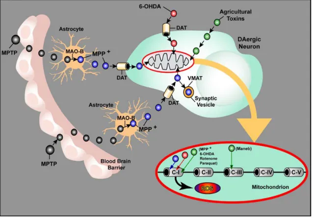
Figure 2 - Molecular and intracellular effects caused by dopaminergic toxins. Once inside the brain,
1-methyl-4-phenyl-1,2,3,6-tetrahydropyridine (MPTP, black vesicles) is taken up by astrocytes and is converted in 1-methyl-4-phenylpyridinium (MPP+, blue vesicles) by the enzyme MAO-B. Then, MPP+ is released to extracellular space and is transported into dopaminergic neurons via dopamine transporter (DAT). Inside neurons, MPP+ can be concentrated in mitochondria, or be sequestrated into synaptic vesicles via vesicular monoamine transporter (VMAT). 6-OHDA (red vesicles) is taken up via DAT and can accumulate in mitochondria. Moreover, agricultural toxins (rotenone, paraquat and maneb, green vesicles) penetrate unspecifically in neurons and accumulate in mitochondria. In mitochondria, MPP+, rotenone and paraquat can inhibit the complex(C)-I of mitochondrial ETC and maneb inhibit the C-III. This inhibition leads to the production of ROS. Adapted from (19).
In this work, I will highlight two toxin-induced PD models, 6-OHDA and MPTP models.
1.2.1 6-OHDA model
6-OHDA is a hydroxylated analogue of dopamine (19). This toxin is selective for catecholaminergic neurons, especially those with a preferential uptake by dopamine transporter (DAT) and noradrenergic transporters (44). Also, 6-OHDA is a putative endogenous toxin, taking into account that it is a product of the dopamine metabolism, and it is the result of hydroxyl radical attack in the presence of excess dopamine (45).
This toxin is hydrophilic and therefore unable to cross the blood-brain barrier, thus it is administered by stereotaxic injections into the SNpc, median forebrain or ST. In particular, the administration into the ST leads to retrograde degeneration of nigrostriatal neurons, which lasts several weeks (46), in contrast to the other forms of administration that lead to a degeneration within 24h (47). Also, its administration can be unilateral or bilateral (19). The unilateral injection is more frequently used, because the bilateral injections induce an elevated death rate or the animals require many nursing care.
7
6-OHDA accumulates in the cytosol, and it can autoxidize forming semiquinone and superoxide radicals (48). 6-OHDA can also decrease striatal GSH and SOD activity (49), leading to increased levels of H2O2 (50). Additionally, the superoxide radical can be subsequently
converted to a more cytotoxic compound, the hydroxyl radical, through interaction with H2O2
(48).
1.2.2 MPTP model
MPTP is a lipophilic substance, which after systemic administration is able to cross the blood-brain barrier. Once inside the blood-brain, is converted to 1-methyl-4-phenyl-2,3-dihydropyridium (MPDP) by the enzyme MAO-B in non-dopaminergic cells, like glial cells and serotonergic neurons. Then, MPDP is oxidized to 1-methyl-4-phenylpyridinium (MPP+), the active toxic
molecule. Subsequently, MPP+ is released to the extracellular space and its cellular uptake
depends on active plasma membrane carrier systems (51). MPP+ has high affinity to the DAT,
as well as noradrenaline and serotonin transporters and can be stored in vesicles via uptake by the vesicular monoamine transporter (VMAT). Inside dopaminergic neurons is able to impair complex I of the mitochondrial ETC resulting in the release of ROS and in the reduction of ATP production (19). These events culminate in an apoptotic degenerative process involving the upregulation of the Bax and the c-Jun N-terminal kinase, the release of cytochrome c and the activation of caspases -3 and -9 (52).
This toxin can be administered by diverse regimens, for example by stereotaxic injection or by gavage, but the most common form is by systemic administration, more specifically by subcutaneous, intravenous, intraperitoneal (i.p.) or intramuscular administration (19). The schedules of administration may induce distinct mechanisms and extent of dopaminergic death.
2 Regulation of transcriptional factors associated with PD
Epigenetic consists in several alterations that can regulate gene expression without changing genotype. These include DNA methylation, which consists in the addiction of methyl groups to the 5’ position of the cytosine residues within CpG dinucleotides, forming heterochromatin
regions, and post-translational histone modifications, which include
acetylation/deacetylation, methylation/demethylation, phosphorylation, ubiquitination, SUMOylation, ADP and ribosylation (53). Dysregulation of these mechanisms can lead to several neurodegenerative diseases, like PD (54). Histone acetylation and deacetylation are mechanisms associated with transcriptional activation and repression, respectively (55) (Figure 3). Histone Acetyltransferases (HATs) are divided in three families: Gcn5-related N-acetyltransferase, MYST and CREB-binding protein (CBP)/p300 (56). They act as transcriptional co-activators, being part of large multisubunit complexes and are recruited to promoters through interacting with DNA-bound activators. The acetylation can be reversed by
8
Histone Deacetylases (HDACs), which are categorized into four classes based upon sequences homology and cofactor dependencies (53).
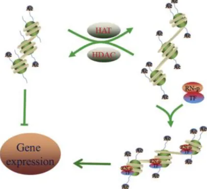
Figure 3 - Mechanisms of acetylation and deacetylation. The HAT and HDACs mediate the acetylation
and deacetylation, respectively. HATs produce a more loosened chromatin, allowing the transcription activation and the HDACs form a heterochromatin structure, repressing the transcription. TF, transcription factor; RN-p, RNA-polymerase; Ac, acetyl group. Adapted from (57).
Methylation can have a positive or negative effect on gene transcription, depending on the target histones (53). Also, the methylation is reversible, with two families of histone demethylases identified, including the amine oxidase domain-containing lysine specific demethylase 1 (LSD1) and Jumonji C domain-containing protein family (58). The LSD1 can be found in repressive (like C-terminal binding proteins (CtBP) and CoREST) and activating (like androgen receptor) complexes.
Dysfunction in the epigenetic machinery has been proposed to play a role in PD etiology. For example, α-synuclein is normally expressed in the nucleus and presynaptic nerve terminals, but increased nuclear targeting is neurotoxic. This nuclear toxicity might result from direct binding to histones, reducing the levels of acetylated histone and general acetylation through interactions with sirtuin (SIRT)2 (a HDAC) (59). Similarly, under oxidative stress conditions, α-synuclein goes to the nucleus, where it binds to the peroxisome proliferator receptor gamma coactivator-1 alpha (PGC1-α) promoter. This binding causes histone deacetylation, lowering PGC1-α expression, which is noxious to mitochondrial function (60). Another example is Nurr1, which is important for the development and maintenance of the dopaminergic neurons and it was found decreased in PD patients. It happens because CoREST together with HDACs, G9a (a histone methylstransferase) and LSD1 can repress Nurr1 transcription (61).
9
2.1 C-terminal binding proteins
CtBPs are transcriptional co-repressors essential for brain development and for the inhibition of many transcriptional factors (62). Mammalian CtBPs are enconded by two major genes,
Ctbp1 and CtBP2, which produce different CtBPs isoforms. Ctbp1 encodes two major proteins
CtBP1-S and CtBP1-L (62). Both isoforms display mostly identical sub-cellular localization and probably share similar functions in the regulation of gene expression and membrane trafficking processes. On the other hand, Ctbp2 encodes three isoforms, CtBP2-S and CtBP2-L are highly homologous to the isoforms of CtBP1 and they act mainly as nuclear transcriptional regulators (62). The third isoform, RIBEYE, is expressed from an alternative promoter, and active only in ribbon synapse containing neurons, like bipolar cells (63).
All isoforms have a hydrophobic cleft, named Pro-X-Asp-Leu-Ser (PXDLS)-binding. This domain is essential to recruit other members of the co-repressor complex, in a PXDLS-depend or independent manner (64). In particular, is crucial for the recruitment of the core co-repressor machinery, which includes HDACs, histone methyltransferases, and transcriptional repressors. Moreover, they also have a RRTGXPPXL sequence (RRT-binding pocket), which is mainly used to bind and recruit members of the co-repressor complex (65). Thus, each CtBP contains two binding sites that can be occupied at the same time by distinct members of the co-repressor complex.
These co-repressors have homology with D-2-hydroxy acid dehydrogenases, which contain a dinucleotide binding site capable of binding to oxidized nicotinamide adenine dinucleotide (NAD+) or reduced nicotinamide adenine dinucleotide (NADH) (66), with the last one being
more effective in stimulating CtBPs binding (67). Indeed, fluorescence resonance energy transfer studies showed a >100-fold higher affinity for NADH than NAD+. The interaction with
NADH, which is increased in hypoxic environments, allows CtBPs to form dimers, increasing the ability of binding to transcriptional repressors (67). Increased levels of intracellular NADH may be found in response to some biological events, like in developing embryos in utero, cellular hypoxia, metabolic diseases, and healthy aging, which can ultimately activate the CtBP-mediated repression of target genes (67,68).
There are small differences in protein sequence of CtBP1 and CtBP2 responsible for different functions. The most evident is a nuclear localization signal (NLS) at the N-terminal of CtBP2, responsible for the nuclear retention of this protein (69) (Figure 4). But there are CtBP2 isoforms without the NLS domain, leading to a cytoplasmic retention.
10
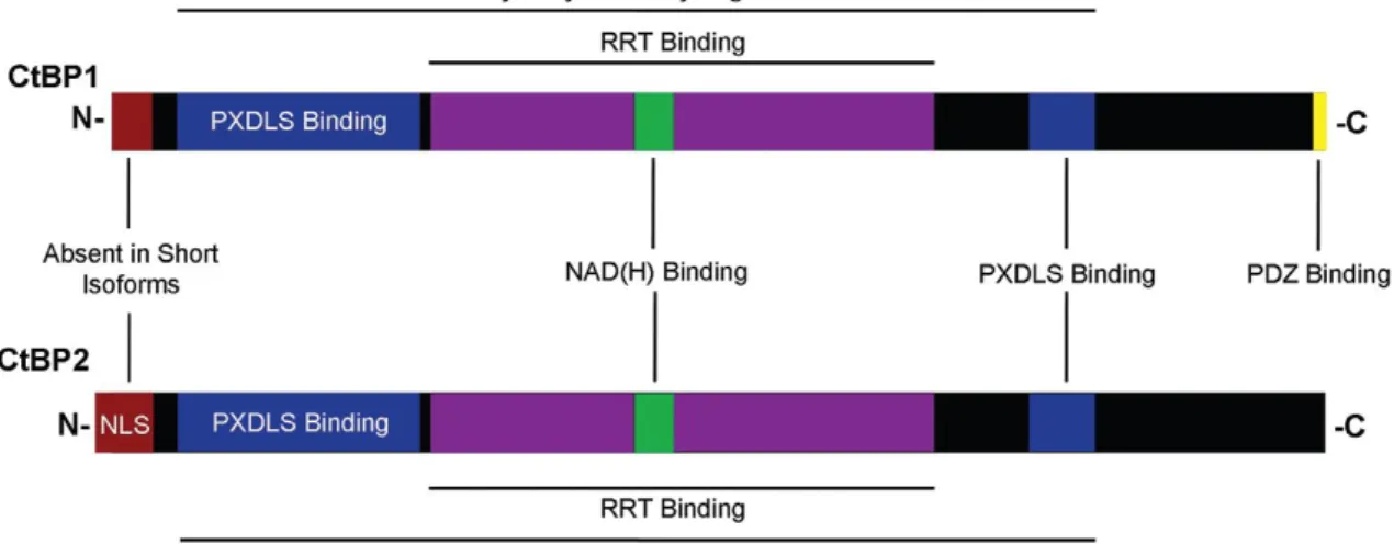
Figure 4 - CtBP1 and CtBP2 protein structures. CtBPs are composed by a PXDLS-binding cleft, a
RRT-binding cleft, and the dehydrogenase domain. The main structural difference between them is the longer N-terminal of CtBP2, which contains an NLS domain. Moreover, CtBP1 have a PDZ binding domain at the C-terminus. Adapted from (62).
CtBP1 have both nuclear and cytoplasmic functions, in the nucleus it can acts as a transcriptional co-repressor (70) and in the cytoplasm can regulate membrane fission (62). In the neurons, CtBP1 is widely expressed in the presynaptic compartment and it is able to interact with presynaptic proteins. In the absence of neuronal activity, CtBP1 is mainly retained in nucleus and represses transcription of genes, like brain derived neurotrophic factor (BDNF), Fos and Arc. After neuronal activity, CtBP1 rapidly stabilizes at presynaptic terminals, through ligation with Basson. So, in this case there is a decrease of nuclear CtBP1 and the transcription of targets genes is increased (71,72). Concluding, neuronal activity may modulate synapto-nuclear distribution (Figure 5) and co-repressor activity of CtBP1 (Figure 6). Furthermore, other mechanisms that have been suggested to regulate CtBPs distribution are neuronal nitric-oxide synthase and p21-activated kinase 1 (Pak1). Both can redirect CtBPs from the nucleus to the cytosol (73,74).
Figure 5 - CtBP1 synapto-nuclear distribution. Representative images showing the synapto-nuclear
distribution of endogeneous CtBP1 in cultured hippocampal neurons. Neurons were stained for Basson to label presynapses and DAPI to label nuclei. Adapted from (71).
11
Figure 6 - Neuronal activity regulates intracellular CtBP1 distribution. Synaptic activity leads to
CtBP1 exit from the nucleus to presynaptic terminals. This translocation depends on elevation of NADH levels. Absence of synaptic activity or inhibition of glycolysis causes nuclear retention of CtBP1 and repression of target genes, like Arc, Fos and BDNF. Adapted from (72).
CtBPs can act as a bridge between DNA-binding proteins and enzymes associated with transcriptional repression, like HDACs (75). CtBPs can also interact with HAT, such as p300, CBP and pCAF and prevents their interaction with chromatin (76). Moreover, CtBPs bind to the human polycomb 2 protein (Pc2), producing a densely packed heterochromatin (77). Ultimately, CtBP-mediated transcriptional regulation may involve SUMOylation of transcriptional factors (78). Also, the Drosophila CtBP and the vertebrate CtBP2 might activate transcription in a gene specific manner (78).
2.1.1 Role of CtBPs on Cell Survival and Proliferation
The first evidence highlighting the relevance of CtBPs for cell survival was that Ctbp1-null mice are viable and about 30% smaller than wild-type and heterozygous type and about one fourth of homozygous mutant mice die at postnatal day 20 (63). On the other side, Ctbp2-null mice exhibit embryonic lethal phenotype. The embryos die by E10.5 and they are smaller and exhibit axial truncations. These truncations are correlated with reduced levels of expression of the T-box transcription factor Brachyury, which modulates mesodermal and neural cell fates during development. These embryos have defects in heart morphogenesis, and delayed development of the forebrain and midbrain. In the axial defect phenotype, the expression of
Brachyury (target gene of Wnt-3a) is low (63), suggesting that CtBP2 may be a regulator of
Wnt-mediated gene expression.
CtBPs can act as apoptosis inhibitors, mediating the repression of several tumor suppressor genes. However, some tumor suppressors target CtBPs to confine their anti-apoptotic activity.
12
This down-regulation of CtBPs results in p53-independent apoptosis (70). A study with mouse embryo fibroblasts revealed that CtBPS can co-repress some pro-apoptotic genes, like p53-effector related to pmp-22 (PERP), p21, Bax, Noxa, caspase-3 and its cleaved substrate, poly(ADP-ribose) polymerase (PARP) (79,80). CtBPs modulate the expression and activities of the Ink4 family. The Ink4 codes for three different cell cycle inhibitors, p16Ink4a, Ink4a/Arf and p15Ink4b (Figure 7) (81). The Ink4a/Arf mediates its tumor suppressive function by stabilizing p53 and by p53-independent mechanisms, however the others two function in the retinoblastoma pathway by inhibiting cyclin-dependent kinase 4 and 6. Also, tumor growth factor β, an activator of p15Ink4b expression via activated SMAD, may mediate its effect by forming an activation complex consisting of zinc finger E-box-binding homeobox 1-SMAD-p300 and acetylation of the CtBP-binding domain resulting in displacement of CtBPs (82,83). CtBPs are regulated by a number of factors, especially by post-translational modifications. For example, phosphorylation targets these proteins for ubiquination and consequently to proteasomal degradation, which can occur under stress conditions (62). Also, adenomatous polyposis coli (APC) may target CtBP1 to proteosomal-dependent degradation, by targeting both β-catenin and CtBP1 simultaneously to inhibit expression of Wnt target genes (84). The previous mechanisms are responsible to targeting CtBPs for degradation and consequently inducing apoptosis (Figure 7). Several biological activities are regulated by Pak1 phosphorylation, like cell survival and can also influence gene expression. Pak1 interacts with CtBP, phosphorylating it and subsequently blocking the CtBPs dehydrogenase activity (74). The loss of activity occurs due to a transient loss of nuclear localization in conjunction with a conformational change, and not due to triggering ubiquitination or degradation of these co-repressors. Another CtBPs regulator is the Pc2, by acting as a SUMO E3 ligase and consequently regulates the localization of these proteins within cell. For example, mutants lacking the SUMOylation consensus sequence have a cytoplasmic distribution (85).
13
Figure 7 – Role of CtBPs in tumorigenesis. Under the increase of NADH the CtBP activity is stimulated,
resulting in dimerization. CtBP can enhance cell proliferation by repressing the activity of cell cycle inhibitors, like p16Ink4a and p15Ink4b. As result of the repression of pro-apoptotic genes, like PERP, Bax and Noxa, CtBP can promote cell survival. However, these CtBP functions can be inhibited by tumor suppressors like homeodomain interacting protein kinase 2 (HIPK2) or Ink4a/Arf or APC, which promote CtBP degradation as a result of phosphorylation and ubiquitination. Adapted from (70). The previously referred mechanisms were observed mostly in cancer conditions. Although there is also evidence that CtBPs can regulate neuronal proliferation and differentiation. For example, in high concentrations of oxygen, CtBPs are excluded from the Hes1 promoter and its expression is maintained, preserving the self-renewing ability of neural progenitors and inhibiting neurogenesis. Furthermore, evidence from the analysis of roof plate phenotypes and molecular analysis of neural stem cell culture suggest that under bone morphogenetic proteins and high oxygen, CtBPs associate with HES1 and repress neuronal differentiation (86). Also, Stankiewicz and colleagues reported that CtBPs undergo caspase-dependent downregulation in primary cerebellar granule neuron exposed to neurotoxins (87). It has been suggested that this dysregulation of CtBPs may be associated with neurodegenerative diseases, such as Huntington disease (88). Also, in brain trauma homeodomain interacting protein kinase 2 (HIPK2) and CtBP2 are increased in the peritrauma brain cortex. This increase was associated with activation and proliferation of astrocytes (89). However, in a neuroinflammatory context, some authors argue that the recruitment of CtBPs to DNA prevent inflammation whereas others reported that these proteins also trigger a pro-inflammatory response (90,91).
So, considering the ability of CtBPs to modulate cell survival and proliferation, it seems that they may be a good target for neurodegenerative diseases, including PD.
15
Chapter 2
Objectives
Some studies have suggested an epigenetic role on neurodegenerative diseases, like PD (54). Indeed, several evidence suggest that CtBPs, transcriptional co-repressors, may modulate proliferation and neuronal survival by down-regulating pro-apoptotic genes (86,87). However, so far, just one report briefly exploit the role of CtBPs on PD (87).
Our main aim was to analyze the expression levels of CtBPs in PD models and to explore their putative effect on dopaminergic survival. To address this main aim three tasks were designed:
To evaluate the protein expression levels of CtBPs in in vitro and in vivo models for
PD;
To characterize the cellular and subcellular localization of CtBP1 and CtBP2;
To analyze the effect of a broad CtBPs antagonist, 4-methylthio-2-oxobutyric acid (MTOB), on dopaminergic survival.
17
Chapter 3
Materials and Methods
1 N27 cell cultures
The immortalized rat mesencephalic dopaminergic cell line (N27 cells) was grown in Roswell Park Memorial Institute (RPMI) 1640 medium (Sigma-Aldrich) containing 2g/L sodium bicarbonate, 10% fetal bovine serum (FBS; Millipore) and 1mL/L of penicillin/streptomycin
(GIBCO), in a humidified atmosphere of 5% CO2 at 37°C. For western-blot experiments, cells
were plated at a density of 1.5x10⁵ cells per plate in 6-well culture plates. For MTT assay
experiments, cells were plated at a density of 0.25x10⁵ cells per plate in 48-well culture
plates and for proliferation assay cells were seeded at a density of 0.5x10⁵ cells per plate in
24-well plates with 10mm glass coverslip.
For western-blot experiments, cells were exposed to 6-OHDA (25 µM or 50µM; Sigma-Aldrich) or MPP+ (30µM or 1mM; Sigma-Aldrich) (92). For the MTT assay, N27 cells were incubated with
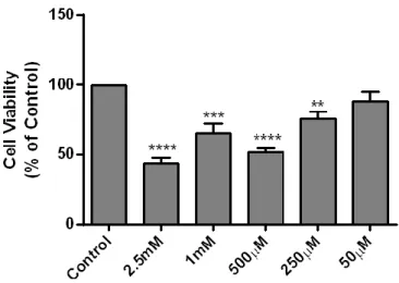
different concentrations of MTOB (2.5mM, 1mM, 500µM, 250µM and 50µM; Sigma-Aldrich) and/or with 6-OHDA (50µM) or MPP+ (1mM). Ultimately, for proliferation assay cells were
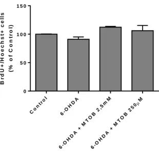
treated with 6-OHDA (50µM) and/or MTOB (2.5mM or 250µM).
1.1 MTT reduction assay
The levels of MTT reduction were measured to assess cell viability. This assay is based on the capacity of metabolically active cells to reduce tetrazolium MTT salt (yellow) in a water-insoluble formazan dye (purple).
After 24h of cell treatments, 0.5mg/mL of MTT (Acros Organics) in Hanks Balanced Salt
Solution (HBSS; 137mM NaCl, 5.36mM KCl, 4.16mM NaHCO3, 0.44KH2PO4, 0.34mM
Na2HPO4.2H2O, 5mM glucose, 1mM sodium pyruvate, 10mM HEPES, pH7.4 ) was added to cells
for 4h at 37°C. Then, the precipitate formed was dissolved in 10% sodium dodecyl sulfate (SDS), transferred to 96-well culture plates and, lastly, colorimetric quantified at the
wavelength of 570nm, using a XMarkTM Microplate Spectrophotometer (Bio-Rad). The
measured absorbance can be directly correlated with the number of viable cells.
1.2 Cell proliferation assay
Proliferation was detected by incubating the cells with Bromodeoxyuridine (BrdU), a thymidine analog that is incorporated during the S-phase of the cell cycle. The immunostaining was performed using an adapted protocol described in (93).
18
Cells were treated with BrdU (50mM; Sigma-Aldrich) for the remaining 2h of cell treatments. After fixation with formalin, 0.3% Triton X-100 was added to cells for 10 minutes. Then, DNA was denatured by HCl 1M at 37°C for 30 minutes. Non-specific binding was prevented by incubating cells in 2% of FBS and 0.3% Triton X-100 solution for 1h at room temperature (RT).
Then, cells were incubated overnight at 4°C with rat monoclonal anti-BrdU (1:100; Serotec),
washed with phosphate buffer saline (PBS; NaCl 140mM, KCl 2.7mM, KH2PO4 1.5mM and Na2HPO4 8.1mM, pH 7.4), and incubated for 1h at RT with the Alexa Fluor 488 donkey anti-rat (1:200; Life Technologies) and Hoechst33342 (2mg/mL; Sigma-Aldrich). Lastly, coverslips were mounted with Fluoroshield Mounting Medium (Abcam) and images were acquired under the magnification of 40x at the Zeiss Axiovert 200 imaging microscope (Axiobserver Z1, Zeiss).
2 Animals
All animals were handled in accordance with institutional animal house, national ethical requirements and in accordance with the European Community guidelines (2010/63/EU). 49 adult male C57BL/6 mice with 2-26 months-old were housed in the same room and in appropriate cages under controlled temperature conditions (~22°C), with a fixed 12h light ⁄ dark cycle and with ad libitum food and water access.
For the western-blot analysis, mice were sacrificed by spinal cord dislocation, the brains collected and the regions of interest (SN and ST) dissected, frozen in liquid nitrogen and stored at -80°C until used.
2.1 MPTP injections
MPTP (Sigma-Aldrich), dissolved in sterile 0.9% NaCl was injected i.p. at 2h intervals, using a dose of 15mg/Kg body weight, to the total dose of 60mg/Kg (94). Animals were sacrificed 7 days after the MPTP intoxication protocol, by spinal cord dislocation, and the brains were dissected and the regions of interest (SN and ST) were removed, frozen in liquid nitrogen and stored at -80°C until be used in western-blot.
2.2 Stereotaxic injections
First, mice were anesthetized with a mixture of ketamine and xylazine (0.5mL of xylazine, 0.9mL of ketamine and 3.5mL of NaCl, 5µL/g of mouse weight) via i.p. Then, animals were placed in the digital stereotaxic frame (51900 Stoelting) and an incision was made in the scalp in order to expose the skull. MTOB (5mM, 2.5mM, 250 µM or 50µM dissolved in sterile 0.9% NaCl; 2µL total volume) or saline solution (sterile 0.9% NaCl) were injected in the right SN (Anterior-posterior (x): +3.0mm, Medial-lateral (y): -1.4mm, Dorso-ventral (z): -4.4mm), with a 10uL Hamilton syringe at a speed of 0.2µL/min. Some mice were also subjected to a stereotaxic injection of 6-OHDA (95) (10µg dissolved in 0.1% of ascorbic acid; 2µL total volume), in the right ST (Anterior-posterior (x): -0.6mm, Medial-lateral (y): -2.0mm, Dorso-ventral (z): -3.0mm).
19
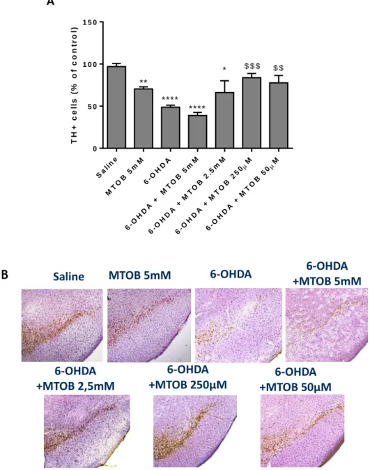
Seven experiment groups were designed: 1) Saline; 2) MTOB 5mM; 3) 6-OHDA; 4) 6-OHDA and MTOB 5mM; 5) 6-OHDA and MTOB 2.5mM; 6) 6-OHDA and MTOB 250µM and 7) 6-OHDA and MTOB 50µM. After intracerebral injection, the incision was sutured and mice were kept warm (37 ºC), until they recovered from surgery. Then, animals were maintained in appropriate cages for 7 days, until euthanized.
2.3 Brain slices preparation
Seven days after the stereotaxic injections, mice were anesthized with a mixture of ketamine and xylazine and an incision through the thoracic midline was made. Immediately after the heart being exposed, a needle was inserted in the left ventricle and the right aorta was cut with a scissor. Then, the transcardial perfusion with 0.9% NaCl was performed until the blood was entirely clear, followed by a perfusion with 4% paraformaldehyde (PFA). Afterwards the brains were removed and were left overnight in 4% PFA at 4°C, following dehydratation in 30% Sucrose at 4°C until they sunk. Then, brains were frozen with liquid nitrogen and were stored at 80°C until used.
For slices preparation, brains were embedded in optimal cutting temperature gel (Bio-Optica) and cut into coronal sections with a thickness of 40µm, from the olfactory bulb towards midbrain, on a freezing cryostat-microtome (Leica CM 3050S, Leica Microsystems). The sections of each animal were collected sequentially in six wells of a 24-well plate, resulting in a 240 µm distance between each brain slice. The slices were kept in anti-freeze solution (30% of ethylene glycol, 30% glycerol, 30% water and 10% phosphate buffer solution (0.2M)) until used for immunohistochemistries.
2.4 TH
+immunohistochemistry
First, slices were rinsed in PBS to remove the anti-freeze solution. Then, brain slices were washed with 0.1% Tween-20 in PBS (PBS-T) for 10 minutes, followed by permeabilization and blocking with 0.1% Triton X-100 and 10% FBS in PBS for 1h. Afterwards, the activity of endogenous peroxidases was inhibited by an incubation with 3% H2O2 for 10 minutes and
protected from light. Lastly, the sections were incubated overnight at 4°C with the mouse anti-TH antibody (1:500, Transduction Laboratories) diluted in 5% FBS in PBS. The day after, slices were incubated with biotinylated goat anti-mouse (1:200, Vector Laboratories) in 1% FBS in PBS, 1h at RT. Then, the slices were incubated for at least 30 minutes at RT with the Avidine/biotine peroxidase complex reagent (Vectasin ABC kit, Vector Laboratories). Finally, sections were incubated with Horseradish Peroxidise and DAB substrate (both from DAKO), for about 5-10 minutes until developing a brown color in the SN region. Sections were mounted on Superfrost slides, dried, and dehydrated with increasing concentrations of ethanol (50%, 75%, 95% and 100%). Then, TH-stained slices were counterstained with Nissl (0.25% Cresyl Violet dissolved in Acetate Buffer) for 4 minutes, quickly washed in tap water, air dried, cleaned with xylene, and ultimately mounted with Entellan (Merck).
20
To count the number of dopaminergic neurons in the SNpc, serial sections of this region were used. This region doesn’t have well-defined limits, so the area corresponding to the SNpc was delineated and the total number of TH+ cells was counted in ipsilateral hemisphere (5
sections). Due to the restricted number of available animals, the contralateral side of some conditions was used as control condition in some experimental groups (3 animals of the 6-OHDA-challenged mice; 6-OHDA+2,5mM MTOB; 6-OHDA+250µM; and 6-OHDA+50µM). No statistical difference was found between the number of TH+ cells found between the
contralateral in the aforementioned groups and the ipsilateral side of saline animals. The images were acquired under the magnification of 10x at the Zeiss Axiovert 200 imaging
microscope (Axiobserver Z1, Zeiss) and the number of TH+ cells was counted using the ImageJ
program.
2.5 Fluorescent immunohistochemistry
Tissue sections were rinsed in PBS to remove the anti-freeze solution, and then, to prevent unspecific binding, were incubated in blocking solution (2% FBS and 0.3% Triton X-100 in PBS) for 2h at RT. Thereafter, slices were incubated in an orbital shaker with the primary antibodies in blocking solution for 3 overnights at 4°C. Next, slices were rinsed with PBS and incubated in an orbital shaker with the respective secondary antibodies (1:1000) in PBS containing 0.3% Triton X-100 for 2h at RT. The antibodies used are listed in the Table 3.1. Lastly, sections were rinsed with PBS and mounted in Fluoroshield Mounting Medium (Abcam). Images were acquired using a Zeiss inverted confocal microscopy (Axiobserver Z1, Zeiss) using an objective with a 40x lens.
N27 cells were fixed with formalin and subjected to the previously described protocol, with the difference that the incubation with primary antibodies (mouse anti-CtBP1 and CtBP2; 1:200; BD Biosciences) only last for 24h.
21
Table 1– Primary and secondary antibodies used for fluorescent immunohistochemistry.
(CtBP1, C-terminal binding protein-1; CtBP2, C-terminal binding protein-2; CD11b, cluster of differentiation molecule 11b; GFAP, glial fibrillary acid protein; NeuN, neuronal nuclei, TH, tyrosine hidroxilase).
3 Western-blot
N27 cells were lysed on ice with RIPA buffer (50mM Tris, pH=8.0, 150mM NaCl, 1% Triton X-100, 0.5% Sodium deoxycholate, 0.1% SDS, and a cocktail of protease inhibitors). After 15 minutes on ice, cells were centrifuged (centrifugation at 11 300rpm during 20 minutes at 4°C), then the supernatant was collected and the total amount of protein concentration was quantified using a Pierce bicinchoninic acid Protein Assay Kit (Thermo Scientific). The brain tissues were mechanically dissociated and lysed on ice in RIPA buffer. Then, the soluble fraction was obtained (centrifugation at 12000rpm, during 20 minutes at 4°C) and, ultimately, the protein concentration was determinated using the previous kit.
After protein quantification, the samples were treated with Loading Buffer (6x concentrated: 350mM Tris, 10% SDS, 30% glycerol, 0.6M DTT, 0.06% bromophenol blue) and boiled for 15 minutes at 100°C.
Then, 40µg of total protein were loaded into each lane of 12% bisacrylamide gel (Nzytech). Proteins were separated by SDS-PAGE electrophoresis in a 100V until the front of the race reach the final of the gel, in a running buffer solution (25mM Tris, 190mM glycine pH=8.3, 0.1% SDS) at RT. Afterwards, proteins were transferred to a polyvinylidene difluoride membrane (Millipore) through semi-dry transfer during 25 minutes at 1.0A, 25V, using Towbin transfer buffer (25mM Tris, 192 glycine pH=8.3, 20% methanol) at RT. After that, membranes
Primary
Antibody Target Dilution Company
Secondary antibody Company Mouse anti-CtBP1 CtBP1 1:1000 BD Biosciences Donkey anti mouse 594 Abcam Mouse anti-CtBP2 CtBP2 1:1000 BD Biosciences Donkey anti mouse 594 Abcam Rat anti-
CD11b Microglial cells 1:1000 Serotec
Donkey anti rat 488
Life Technologies Rabbit
anti-GFAP Astrocytes 1:200 DAKO
Donkey anti rabbit 647
Life Technologies Rabbit
anti-NeuN Neuronal cells 1:500 Cell Signaling
Donkey anti rabbit 647 Life Technologies Rabbit anti-TH Dopaminergic neurons 1:1000 Santa Cruz Biotechnology Donkey anti rabbit 647 Life Technologies
22
were blocked with 5% non-fat milk (Regilait) in Tris buffer saline solution – Tween 20 (TBS-T; 20 mM Tris, 137 mM NaCl solution and 0.1% Tween 20) for 1h at RT. Membranes were then incubated overnight at 4°C with mouse anti-CtBP1 (1:2500; 48kDa; BD Bioscience), anti-CtBP2 (1:2500; 48kDa; BD Bioscience) or anti-GAPDH (housekeeping; 1:5000; 37kDa; Millipore) antibodies and further incubated with the goat anti-mouse antibody conjugated with horseradish peroxidase (1:5000 Santa Cruz Biotechnology) at RT for 1h. After the antibody incubation, the membranes were incubated with Luminata Crescendo Western HRP Substrate (Millipore) for 5 minutes. Protein bands were detected using the ChemiDocTM MP Imaging
System (Bio-Rad) and quantified by densitometry analyses, using the Image Lab 5.1 software (Bio-Rad Laboratories).
4 Statistical analysis
All data are expressed as mean ± S.E.M. of at least three independent experiments, performed at least in triplicate (in vitro) or at least three different animals (in vivo), with the exception of the condition 6-OHDA+2,5mM, used for immunohistochemistry. Statistical analysis was performed using one-way ANOVA followed by the Dunnett’s multiple comparisons test. Values of P<0.05 were considered significant. All statistical analysis were made using the GraphPad Prism 6.0 Software (GraphPad Sotware Inc.).
23
Chapter 4
Results
1 Expression of CtBP1 and CtBP2 in the SN and in the ST of
healthy mice
To date, the characterization of CtBPs expression in the SN and ST of adult and aged mice as well as in distinct cell phenotypes, was not explored. So, the first aim of this work was analyze the regional (ST and SN) and subcellular (microglia, astrocyte or neuronal) expression of CtBPs.
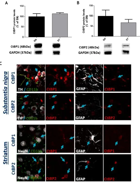
As shown in figure 8A and B, no statistical differences were found regarding the protein expression levels of both CtBP1 (meanSN= 100.0±14.1; meanST= 114.0±5.0; n=3) and CtBP2
(meanSN= 100.0±29.3; meanST= 67.2±16.3; n=3) in the ST and SN of saline mice.
Then, to disclose the specific subcellular expression, co-labelings against CtBP1 or CtBP2 and microglia (CD11b), astrocytes (GFAP), dopaminergic neurons (TH) and neurons (NeuN) were analyzed in the SN and ST of saline animals (Figure 8C). In the SN, CtBP1 is expressed in the nucleus of almost every TH+ cell and in some ramifications of CD11b+ and GFAP+ cells. On the
other hand, CtBP2 is expressed almost exclusively in the nuclei of all cell types analyzed in this region (TH, CD11b and GFAP). CtBP2 is also expressed in almost every TH+ cell. In the ST,
some nuclear co-localization was found between NeuN+ and CtBP1 or CtBP2. In both CD11b+
and GFAP+ cell populations, CtBP1 was found in the cytoplasm (ramifications) whereas CtBP2
24
C
Figure 8 - Expression levels and cellular localization of CtBPs in the ST and SN of adult mice.
Graphs depicts the percentage of (A) CtBP1 and (B) CtBP2 in the SN and ST of wild-type C57BL/6J adult mice. Protein expression was normalized to GAPDH. Data are expressed as percentage of mean ± SEM (n = 3). Protein expression in the SN was set to 100%. Bellow the graph, CtBP1 (48kDa), CtBP2 (48kDa) and GAPDH (37kDa) western blots are shown. Statistical analysis was performed using one-way ANOVA, followed by the Dunnett’s multiple comparison test. (C) Representative confocal digital images of expression of CtBP1 and CtBP2 in the SN and ST of wild-type C57BL/6J adult mice. Blue arrows highlight cells with double labeling for a neuronal (TH, NeuN) or glial (CD11b or GFAP) marker and CtBP1 or CtBP2.
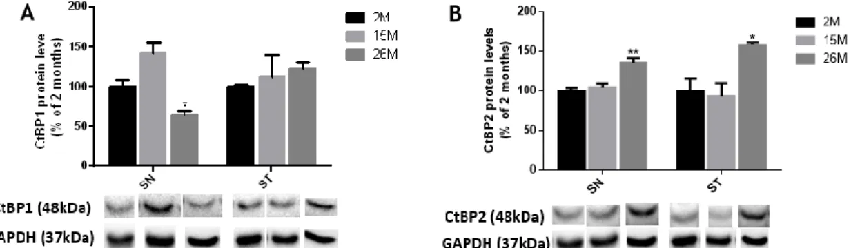
The nigrostriatal pathway is sensitive to several alterations that may occur during aging and this feature is a major risk factor for the development of neurodegenerative diseases (8). Then, to disclose a putative effect of aging on CtBPs expression, we performed western-blot in the SN and ST of healthy mice with different ages (2, 15 and 26 months).
As shown in the Figure 9A, CtBP1 expression levels in the SN increased at 15 months
(mean15M=142.6±13.9, non-significant), while decreased at 26 months, when compared with 2
25
months-old mice (mean2M=100.0±9.1; mean26M=63.8±6.7; n=3). In contrast, no statistical
differences were found regarding the expression of CtBP1 in the ST of animals at different ages (Figure 9A; mean2M=100.0±2.1; mean15M=112.3±27.1; mean26M=122.6±8.0; n=3). Regarding
CtBP2, an increased expression was found both in the SN (mean26M=135.9±5.7, n=3) and the ST
(mean26M=158.3±3.2, n=3) of 26 months-old mice as compared with 2 months-old mice, as
shown in Figure 9B.
Figure 9 - Protein expression levels of CtBPs during aging. Bargraphs depicts the expression of (A)
CtBP1 and (B) CtBP2, in the SN and ST of young-adults (2months), adults (15 months) and old mice (26 months). Protein expression was normalized to GAPDH. Data are expressed as mean ± SEM (n = 3). Protein expression in 2 months-old was set to 100%. Bellow the graph, CtBP1 (48kDa), CtBP2 (48kDa) and GAPDH (37kDa) western blots are shown. Statistical analysis was performed using one-way ANOVA, followed by the Dunnett’s multiple comparison test. *P<0.05, **P<0.01 when compared to 2 months mice.
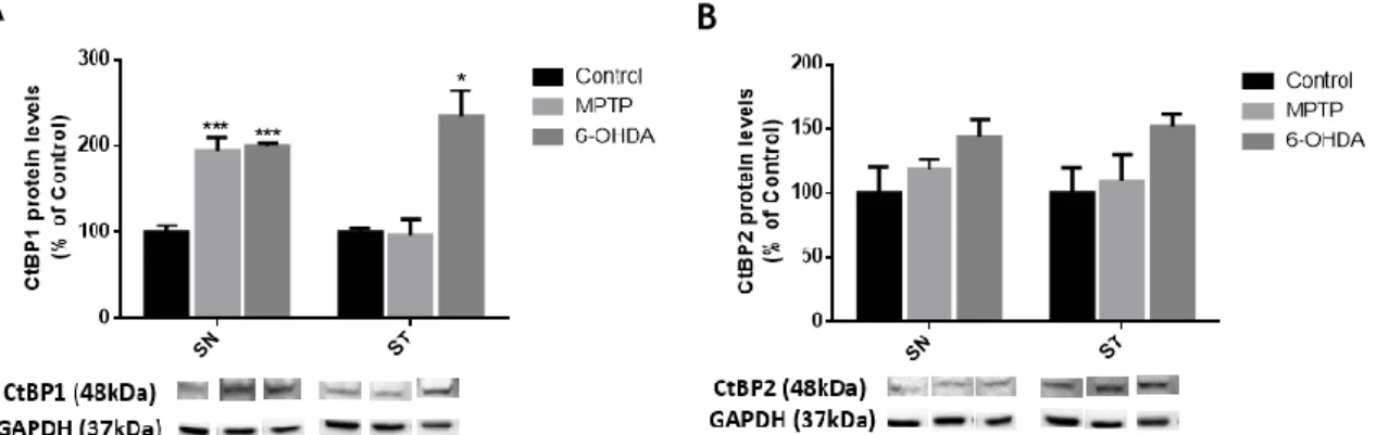
2 CtBP1 expression levels are increased in in vivo mouse models
for PD
Next, we analyzed the expression of CtBPs in the SN and ST of 6-OHDA and MPTP lesioned mice.
As shown in Figure 10A, CtBP1 expression levels are significantly increased in the SN of
6-OHDA and MPTP challenged mice (meanMPTP=1945.0±14.5; mean6-OHDA=199.8±3.2; n=3), and in
the ST of 6-OHDA-challenged mice (mean6-OHDA=233.8±30.5, n=3).
Regarding CtBP2, a slight non-significant increased expression was found in both the SN and ST of 6-OHDA lesioned mice (in SN: meancontrol=100.0±20.2; meanMPTP=118.5±7.6; mean
6-OHDA=143.5±13.5; and in ST: meancontrol=100.0±19.5; meanMPTP=109.7±20.6; mean
6-OHDA=151.9±9.0; n=3; Figure 10B).