Hovenia dulcis
Thunb. pseudofruits as functional foods: Phytochemicals
and bioactive properties in different maturity stages
Patricia Morales
a,b,⇑, Helayne Aparecida Maieves
a,c, Maria Inês Dias
b,d, Ricardo C. Calhella
b,
María Cortes Sánchez-Mata
a, Celestino Santos-Buelga
e, Lillian Barros
b,d, Isabel C.F.R. Ferreira
b,⇑ aDpto. Nutrición y Bromatología II, Facultad de Farmacia, Universidad Complutense de Madrid (UCM), Plaza Ramón y Cajal, s/n, 28040 Madrid, SpainbMountain Research Centre (CIMO), ESA, Polytechnic Institute of Bragança, Campus de Santa Apolónia, 1172, 5300-253 Bragança, Portugal cFaculdade de Nutrição-Gastronomia, Universidade Federal de Pelotas, Av. Gomes Carneiro, 01 Campus Anglo, Porto, 96010-610 Pelotas, RS, Brazil
dLaboratory of Separation and Reaction Engineering, Laboratory of Catalysis and Materials (LSRE-LCM), Polytechnic Institute of Bragança, Campus de Santa Apolónia, 1134, 5301-857 Bragança, Portugal
eGrupo de Investigación en Polifenoles (GIP-USAL), Facultad de Farmacia, Universidad de Salamanca, Campus Miguel de Unamuno, 37007 Salamanca, Spain
a r t i c l e
i n f o
Article history:
Received 13 July 2016
Received in revised form 23 November 2016 Accepted 2 December 2016
Available online 18 December 2016
Keywords:
Hovenia dulcisThunb. Pseudofruits Phytochemicals
Antioxidant/antitumor/antimicrobial capacity
Ripening process
a b s t r a c t
Pseudofruits ofHovenia dulcis,at five different ripening stages, were evaluated in order to characterize their bioactive compounds (phenolic compounds, tocopherols and fatty acids), and determine their bio-functionality (antitumor, antimicrobial and antioxidant activity). Epi(gallochatechin) was the main phe-nolic compound (24.7 mg/g, Hd05),a-tocopherol was the main isoform, increasing its content with the maturity process (5.43 mg/100 g, Hd04), C18:2 was the main polyunsaturated fatty acid (35.5%, Hd04), while C16:0 was the predominant saturated fatty acids (38%, Hd01). The most immature stages (Hd01-Hd02) showed antitumor activity against all tested tumor cell lines, mainly against NCI-H460; also Hd01 and Hd02 presented the highest antimicrobial activity, mainly againstPseudomonas aeruginosaand Staphylococcus aureus. This underutilized product demonstrated to be a rich source of phenolic com-pounds and tocopherols showing an interesting activity in immature stages (unfit for consumptionin nat-ura) and could be use as alternative bioactive ingredient for functional foods, dietary supplements or nutraceuticals.
Ó2016 Elsevier Ltd. All rights reserved.
1. Introduction
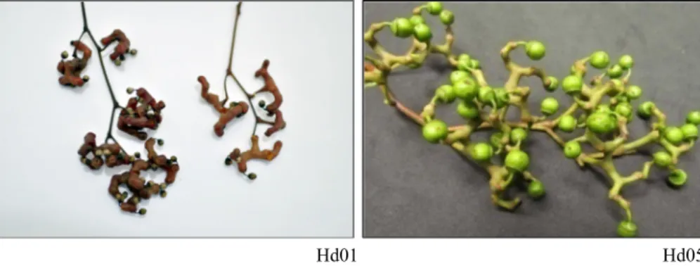
Pseudofruits are stems from a single flower that, as a result of fertilisation, have the development of an organ in addition to the accessory ovary (Chitarra & Chitarra, 2005). In the case ofHovenia dulcis Thunb., traditionally known as rasing tree, the mature peduncle, that is the edible part, was commonly and erroneously called fruit (Fig. 1). Tasty pulp of ripe pear aroma reminiscent of peduncle presents a good acceptance for human consumption due to the high levels of sugars, especially sucrose (Carvalho, 1994; Maieves, Ribani, Morales, & Sánchez-Mata, 2015a).
Hovenia dulcis pseudofruit, whose popular name is Japanese grape, has a long history as a food supplement and traditional
med-icine in Japan, China and Korea, but it is quite unknown and scar-cely used in Western countries (Hyun, Eom, Yu, & Roitsch, 2010; Rigatto, Pereira, Mattos, & Schaitza, 2001). It is cultivated in China, invasive in South American rainforests, being widely distributed in Brazil, and Tanzania, and has been introduced as a rare ornamental in different countries including the USA, Australia, New Zealand and Central Africa (Hung et al., 2010). According toHyun et al. (2010), in East Asia,H. dulcishas long been used in traditional her-bal medicine, being traditionally employed in the treatment of liver diseases and detoxification after alcoholic poisoning; also, in ancient Chinese medicine, its fruits and peduncles have been used as a febrifuge and administered to treat parasitic infections, as antispasmodic, laxative and diuretic agent. Seeds have been used as a diuretic being also useful in alcohol intoxication.
Previous studies have revealed further pharmaceutical applica-tions ofH. dulcisbased on its anti-inflammatory effects (inhibition of KappaB-alpha phorylation and nuclear translocation of nuclear factor-KappaB), hepatoprotective effect (preventive effect on D-GalN/LPS-induced liver injury) and alcohol detoxification effect, among others (Hase et al., 1997; Ji, Yang, & Li, 2000). In this way,
http://dx.doi.org/10.1016/j.jff.2016.12.003
1756-4646/Ó2016 Elsevier Ltd. All rights reserved.
⇑ Corresponding authors at: Dpto. Nutrición y Bromatología II, Facultad de Farmacia, Universidad Complutense de Madrid (UCM), Plaza Ramón y Cajal, s/n, 28040 Madrid, Spain (P. Morales), Mountain Research Centre (CIMO), ESA, Polytechnic Institute of Bragança, Campus de Santa Apolónia, 1172, 5300-253 Bragança, Portugal (I.C.F.R. Ferreira).
E-mail addresses:[email protected](P. Morales),[email protected]
(I.C.F.R. Ferreira).
Contents lists available atScienceDirect
Journal of Functional Foods
different studies have demonstrated that polysaccharides from the peduncles ofH. dulcishave a significant hepatoprotective effect on acute alcohol-induced liver injury in mice via its antioxidant capacity. Its phenolic compounds have been shown to induce pro-tective effects on glutamate-induced neurotoxicity in HT22 cells, which was related with free radical scavenging activities (Cha, Lee, Lee, & Park, 2004; Kiyoshi, 1987).
The nutritional profile and rheological properties of this pseud-ofruit have been previously studied (Maieves et al., 2015a, 2015b, 2016). Furthermore, the types of phenolic compounds (in terms of total contents) inH. dulcispeduncles have been already investi-gated (Maieves et al., 2015b). However, the information about the individual phenolic compounds in what concerns to the effects of maturation process is scarce. Therefore, the main objective of this work was to analyse the pseudofruits ofH. dulcisin different stages of maturation, in order to characterize some bioactives such as phenolic compounds, tocopherols and fatty acids, as well as to determine its biofunctionality in terms of antitumor activity in dif-ferent human tumor cell lines, antimicrobial properties, and antioxidant capacity.
2. Material and methods
2.1. Standards and reagents
Methanol was of analytical grade purity and supplied by Prona-lab (Lisbon, Portugal). HPLC-grade acetonitrile, n-hexane and ethyl acetate were obtained from Merck KgaA (Darmstadt, Germany) and Lab-Scan (Lisbon, Portugal), respectively. Formic and acetic acids were purchased from Prolabo (VWR International, Fontenay-sous-Bois, France). The fatty acids methyl ester (FAME) reference standard mixture 37 (standard 47885-U) as well as other individual fatty acid isomers, tocopherol standards (a,b,
c
anddisoforms), glucose, fructose, sucrose, organic acid standards (L (+)-ascorbic, oxalic, malic, citric and quinic acids), acetic acid, for-mic acid, ellipticine, sulphorhodamine B (SRB), trypan blue, tri-chloroacetic acid (TCA), 2,3,5-triphenyltetrazolium chloride (TTC) and chloramphenicol were purchased from Sigma (St. Louis, MO, USA). Phenolic compound standards (catechin, naringenin, quercetin-3-O-rutinoside, quercetin-3-O-glucoside, taxifolin, isorhamentin-3-O-rutinoside) were from Extrasynthese (Genay, France). Racemic tocol in n-hexane, 50 mg/mL, was purchased from Matreya (Plesant Gap, PA, USA). The 2,2-diphenyl-1-picrylhydrazyl (DPPH) used for the antioxidant capacity evaluation was obtained from Alfa Aesar (Ward Hill, MA, USA). Fetal bovine serum (FBS) L-glutamine, Hank’s balanced salt solution (HBSS), trypsin-EDTA (ethylenediaminetetraacetic acid), penicillin/strepto-mycin solution (100 U/mL and 100 mg/mL, respectively)), RPMI-1640 and DMEM media were from Hyclone (Logan, Utah, USA). Water was treated in a Milli-Q water purification system (TGI Pure
Water Systems, Greenville, SC, USA). Mueller Hinton Broth from Difco (New Jersey, NY, USA).
2.2. Plant material and sample preparation
The pseudofruits ofHovenia dulcisThunb.were collected for five consecutive months, in trees situated in the neighbourhood of Jar-dim das Américas, Curitiba-PR-Brazil, under the coordinates (S) 25° 20 56 and (W) 49°13 57. The most immature peduncles (Hd01) were collected in February, corresponding to a maturity degree (MD) of 0.52 (calculated as % soluble sugars/titratable acidity expressed as % tartaric acid). Then peduncles were collected approximately each month (Hd02, MD = 0.64; Hd03, MD = 4.61; Hd04, MD = 9.17; Hd05, MD = 8.31). It should be noted that MD was used to better characterize the stage of each sample, however, it may not have the habitual significance, since this product is not a fruit, and thus its metabolism is expected to be different from real fruits. Stages Hd01 is the post-anthesis development, not suitable for food consumption (the most immature fruit), along with the stages Hd02 and Hd03. The full maturation phase with onset of senescence, corresponds to the stages Hd04 and Hd05 (the most mature fruit), which are suitable for fresh consumption.
Each sample was constituted by about 800–1000 g of pedun-cles, collected from at least two different trees, and a composite sample for each stage was prepared by mixing all the pseudofruits harvested. They were washed in water and left for 10 min under concentration of 200 ppm sodium hypochlorite, then rinsed and freeze-dried (L101-Liotop, Brazil-São Carlos-SP). Finally the sam-ples were stored at20°C until analysed. Determinations were performed in freeze-dried samples.
The hydromethanolic extraction was performed using the dry plant material (1 g) stirring with 30 mL of methanol:water (80:20, v/v) at 25°C and 150 rpm for 1 h, and filtered through Whatman No. 4 paper. The residue was then extracted with one additional 30 mL portion of the hydroalcoholic mixture. The com-bined extracts were evaporated at 35°C under reduced pressure (rotary evaporator Büchi R-210, Flawil, Switzerland) and then fur-ther lyophilized (FreeZone 4.5, Labconco, Kansas, USA) for furfur-ther use in the phenolic compounds identification and to evaluate the bioactive properties (antioxidant, antitumor and antimicrobial assays).
2.3. Bioactive compounds
2.3.1. Phenolic compounds
Prior to the analysis, the extracts were dissolved in methanol/ water 80:20 v/v (5 mg/mL), filtered through a 0.22
lm Whatman
syringe filter and transferred to amber color vials. RP-HPLC analy-sis was performed to identified the phenolic compounds by using an Agilent 1100 series (Hewlett-Packard 1100, AgilentHd01
Hd05
gies, Santa Clara, CA, USA) as described byBarros et al. (2013). Dou-ble online detection was carried out in a PDA using 280 nm and 370 nm as preferred wavelengths, as well as a mass spectrometer API 3200 Qtrap (Applied Biosystems, Darmstadt, Germany) equipped with an ESI source and a triple quadrupole-ion trap mass analyzer that was controlled by the Analyst 5.1 software. Phenolic compounds were separated using a Spherisorb S3 ODS-2 C18
col-umn (3
lm, 4.6
150 mm, Waters, Milford, MA, EUA) at 35°C, with 0.1% formic acid in water (A) and acetonitrile (B), following the elution gradient: 15% B (5 min), 15% B to 20% B (5 min), 20–25% B (10 min), 25–35% B (10 min), 35–50% B (10 min), and re-equilibration of the column (50–15% B, 15 min), using a flow rate of 0.5 mL/min. Phenolic compounds were identified by com-parison with standard compounds, when available, or tentatively identified using reported information from literature. Quantifica-tion was based on calibraQuantifica-tion curves of available standards, using the peak area recorded at 280 and 370 nm, when commercial standards were not available, quantification was performed using phenolic compounds from the same group. Results were expressed as mg per g of extract.2.3.2. Fatty acids
Fat was obtained after Soxhlet extraction and fatty acids were determined after the following methylation procedure: fatty acids were methylated with 5 mL of methanol:sulphuric acid:toluene 2:1:1 (v:v:v), in a bath at 50°C, 160 rpm over night; then 3 mL of deionised water was added to obtain phase separation; the FAME were recovered with 3 mL of diethyl ether by shaking in a vortex, and the upper phase was transferred to a vial with Teflon contain-ing sodium sulphate anhydrous, in order to eliminate the water and before injection the sample was filtered with 0.22
lm nylon
filter from Milipore as described by the authors (Morales, Barros, Ramírez-Moreno, Santos-Buelga, & Ferreira, 2015). Fatty acids characterization were determined by gas–liquid chromatography with flame ionization detection (GC-FID) with a DANI model GC 1000 instrument (Milan, Italy) equipped with a split/splitless injec-tor, a flame ionization detector (FID at 260°C) and a Macherey-Nagel (Duren, Germany) column (30 m0.32 mm i.d., 0.25lm,
50% cyanopropyl-methyl-50% phenylmethylpolysiloxane). The oven temperature program was as follows: the initial temperature of the column was 50°C, held for 2 min, then a 30°C/min ramp to 125°C, 5°C/min ramp to 160°C, 20°C/min ramp to 180°C, 3°C/min ramp to 200°C, 20°C/min ramp to 220°C and held for 15 min. The carrier gas (hydrogen) flow-rate was 4.0 mL/min (0.61 bar), measured at 50°C. Split injection (1:40) was carried out at 250°C. Fatty acid identification was made by comparing the relative retention times of FAME peaks from samples with standards. The results were recorded and processed using Clarity Software (DataApex, Prague, The Czech Republic) and expressed in relative percentage of each fatty acid.2.3.3. Tocopherols
Tocopherols were determined by HPLC-FL, using a fluorescence detector (FP-2020; Jasco; Easton, MD, USA) programmed for exci-tation at 290 nm and emission at 330 nm, separation was per-formed with a Polyamide II (2504.6 mm) normal-phase column from YMC Waters operating at 30°C. The mobile phase used was a mixture of n-hexane and ethyl acetate (70:30, v/v) at a flow rate of 1 mL/min. Extraction procedure was performed fol-lowing the procedure described by the authors (Morales et al., 2015), BHT (butylhydroxytoluene) solution in hexane (10 mg/mL; 100
lL) and internal standard (IS) solution in hexane (tocol;
2.0lg/mL; 250
lL) were added to the sample (500 mg) prior to
the extraction procedure. Samples were homogenised with metha-nol (4 mL) by vortex mixing (1 min). Subsequently, hexane (4 mL) was added and vortex mixed again for 1 min. After that, saturatedNaCl aqueous solution (2 mL) was added, the mixture was homo-genised (1 min), centrifuged (Centurion K24OR-2003 refrigerated centrifuge, 5 min, 6200 rpm) and the clear upper layer was care-fully transferred to a vial containing anhydrous sodium sulphate. The residue was re-extracted twice with hexane. The combined extracts were evaporated to dryness under a nitrogen stream, redissolved in 2 mL of n-hexane, filtered through a 0.22
lm
disposable LC filter disk, transferred into a dark injection vial and analysed by HPLC. The compounds were identified by chromato-graphic comparisons with authentic standards. Quantification was based on the fluorescence signal response of each standard, using the IS (tocol) method and by using calibration curves obtained from commercial standards of each compound. The results were expressed as mg per 100gof dry weight.2.4. Bioactive properties
The extracts were re-dissolved in methanol/water 80:20 v/v (concentration of 10 mg/mL) and water (concentration of 8 mg/mL), for antioxidant and antitumor assays respectively, and stored at 4°C (2 days) for further use. For antimicrobial assay the extracts were diluted in water, with an end concentration of 100
lg/mL.
2.4.1. Antioxidant capacity assays
2.4.1.1. DPPH radical-scavenging activity. DPPH radical-scavenging activity was performed using an ELX800 Microplate Reader (Bio-Tek Instruments, Inc., Winooski, VT, USA) and calculated as a per-centage of DPPH discolouration using the formula: [(ADPPH-AS)/ ADPPH]100, where AS is the absorbance of the solution contain-ing the sample at 515 nm, and ADPPH is the absorbance of the DPPH solution, according to Morales et al. (2015). The reaction mixture in each one of the 96-wells consisted of one of the differ-ent concdiffer-entrations (range from 0.078 to 10 mg/mL of extract) of the extracts (30
lL) and aqueous methanolic solution (80:20 v/v,
270lL) containing DPPH radicals (6
105mol/L), and left to stand for 60 min in the dark. The extract concentration providing 50% of radicals scavenging activity (EC50) was calculated from thegraph of radical-scavenging activity (RSA) percentage against extract concentration. Trolox was used as positive control.
2.4.1.2. Reducing power. Reducing power was evaluated by the capacity to convert Fe3+ into Fe2+, measuring the absorbance at
690 nm in the microplate reader mentioned above following the procedure described byMorales et al., 2015). Different concentra-tions of the extracts (0.5 mL) were mixed with sodium phosphate buffer (200 mmol/L, pH 6.6, 0.5 mL) and potassium ferricyanide (1% w/v, 0.5 mL). The mixture was incubated at 50°C for 20 min, and trichloroacetic acid (10% w/v, 0.5 mL) was added. The mixture (0.8 mL) was poured in the 48-wells, as also deionised water (0.8 mL) and ferric chloride (0.1% w/v, 0.16 mL), and the absor-bance was measured at 690 nm in the Microplate Reader described above. The extract concentration providing 0.5 of absorbance (EC0.5: 50% of the maximal absorbance) was calculated from the
graph of absorbance at 690 nm against extract concentration. Tro-lox was used as positive control.
2.4.1.3. Inhibition of b-carotene bleaching inhibition. Inhibition of
b-carotene bleaching was evaluated through the b-carotene/ linoleate assay; the neutralization of linoleate free radicals avoids
(40 mg), Tween 80 emulsifier (400 mg), and distilled water (100 mL) were added to the flask with vigorous shaking. Aliquots (4.8 mL) of this emulsion were transferred into different test tubes containing different concentrations of the extracts (0.2 mL). The tubes were shaken and incubated at 50°C in a water bath. As soon as the emulsion was added to each tube, the zero time absorbance was measured at 470 nm. The extract concentration providing 50% antioxidant capacity (EC50) was calculated by interpolation from
the graph of b-carotene bleaching inhibition percentage against extract concentration. Trolox was used as positive control.
2.4.2. Antitumor activity and hepatotoxicity
Four human tumor cell lines were used: MCF7 (breast adenocar-cinoma), HCT15 (colon caradenocar-cinoma), HeLa (cervical carcinoma) and HepG2 (hepatocellular carcinoma). Cells were routinely maintained as adherent cell cultures in RPMI-1640 medium containing 10% heat-inactivated FBS and 2 mM glutamine (MCF7 and HCT15) or in DMEM supplemented with 10% FBS, 2 mM glutamine, 100 U/ mL penicillin and 100 mg/mL streptomycin (HeLa and HepG2 cells), at 37°C, in a humidified air incubator containing 5% CO2. Each cell line was plated at an appropriate density (7.5103cells/well for MCF7 and HCT15 or 1.0104cells/well for HeLa and HepG2) in 96-well plates. Sulphorhodamine B assay was performed according to a procedure described by the authors (Carocho et al., 2015). Ellipticine was used as positive control.
To check the possibility of damaging non-tumor liver cells, hep-atotoxicity was evaluated using a cell culture (PLP2) prepared from a freshly harvested porcine liver obtained from a local slaughter-house, according to an established procedure (Abreu et al., 2011). Cell culture was continued with direct monitoring every 2–3 days using a phase contrast microscope. Before confluence was reached, cells were sub-cultured and plated in 96-well plates at a density of 1.0104cells/well, and cultivated in DMEM medium with 10% FBS, 100 U/mL penicillin and 100
lg/mL streptomycin. Ellipticine
was used as positive control. The results were expressed in GI50values (sample concentration that inhibited 50% of the net cell growth).
2.4.3. Antimicrobial activity
The Minimum inhibitory concentration (MIC) was determined based on the methodology described in the document National Committee for Clinical Laboratory Standards (NCCLS - National Committee For Clinical Laboratory Standards, 1997) and consisted in distributing 160
lL of Müller-Hinton Broth (MH) in each well of
a dilution microplate with 96 wells, arranged in 12 columns (1–12) and 8 lines (A-H). Each one of these boards was the analysis of one at the following microorganism:Escherichia coli(25922), Entero-coccus faecalis(29212), Pseudomonas aeruginosa(27853), Staphylo-coccus epidermidis (12228) and Staphylococcus aureus (25932). Serial dilutions of the sample extracts were processed and 100lL
distributed into each hole of the microplate, sterile, of 96 wells. Then 5lL of bacterial inoculants containing 5
105UFC/mL of each bacterium were added, and the plates were incubated in aer-obic medium at 36°C ± 1°C for 24 h. After that, 10lL of 5% (
v:v) 2,3,5-triphenyltetrazolium chloride solution (TTC) in methanol were added, to each well of the microplates used, which were taken for incubation in aerobic conditions during 30 min at 36°C ± 1°C, to check for bacterial growth. Over the course of 2 h the wells which showed activity remain colourless after adding TTC (an oxidation-reduction indicator used to differentiate metabolically active tissues of those not active, mainly cell viabil-ity), while the wells in which there was microbial growth blush of red (Duarte, Figueira, Sartoratto, Rehder, & Delarmelina, 2005; Gabrielson et al., 2002). Chloramphenicol was used, at 30lg/mL,
as standard antimicrobial. The determination of MIC, was defined as the lowest concentration of the extract, able to preventmicro-bial growth, that is, on presence or absence of turbidity, being agreed the presence of turbidity and bacterial growth and absence as no bacterial growth. Thus, it was considered the lowest concen-tration (lg/mL extract) of the extract that can provide a complete inhibition of growth.
2.5. Statistical analysis
Three independent samples were analysed for each maturity degree and all the extraction and assays were carried out in tripli-cate. The results for each sample were expressed as mean val-ues ± standard deviation (SD) and were analysed using one-way analysis of variance (ANOVA) followed by Tukey’s HSD Test with p = 0.05. Analyses were carried out using IBM SPSS Statistics for Windows, version 23.0 (IBM Corp., Armonk, New York, USA).
3. Results and discussion
3.1. Phytochemical characterization of Hovenia dulcis pseudofruits
3.1.1. Hydrophilic compounds
There is little information about the phenolic composition of Hovenia dulcis. The presence of four flavonoids, i.e., myricetin, quercetin, ampelopsin (dihydromyricetin) and taxifolin (dihydro-quercetin) was described by Park, Kim, Rehman, Na, and Yoo (2015)in the fruit-stalk ofHovenia dulcis.Myricetin was also iso-lated fromHovenia dulcisfruits (Guo et al., 2015), as well as dihy-dromyricetin from fruits (Pinto et al., 2014; Yoo, Mun, & Kim, 2006) and leaves (Chaturvedula & Ruo, 2013). Various flavonol glycosides derived from kaempferol and quercetin have also been identified in the leaves (Cho, Hyun, Moon, & Park, 2013), and up to eigth pheno-lic compounds, including phenopheno-lic acids (vanilpheno-lic, ferupheno-lic and trihy-drobenzoic acid), flavan-3-ols (catechin and afzelechin), aromadendrin (a dihydroflavonol), 3,5-dihydroxystilbene, and methyl vanillate have been found inH. dulcisstem bark (Li et al., 2005).
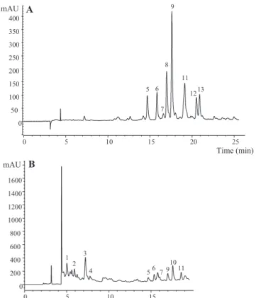
In the present study, thirteen individual phenolic compounds were detected and tentatively identified in the extracts prepared from Hovenia dulcispeduncles (Table 1; Fig. 2): three flavan-3-ols, eight flavonflavan-3-ols, one flavanone and one dihydrochalcone.
Peaks 1, 2 and 4 corresponded to flavan-3-ols. Peak 4 was pos-itively identified as (+)-catechin by comparison with a commercial standard, whereas peak 1, the major compound found in samples Hd02, Hd04 and Hd05, was assigned as (epi)gallocatechin based on its absorption and mass spectra. The pseudomolecular ion of peak 2 [M-H]atm/z593 was coherent with a proanthocyanidin (PA) dimer consisted of a gallocatechin (GC) and a catechin (C). The observation of an MS2 fragment ion at m/z 423 (
170 mu) from the Retro Diels-Alder (RDA) cleavage of a GC suggested that this was located in the upper subunit of the dimer, as the RDA has been indicated to occur preferentially in that unit (de Pascual et al., 2000; Friedrich, Eberhardt, & Galensa, 2000; Gu et al., 2003). Nevertheless, mass spectra do not allow to conclude about the precise identity of the units, whether (gallo)catechin or epi(gallo)catechin, so that peak 2 was tentatively identified as a prodelphinidin dimer (GC-C).
Peak 3 presented a pseudomolecular ion [M-H]at m/z 595 releasing MS2 fragment ions corresponding to losses of 90 and
sub-stituents, peak 3 was tentatively assigned as a C -dihexosyl-trihydroxyflavanone.
Peaks 5–9 and 11–13 showed absorption spectra characteristic of flavonols, which represented the largest group of phenolic com-pounds found inH. dulcissamples. The absorption spectrum of peak 5 pointed to a flavonol and a compound with the same pseudo-molecular ion ([M-H]atm/z431) was found byCho et al. (2013) inH. dulcisleaves and identified as kaempferol-3-O-rhamnoside; nevertheless, the fragmentation pattern of the compound obtained in our case was not coherent with that identity, with no fragments that could be associated to the aglycone. Thus, the identity of peak 5 remains controversial being just designed as an unknown flavo-noid. Peaks 11 (quercetin-3-O-rutinoside) and 12 (quercetin-3-O -glucoside) were positively identified by comparison of their reten-tion, mass and UV–vis characteristics with commercial standards. Similarly, peaks 6 ([M-H]- atm/z625) and 7 ([M-H]- atm/z479), yielding a unique MS2fragment atm/z317 (myricetin) from the loss
of 308 mu and 162 mu, respectively, were tentatively assigned to myricetin-O-rutinoside and myricetin-O-glucoside. Peaks 8 and 9 possessed the same pseudomolecular ion [M-H]atm/z771 releas-ing a unique MS2fragment ion atm/z301 associated to quercetin,
coherent with the loss of two hexosyl and one deoxyhexosyl resi-dues. The fact that the three sugar moieties are released simultane-ously to produce one daughter ion suggested that they were forming a trisaccharide. Although no information could be obtained about the nature and location of the sugar residues, they were ten-tatively identified as two possible quercetin-O -rutinosyl-glucosides. The presence of a kaempferol-3-O-rutinosyl-glucoside
(i.e., 3-O-a-L-rhamnopyranosyl-(1?6)-O-b-D-glucopyranosyl-(1? 2)-O-b-D-glucopyranoside) was previously reported in the leaves ofH. dulcisbyCho et al. (2013). Similar substitution pattern was observed for peak 13 ([M-H]atm/z785), although releasing a frag-ment atm/z315, corresponding to a methylquercetin, so that the compound was tentatively assigned as isorhamnetinO -rutinosyl-glucoside.
Finally, peak 10 ([M-H]atm/z597) also presented a fragmen-tation pattern consistent with C-glycosyl moieties. A compound with similar absorption and mass spectrum was identified as the dihydrochalcone phloretin-30,50-di-C-b-glucoside inCitrusspecies (Barreca, Bellocco, Caristi, Leuzzi, & Gattuso, 2011; Roowi & Crozier, 2011) andCyclopia subternata(de Beer et al., 2012), iden-tity was also tentatively assumed in our case.
Quantitatively,H. dulcispseudofruits in the last maturity stage (Hd05) presented the highest total phenolic compounds, mainly due to the presence of (epi)catechin, followed by Hd01, sample that gave the highest content of all the remaining phenolic compounds detected. The astringency of unripe or immature pseudofruits is a consequence of the presence of intermediate molecular weight tannins, mainly ‘‘condensed or hydrolysables tanning agents” (mostly catechins and proanthocyanidins), that reduce its content during ripening process, mainly due to complex-ation (with fiber or proteins) and polymeriscomplex-ation processes (Belitz, Grosh, & Schieberle, 2009; Ozawa, Lilley, & Haslam, 1987), this fact was observed in persimmon fruits and dwarf cashew pseudofruits (de Figueiredo, Lajolo, Alves, & Filgueiras, 2002; Del Bubba et al., 2009). This could also explain the reduction in catechins Table 1
Retention time (Rt), wavelengths of maximum absorption (kmax), mass spectral data, tentative identification and quantification (mg/g extract) of phenolic compounds inHovena dulcisThunb. pseudofruits at different maturity stages (means ± SD, n = 9).
Peak Rt (min)
kmax(nm) Molecular
ion [M-H](m/z)
MS2(m/z) Tentative
identification
Hd01 Hd02 Hd03 Hd04 Hd05
1 5.0 278 305 219(37), 177(16), 167(36), 137(32), 125(65)
(Epi)gallocatechin1 2.9 ± 0.3 3.8 ± 0.1 1.1 ± 0.1 5.5 ± 0.1 21.3 ± 0.4
2 6.0 276 593 423(78), 305(100), 289(27) Prodelphinidin dimer (GC-C)a,1
1.60 ± 0.04 1.45 ± 0.01 0.9 ± 0.1 1.3 ± 0.1 1.40 ± 0.01 3 7.2 293,342sh 595 577(8), 505(18), 487(8), 475
(33), 457(19), 415(25), 385 (54), 355(53), 343(3), 313(8)
Trihydroxyflavanone di-C-glucoside2
3.3 ± 0.2 2.6 ± 0.2 3.17 ± 0.01 1.66 ± 0.01 1.2 ± 0.1
4 7.8 280 289 245(10), 203(9), 187(6), 161 (4), 137(22)
(+)-Catechin1 0.8 ± 0.1 0.7 ± 0.1 0.22 ± 0.01 nd nd
5 14.7 356 431 385(4), 367(20), 221(8), 205 (100), 187(13), 161(4), 131 (11)
Unknown flavonoid3 0.46 ± 0.01 0.01 ± 0.01 0.27 ± 0.02 tr tr
6 15.8 354 625 317(100) Myricetin-O -rutinoside3
0.48 ± 0.01 tr 0.17 ± 0.02 tr 0.14 ± 0.03 7 16.5 344 479 317(100) Myricetin-O
-glucoside4
tr nd tr tr 0.22 ± 0.01 8 16.9 356 771 301(100) Quercetin-O
-rutinosyl-glucoside3
1.13 ± 0.04 0.10 ± 0.01 0.61 ± 0.02 0.14 ± 0.01 0.11 ± 0.02 9 17.6 356 771 301(100) Quercetin-O
-rutinosyl-glucoside3
2.76 ± 0.02 0.22 ± 0.01 1.11 ± 0.03 0.50 ± 0.04 0.51 ± 0.02 10 18.6 284,336sh 597 579(13), 507(14), 489(17),
477(67), 459(29), 417(67), 387(95), 357(100), 315(16)
Phloretin-30,50-di-C
-b-glucoside5
1.5 ± 0.1 0.46 ± 0.03 0.95 ± 0.01 nd nd
11 19.1 356 609 301(100) Quercetin-3-O -rutinoside3
0.88 ± 0.00 tr 0.28 ± 0.03 tr 0.04 ± 0.03 12 20.5 352 463 301(100) Quercetin-3-O
-glucoside4
0.37 ± 0.00 tr 0.050 ± 0.001 tr tr 13 20.9 356 785 315(100) Isorhamnetin-O
-rutinosyl-glucoside6
0.50 ± 0.01 tr 0.03 ± 0.01 tr tr
Total phenolics compounds
16.6 ± 0.2b 9.3 ± 0.4c 9.0 ± 0.3c 9.2 ± 0.1c 24.7 ± 0.3a
a C: (epi)catechin, GC: (epi)gallocatechin. tr-taces; nd-not detected. Phenolic compounds standards used for the quantification: 1 - catechin (y = 131.65x + 4.11; R2= 0.999),
2 - naringenin (y = 539.98x + 161.46; R2= 0.998), 3 - quercetin-3-O-rutinoside (y = 280.87x + 373.73; R2= 0.999), 4 - quercetin-3-O-glucoside (y = 336,36x + 358,06;
R2= 0.999), 5 - taxifolin (y = 224.31x + 148.41; R2= 0.999) and 6-isorhamentin-3-O-rutinoside (y = 284.12x + 67.05; R2= 0.999). In each row, letters mean significant
(flavan-3-ols) in the samples studied herein. Moreover, other important effect observed during pseudofruits maturity was the increase in (epi)gallocatechin (caracteristic compound present in black tea) which is normally synthesized after flavonols oxidation (polyphenoloxidase reaction) and polymerization process (Belitz et al., 2009), this fact could also explain the reduction in catechin and flavonols content (mainly quercetin derivatives) during ripen-ing process.
3.1.2. Lipophilic compounds
Fatty acid and tocopherol profiles of the analysed pseudofruits are shown inTable 2. Saturated fatty acids (SFA) were the predom-inant lipid fraction at all maturity stages (average values of 44.45 – 58.60%), being palmitic acid (C16:0) the predominant one. The highest relative percentage was observed in the most immature stages (Hd01), decreasing the content along the ripening process.Ding, Liang, and Teng (1997)also reported SFA as predom-inated fatty acids (87.46%) inH. dulcisseeds. Polyunsaturated fatty acids (PUFA) fraction with linoleic acid as the major one (LA, C18:2n6) increased during ripening process, being observed the highest relative percentages in Hd04 (PUFA: 51.70%; LA: 35.90%), which is consider as mature being the most suitable moment for consumption. In an overview, MUFA were the lowest lipids present, while both SFA and PUFA showed similar concentrations. Total tocopherols increased during ripening process, as hap-pened with fatty acids, being the highest values observed in Hd04 (5.66 mg/100 g dw; Table 2). All forms were identified in the studied peduncles, exceptd-tocopherol in Hd05 pseudofruits.
a-Tocopherol was the major form in all ripening stages, being
the highest value observed in Hd04 (5.43 mg/100 g dw). To the authors best of knowledge, this is the first report about tocopherols content inH. dulcispeduncles.3.2. Bioactive properties of Hovenia dulcis pseudofruits
3.2.1. Antioxidant activity
Antioxidant mechanisms in biological tissues are extremely complex, and there is not one method that can provide unequivo-cal results (Carocho & Ferreira, 2013). Thus, the antioxidant capac-ity of H. dulcis peduncles (hydromethanolic extracts) was evaluated by three differentin vitroassays. DPPH scavenging activ-ity and reducing power (Ferricyanide/Prussian blue assay) were used to evaluate total antioxidant capacity, while b-carotene/ linoleate assay was applied to determine the inhibition of lipid per-oxidation process. The evolution of antioxidant capacity during ripening process can be observed in Table 3. Regarding total antioxidant capacity, the most immature peduncles (Hd01) pre-sented higher antioxidant properties (lower EC50values)
compar-ing with the peduncles at mature stages, mainly Hd04. This could probably be explained by the presence of the highest pheno-lic compounds concentration, with the exception of (epi)gallocate-chin, which was higher in Hd05, than in the maturity stage (Hd01). A trend of decreasing (from Hd01 to Hd04) and increasing (from Hd04 to Hd05) in the antioxidant capacity was detected using these methods. To the authors best knowledge, this is the first report of lipid peroxidation inhibition process inH. dulcis pseud-ofruits, the mature peduncle (Hd04) presented the highest antiox-idant capacity comparing with the other ripening stages; probably, this tendency could be related with the increasing content of PUFA and tocopherols observed during the ripening process. Since PUFA and particularly tocopherols are involved in lipid peroxidation pro-cess. The most important biological role of vitamin E is to protect PUFA, other components of cell membranes, and LDL, from oxida-tion by free radicals, and it is particularly effective in preventing lipid peroxidation, limiting the accumulation of high levels of products derived from this process that are associated with numer-ous diseases and clinical conditions. Its action as an antioxidant is due to the donation of a hydrogen atom to peroxyl radicals of unsaturated lipid molecules, forming a hydroperoxide and a toco-pheroxyl radical, which reacts with peroxyl radicals forming more stable adducts (Duthie, 1993; Nogala-Kalucka, Kupczyk, Polewski, Siger, & Dwiecki, 2007).
A similar trend was observed in the same samples using other different antioxidant assays (ABTS and FRAP methods), as reported byMaieves et al. (2015b). In that study total antioxidant capacity decreased from Hd01 to Hd03, being closely related with total polyphenol contents (evaluated as families of compounds).
Hu, Lee, and Wang (2010)evaluated the DPPH scavenging activ-ity of maturedH. dulcisfruit extracts obtaoned using different sol-vents (water, hot water, methanol and ethyl acetate), and reported 12.72% at 100
lg/mL for the methanolic extract.
Basavegowda, Idhayadhulla, and Lee (2014)also described interesting values for DPPH and FRAP assays in Golden nanoparticles made fromH. dulcis fruits hot aqueous extract.Other authors also reported antioxidant activity of other botan-ical parts ofH. dulcis;Cho et al. (2013), described DPPH values of leaves extracts obtained using different solvents (e.g., methanol, ethanol, hot water, n-hexane) and their corresponding isolated compounds, being the ethanolic and methanolic extracts the ones with better results (SC50(50% DPPH free radical scavenging
con-centration) was 83 and 112
lg/mL, respectively). While,
Li et al. (2005)reported DPPH and ABTS values ofH. dulcisstem extracts (e.g., methanol, water, ethyl acetate), and the best results were obtained with the ethyl acetate stem extract (IC50= 18.3 and4.9
lg/mL for DPPH and ABTS assays, respectively).
3.2.2. Antitumor activity
Table 3presents the antitumor and hepatotoxic activity of the hydromethanolic extracts ofH. dulcisat different stages of
matura-A
B
Time (min)
0 5 10 15 20 25
mAU
0 50 100 150 200 250 300 350 400
5 6
7 8
9
11
1213
Time (min)
0 5 10 15
mAU
0 200 400 600 800 1000 1200 1400 1600
1 2
3
4 567 9
10 11
tion. Ellipticine, a very strong antitumor compound, which interca-lates with DNA and inhibits topoisomerase II was used as a positive control, although it has the important disadvantage of being highly toxic also for non-tumor cells. The tested human tumor cell lines of breast, colon, cervical, and liver carcinoma are some of the most used for primary screening of antitumor potential. Only the most immature stages showed antitumor activity against all the tested cell lines; the lowest GI50 values were observed with Hd01 for
HCT15 (colon carcinoma) and HepG2 (hepatocellular carcinoma) (78.58 and 82.34
lg/mL, respectively). None of the extracts
showed inhibition towards the non-tumor liver primary culture(PLP2), corroborating that the use of these pseudofruits is safe at this level.
Other authors reported antitumor activity of H. dulcis fruits and leaves against different tumor cell lines. Lee et al. (1999)
reported that ethanol extract of H. dulcis fruits exhibited a potent growth inhibitory activity of Hep3B and MCF-7 cell lines, whereas this extract did not show considerable cytotoxicity on HEL299 cell lines. Castro et al. (2002) reported that ethanol extract of H. dulcis pseudofruits possessed high degree of selectivity against SP2/0 mouse myeloma and BW lymphoma cell in vitro.
Table 2
Fatty acids profile (relative percentage) and tocopherols content (mg/100 g dw) ofHovenia dulcisThunb. pseudofruits at different maturity stages (mean ± SD, n = 9).
Hd01 Hd02 Hd03 Hd04 Hd05
Fatty acids profile (relative percentage)
C6:0 0.30 ± 0.01 0.100 ± 0.001 0.100 ± 0.001 0.25 ± 0.03 0.20 ± 0.01 C8:0 0.45 ± 0.05 0.35 ± 0.04 0.25 ± 0.05 0.20 ± 0.06 0.15 ± 0.03 C10:0 0.45 ± 0.05 0.40 ± 0.01 0.25 ± 0.05 0.200 ± 0.001 0.15 ± 0.03 C12:0 0.85 ± 0.05 0.70 ± 0.01 0.65 ± 0.05 0.50 ± 0.01 0.40 ± 0.01
C13:0 nd nd 0.100 ± 0.001 nd 0.100 ± 0.001
C14:0 1.2 ± 0.1 1.15 ± 0.05 1.2 ± 0.1 1.1 ± 0.1 0.80 ± 0.10 C15:0 0.50 ± 0.01 0.40 ± 0.01 0.50 ± 0.01 0.70 ± 0.07 0.40 ± 0.06 C16:0 38 ± 1 31.95 ± 0.05 30.85 ± 0.05 29.7 ± 0.1 30.8 ± 0.3 C16:1 0.25 ± 0.05 0.70 ± 0.01 0.70 ± 0.07 0.80 ± 0.01 0.85 ± 0.09 C17:0 0.8 ± 0.1 0.70 ± 0.01 0.65 ± 0.05 0.60 ± 0.01 0.55 ± 0.05 C18:0 6.6 ± 0.5 5.8 ± 0.4 5.1 ± 0.4 4.55 ± 0.05 4.75 ± 0.05 C18:1n9 6.2 ± 0.2 5.95 ± 0.05 3.1 ± 0.2 3.10 ± 0.01 4.9 ± 0.3 C18:2n6 23 ± 2 30.05 ± 0.05 35.4 ± 0.2 35.9 ± 0.3 35.2 ± 0.3 C18:3n3 12 ± 1 14.9 ± 0.2 15.1 ± 0.1 15.5 ± 0.1 13.8 ± 0.3 C20:0 1.6 ± 0.2 1.1 ± 0.1 1.05 ± 0.05 1.10 ± 0.01 1.10 ± 0.01
C20:1 nd 0.30 ± 0.06 nd nd nd
C20:3n3 + C21:0 0.45 ± 0.05 0.35 ± 0.05 0.25 ± 0.05 0.30 ± 0.01 0.30 ± 0.01 C22:0 5.5 ± 0.6 3.1 ± 0.1 2.9 ± 0.2 3.45 ± 0.05 3.6 ± 0.1 C23:0 nd 0.40 ± 0.01 0.35 ± 0.04 0.40 ± 0.01 0.30 ± 0.01 C24:0 2.75 ± 0.05 1.65 ± 0.05 1.7 ± 0.1 1.70 ± 0.01 1.85 ± 0.05 SFA 59 ± 3 47.8 ± 0.3 45.7 ± 0.6 44.5 ± 0.4 45.1 ± 0.2 MUFA 6.4 ± 0.1 6.95 ± 0.05 3.7 ± 0.3 3.90 ± 0.01 5.7 ± 0.4 PUFA 35 ± 3 45.3 ± 0.2 50.7 ± 0.2 51.7 ± 0.4 49.2 ± 0.5 n6/n3 1.91 ± 0.05 1.98 ± 0.02 2.30 ± 0.01 2.27 ± 0.01 2.50 ± 0.03
Tocopherols (mg/100 g dw)
a- tocopherol 0.183 ± 0.005a 2.71 ± 0.02b 4.45 ± 0.02d 5.43 ± 0.06e 4.06 ± 0.03c
b-tocopherol 0.038 ± 0.005a 0.104 ± 0.009d 0.038 ± 0.002a 0.048 ± 0.002b 0.088 ± 0.003c c-tocopherol 0.023 ± 0.003a 0.074 ± 0.004b 0.089 ± 0.001c 0.070 ± 0.001b 0.069 ± 0.004b
d-tocopherol 0.052 ± 0.009b 0.064 ± 0.006c 0.037 ± 0.002a 0.060 ± 0.004c nd Total tocopherols 0.30 ± 0.01a 2.96 ± 0.01b 4.7 ± 0.1d 5.7 ± 0.1e 4.24 ± 0.05c nd - non-detected; dw - dry weight.
In each row, letters mean significant differences between stages of maturation (p <0.05).
Table 3
Antioxidant and antitumor properties of the hydromethanolic extracts obtained fromHovenia dulcisThunb. pseudofruits at different maturity stages (mean ± SD, n = 9).
Hd01 Hd02 Hd03 Hd04 Hd05
Antioxidant activity (EC50, mg/mL)
DPPH scavenging activity 0.14 ± 0.004a 0.24 ± 0.02b 1.6 ± 0.2c 3.58 ± 0.04e 3.0 ± 0.1d Ferricyanide/Prussian blue assay 0.13 ± 0.001a 0.20 ± 0.003b 1.04 ± 0.02c 1.89 ± 0.02e 1.21 ± 0.004d
b-carotene bleaching inhibition 0.96 ± 0.04d 0.53 ± 0.01c 0.26 ± 0.01b 0.140 ± 0.001a 0.28 ± 0.01b
Antitumor activity (GI50values,lg/mL)
MCF-7 (breast carcinoma) 247 ± 5a 344 ± 5b >400 >400 >400 NCI-H460 (non-small cell lung cancer) 79 ± 5a 110 ± 9b >400 >400 >400 HeLa (cervical carcinoma) 274 ± 6a 323 ± 21b >400 >400 >400 HepG2 (hepatocellular carcinoma) 82 ± 2a 216 ± 0.1b >400 >400 >400
Hepatotoxicity (GI50values,lg/mL)
PLP2 >400 >400 >400 >400 >400
In each column different letters mean significant differences between stages of maturation (p <0.05). The antioxidant activity was expressed as EC50values, what means that
higher values correspond to lower reducing power or antioxidant potential. EC50: Extract concentration corresponds to 50% of antioxidant activity or 0.5 of absorbance in
reducing power assay (Ferricyanide/Prussian blue assay). Trolox EC50values: 41lg/mL (reducing power), 42lg/mL (DPPH scavenging activity), 18lg/mL (b-carotene
bleaching inhibition). GI50values correspond to the sample concentration achieving 50% of growth inhibition in human tumor cell lines or in liver primary culture PLP2.
3.2.3. Antimicrobial activity
According to the World Health Organisation (WHO, 2014) the bacterial infections which contribute most to human disease are also those in which emerging and microbial resistance is most evident, such as, diarrheal diseases, respiratory tract infections, meningitis, and hospital-acquired (nosocomial) infections. The following are some common examples of bacteria species; penicillin-resistant Streptococcus pneumoniae, methicillin-resistant Staphylococcus aureus, and multi-resistant Salmonellae and the resistant Escherichia Coli, among others. All of them responsible of relevant illness such as bloody diarrhea, abdominal cramps, nausea, vomiting, and fever are the symptoms caused by E. coliO157:H7 haemorrhagic colitis. Other important bacteria’s commonly responsible of food intoxication areStaphylococcus aur-eus, whose enterotoxin include diarrhea, abdominal cramps, nau-sea, and vomiting (de Camargo, Regitano-d’Arce, Gallo, & Shahidi, 2015).
To the authors best knowledge, antimicrobial studies ofH. dulcis pseudofruits are scarce; onlyCho, Moon, and Park (2000), Cho,
Moon, Eun, Chung, and Park (2004) and Basavegowda et al.
(2014) reported antimicrobial activity of pseudofruits extracts. Other authors reported antiparasitic and antimicrobial activity of different extracts from other botanical parts ofH. Dulcis. Therefore,
Gadelha et al. (2005)reported that the dichloromethane fraction from the methanol extract ofH. dulcisleaves inhibited the growth ofGiardia lambliatrophozoites (IC50= 12
lg/mL). Also,
Castro et al. (2002)reported that the activity of the aqueous extracts of fruit stalks and methanolic extracts of leaves from seedlings ofH. dulcis could inhibit the multiplication of Trypanosoma cruzi. Moreover, the hot water extracts from leaves and stems ofH. dulcis have shown antimicrobial activity against positive and Gram-negative bacteria, and yeast (Cho, Moon, Eun, Chung, & Park, 2004; Cho, Moon, & Park, 2000).In the present study, pseudofruits phenolic extracts were eval-uated against different Gram positive and Gram negative bacteria, namelyEscherichia coli, Enterococcus faecalis, Pseudomonas aerugi-nosa, Staphylococcus epidermidis and Staphylococcus aureus (Table 4).In general terms, the antimicrobial activity in the anal-ysed samples was severely influenced by the maturation process. The most immature stages (Hd01 and Hd02) presented higher activity (lower MIC values), as observed forStaphylococcus epider-midis, Sthaphylococcus aureus and Pseudomonas aeruginosa, although no growth inhibition was observed forEnterococcus fae-calisandEscherichia coli. On the other hand, the highest MIC values were observed with Hd04 for all tested bacteria. The stage of mat-uration Hd03 only inhibited the growth inhibition ofEnterococccus faecalis, at a concentration of 50
lg/mL.
Numerous research groups have sought to elucidate the antibacterial mechanisms of action of selected flavonoids. The activity of quercetin, for example, has been at least partially attrib-uted to inhibition of DNA gyrase. It has also been proposed that sophoraflavone G and (-)-epigallocatechin gallate inhibit cytoplas-mic membrane function, and that licochalcones A and C inhibit energy metabolism. Other flavonoids whose mechanisms of action
have been investigated include robinetin, myricetin, apigenin, rutin, galangin, 2,4,20-trihydroxy-50-methylchalcone and lon-chocarpol A (Cushnie & Lamb, 2005). The antiomicrobial activity of the evaluated extract could be related with the flavonoids con-tent. From the obtained results, it can be seen that the better antibacterial activity, mainly againstPseudomonas aeruginosaand Staphylococcus aureus,was observed in Hd01 sample (the most immature pseudofruit stage), which present the highest catechin and quercetin derivatives content. Otherwise, forEnterococcus fae-calis, the better antibacterial results was observed in the most mature pseudofruit stage (Hd05) which present the highest (Epi)-gallocatechin content (21.3 mg/g extract).
Our results were according with those reported byCho et al. (2000)inH. dulcispseudofruits extract, againstStaphylococcus aur-eus,Staphylococcus epidermidis,Bacillus subtilis,Micrococcus luteus, Pediococcus damnosus, Escherichia coli, Pseudomonas aeruginosa andSalmonella typhi.
Generally, phenolic content tends to be higher in immature fruits since they are utilized as secondary metabolites for defence mechanisms of the plants, and thus they protect young fruits and allow their maturation and reproduction function (Fennema, 1996). Different compounds have been previously reported in the methanolic extracts ofH. dulcisto provide antimicrobial effects. In this way, extracts fromH. dulcis leaves showed antimicrobial activity against Staphylococcus aureus and Escherichia coli (Cho et al., 2004), being 3(Z)-dodecenedioic acid the antimicrobial-active compound isolated in these leaves. Furthermore,Cho et al. (2000)described that the methanolic fraction of hot-water extract of H. dulcis showed antioxidant and antimicrobial properties. Vanillic acid and ferulic acid were isolated from this fraction, and exhibited antimicrobial activity against Gram negative and Gram positive bacteria, and yeasts.
4. Conclusions
This is the first study on individual phenolic compounds, fatty acids and tocopherols, as well as antitumor and antibacterial activ-ity ofH. dulcispseudofruits through the maturation process. This underutilized product demonstrated to be a rich source of these bioactive compounds showing antioxidant, antitumor and antibac-terial properties with an interesting activity in the immature stages (Hd01 and Hd02). This study supports the potential applica-tion ofH. dulcispeduncles as alternative bioactive ingredients for functional foods, dietary supplements or nutraceuticals.
Acknowledgements
The authors are grateful to the Foundation for Science and Tech-nology (FCT, Portugal) and FEDER under Programe PT2020 for financial support to CIMO (UID/AGR/00690/2013), LSRE (Project UID/EQU/50020/2013), H. Maieves (CAPES/PDSE and BEX 3480/13-5), L. Barros (SFRH/BPD/107855/2015), R.C. Calhelha (SFRH/BPD/BPD/68344/2010) and I. Dias (SFRH/BD/84485/2012) grants.
Table 4
Antibacterial activity (MIC values,lg/mL) of the hydromethanolic extracts prepared fromHovenia dulcisThunb. pseudofruits at different stages of maturation (mean ± SD, n = 9).
Tested bacteria Hd01 Hd02 Hd03 Hd04 Hd05
Enterococcus faecalis ni ni 50 25 12.5
Pseudomonas aeruginosa 3.12 6.25 ni 25 12.5
Streptococcus epidermidis 25 25 ni 50 25
Staphylococcus aureus 6.25 6.25 ni 25 25
Escherichia coli ni ni ni 50 25
ni - no inhibition; MIC - minimal inhibitory concentration.
References
Abreu, R. M. V., Ferreira, I. C. F. R., Calhelha, R. C., Lima, R. T., Vasconcelos, M. H., & Adega, F. (2011). Anti-hepatocellular carcinoma activity using human HepG2 cells and hepatotoxicity of 6-substituted methyl 3-aminothieno[3,2-b]pyridine-2-carboxylate derivatives:In vitro evaluation, cell cycle analysis and QSAR studies.European Journal of Medical Chemistry, 46, 5800–5806.
Barreca, D., Bellocco, E., Caristi, C., Leuzzi, U., & Gattuso, G. (2011). Kumquat (Fortunella japonica Swingle) juice: Flavonoid distribution and antioxidant properties.Food Research International, 44, 2190–2197.
Barros, L., Pereira, E., Calhelha, R. C., Dueñas, M., Carvalho, A. M., Santos-Buelga, C., et al. (2013). Bioactivity and chemical characterization in hydrophilic and lipophilic compounds ofChenopodium ambrosioides L..Journal of Functional Foods, 5, 1732–1740.
Basavegowda, N., Idhayadhulla, A., & Lee, Y. R. (2014). Phyto-synthesis of gold nanoparticles using fruit extract of Hovenia dulcis and their biological activities. Industrial Crops and Products, 52, 745–751.
Belitz, H. D., Grosh, W., & Schieberle, P. (2009).Food chemistry(4th ed.). Berlin Heidelberg: Springer-Verlag.
Carocho, M., Barros, L., Calhelha, R. C., C´iric´, A., Sokovic´, M., Santos-Buelga, C., et al. (2015).Melissa officinalis L. decoctions as functional beverages: A bioactive approach and chemical characterization.Food & Function, 6, 2240–2248.
Carocho, M., & Ferreira, I. C. F. R. (2013). A review on antioxidants, prooxidants and related controversy: Natural and synthetic compounds, screening and analysis methodologies and future perspectives. Food and Chemical Toxicology, 51, 15–25.
Carvalho, P. E. R. (1994).Ecologia, silvicultura e usos da uva-do-japão (hoveniadulcis thunberg)(pp. 24–65). Colombo: EMBRAPA Florestas.
Castro, T. C., Pelliccione, V. L. B., Figueiredo, M. R., Soares, R. O. A., Bozza, M. T., Viana, V. R. C., et al. (2002). Atividade antineoplásica e tripanocida deHovenia dulcis Thunb. cultivada in vivo e in vitro.Revista Brasileira de Farmacognosia, 12, 96–99.
Cha, B. C., Lee, E. H., Lee, E., & Park, H. H. (2004). Activity of glutathione S-transferase and effect of alcohol decomposition on the fruit ofHovenia dulcisThunb.Yakhak Hoeji, 48, 213–217.
Chaturvedula, V. S. P., & Ruo, H. (2013). Isolation and NMR spectral studies of dihydromyricetin.Journal of Pharmacognosy and Phytochemistry, 2, 113–115.
Chitarra, M. I. F., & Chitarra, A. B. (2005).Pós-colheita de frutos e hortaliças: Fisiologia e manuseio(2nd ed.). Lavras: UFLA.
Cho, J. Y., Hyun, S. H., Moon, J. H., & Park, K. H. (2013). Isolation and structural determination of a novel flavonol triglycoside and 7 compounds from the leaves of oriental raisin tree (Hovenia dulcis) and their antioxidative activity. Food Science and Biotechnology, 22, 115–123.
Cho, J. Y., Moon, J. H., Eun, J. B., Chung, S. J., & Park, K. H. (2004). Isolation and Characterization of 3(Z)-Dodecenedioic Acid as an Antibacterial Substance from Hovenia dulcisThunb.Food Sciences and Biotechnology, 13, 46–50.
Cho, J. Y., Moon, J. H., & Park, K. H. (2000). Isolation and identification of 3-methoxy-4-hydroxybenzoic acid and 3-methoxy-4-hydroxycinnamic acid from hot water extracts ofHovenia dulcisThunb and confirmation of their antioxidative and antimicrobial activity.KoSFoST, 32, 1403–1408.
Cushnie, T. P., & Lamb, A. J. (2005). Antimicrobial activity of flavonoids.International Journal of Antimicrobial Agents, 26(5), 343–356.
Cuyckens, F., & Claeys, M. (2004). Mass spectrometry in the structural analysis of flavonoids.Journal of Mass Spectrometry, 39, 1–15.
de Beer, D., Schulze, A. E., Joubert, E., de Villiers, A., Malherbe, C. J., & Stander, M. A. (2012). Food ingredient extracts ofCyclopia subternata(Honeybush): Variation in phenolic composition and antioxidant capacity.Molecules, 17, 14602–14624.
de Camargo, A. C., Regitano-d’Arce, M. A. B., Gallo, C. R., & Shahidi, F. (2015). Gamma-irradiation induced changes in microbiological status, phenolic profile and antioxidant activity of peanut skin.Journal of Functional Foods, 12, 129–143.
de Figueiredo, R. W., Lajolo, F. M., Alves, R. E., & Filgueiras, H. A. C. (2002). Physical– chemical changes in early dwarf cashew pseudofruits during development and maturation.Food Chemistry, 77(3), 343–347.
de Pascual-Teresa, S., Santos-Buelga, C., & Rivas-Gonzalo, J. G. (2000). Quantitative analysis of flavan-3-ols in Spanish foodstuffs and beverages. Journal of Agricultural and Food Chemistry, 48, 5331–5337.
Del Bubba, M., Giordani, E., Pippucci, L., Cincinelli, A., Checchini, L., & Galvan, P. (2009). Changes in tannins, ascorbic acid and sugar content in astringent persimmons during on-tree growth and ripening and in response to different postharvest treatments. Journal of Food Composition and Analysis, 22(7), 668–677.
Ding, L. S., Liang, Q. L., & Teng, Y. F. (1997). Study on flavonoids in seeds ofHovenia dulcis.Acta Pharmaceutica Sinica, 32, 600–602.
Duarte, M. C. T., Figueira, G. M., Sartoratto, A., Rehder, V. L. G., & Delarmelina, C. (2005). Anti-Candida activity of Brazilian medicinal plants. Journal of Ethnopharmacology, 97, 305–311.
Duthie, G. G. (1993). Lipid peroxidation.European Journal of Clinical Nutrition, 47 (11), 759.
Fennema, O. R. (1996).Food chemistry(3rd ed.). New York: Marcel Dekkan.
Ferreres, F., Silva, B. M., Andrade, P. B., Seabra, R. M., & Ferreira, M. A. (2003). Approach to the study of C-glycosyl flavones by ion trap HPLC-PAD-ESI/MS/MS: Application to seeds of quince (Cydonia oblonga).Phytochemical Analysis, 14, 352–390.
Friedrich, W., Eberhardt, A., & Galensa, R. (2000). Investigation of proanthocyanidins by HPLC with electrospray ionization mass spectrometry. European Food Research and Technology, 211, 56–64.
Gabrielson, J., Hart, M., Jarelöv, A., Kuhn, I., Mckenzie, D., & Möllby, R. (2002). Evaluation of redox indicators and the use of digital scanners and spectrophotometer for quantification of microbial growth in microplates. Journal of Microbiological Methods, 50, 63–73.
Gadelha, A. P. R., Vidal, F., Castro, T. M., Lopes, C. S., Albarello, N., Coelho, M. G. P., et al. (2005). Susceptibility of Giardia lamblia to Hovenia dulcis extracts. Parasitology Research, 97(5), 399–407.
Gu, L., Kelm, M. A., Hammerstone, J. F., Zhang, Z., Beecher, G., Holden, J., et al. (2003). Liquid chromatographic/electrospray ionization mass spectrometric studies of proanthocyanidins in foods.Journal of Mass Spectrometry, 38, 1272–1280.
Guo, J., Meng, Y., Zhao, Y., Hu, Y., Ren, D., & Yang, X. (2015). Myricetin derived from Hovenia dulcisThunb. ameliorates vascular endothelial dysfunction and liver injury in high choline-fed mice.Food & Function, 6, 1620–1634.
Hase, K., Ohsugi, M., Xiong, Q., Basnet, P., Kadota, S., & Namba, T. (1997). Hepatoprotective effect of Hovenia dulcis THUNB. on experimental liver injuries induced by carbon tetrachloride or D-galactosamine/ lipopolysaccharide.Biological and Pharmaceutical Bulletin, 20(4), 381–385.
Hu, W., Lee, K. Y., & Wang, M. H. (2010). Antioxidant activities of various extracts of Hovenia dulcisThunb fruits.Korean Journal of Plant Resources, 23(3), 207–213.
Hung, R. J., Yazdani, U., Yoon, J., Wu, H., Yang, T., Gupta, N., et al. (2010). Mical links semaphorins to F-actin disassembly.Nature, 463, 823–827.
Hyun, T. K., Eom, S. H., Yu, C. Y., & Roitsch, T. (2010).Hovenia dulcis– An Asian traditional herb.Planta Medica, 76, 943–949.
Ji, Y., Yang, P., & Li, J. (2000). Preventive effect of Hovenia dulcis Thunb on alcohol-induced liver injury.Pharmacology and Clinics of Chinese Material Medica, 6, 9–20.
Kiyoshi, S. (1987). Effect of water extracts of crude drugs in decreasing blood alcohol concentration in rats.Chemical Pharmacology Bulletin, 35, 4597–4604.
Lee, M. K., Kim, Y. G., An, S. W., Kim, M. H., Lee, J. H., & Lee, H. Y. (1999). Biological activities ofHovenia dulcisThunb.Korean Journal of Medicinal Crop Science, 7, 185–192.
Li, G., Min, B. S., Zheng, C., Lee, J., Oh, S. R., Ahn, K. S., et al. (2005). Neuroprotective and free radical scavenging activities of phenolic compounds fromHovenia dulcis.Archives of Pharmacal Research, 28, 804–809.
Maieves, H. A., Bosmuler Züge, L. C., de Paula Scheer, A., Ribani, R. H., Morales, P., & Sánchez-Mata, M. C. (2016). Physical properties and rheological behavior of pseudofruits of Hovenia dulcis Thunb. in different maturity stages.Journal of Texture Studies.http://dx.doi.org/10.1111/jtxs.12199.
Maieves, H. A., López-Froilán, R., Morales, P., Pérez-Rodríguez, M. L., Ribani, R. H., Cámara, M., et al. (2015b). Antioxidant phytochemicals ofHovenia dulcisThunb. peduncles in different maturity stages. Journal of Functional Foods, 18, 1117–1124.
Maieves, H. A., Ribani, R. H., Morales, P., & Sánchez-Mata, M. C. (2015a). Evolution of the nutritional composition ofHovenia dulcisThunb. pseudofruit during the maturation process.Fruits, 70, 181–187.
Morales, P., Barros, L., Ramírez-Moreno, E., Santos-Buelga, C., & Ferreira, I. C. F. R. (2015). Xoconostle fruit (Opuntia matudaeScheinvar cv. Rosa) by-products as potential functional ingredients.Food Chemistry, 185, 289–297.
NCCLS - National Committee For Clinical Laboratory Standards (1997). Method for Broth Dilution Antifungical Susceptibility Testing of Yeast; Approved Standard. Villanova, v. 17, p. 28. M27-A.
Nogala-Kalucka, M., Kupczyk, B., Polewski, K., Siger, A., & Dwiecki, K. (2007). Influence of native antioxidants on the formation of fatty acid hydroperoxides in model systems.European Journal of Lipid Science and Technology, 109(10), 1028–1037.
Ozawa, T., Lilley, T. H., & Haslam, E. (1987). Polyphenol interactions: Astringency and the loss of astringency in ripening fruit.Phytochemistry, 26(11), 2937–2942.
Park, J. S., Kim, I. S., Rehman, S. U., Na, C. S., & Yoo, H. H. (2015). HPLC determination of bioactive flavonoids in Hovenia dulcis fruit extracts. Journal of Chromatographic Science, bmv114.
Pinto, J. T., Oliveira, T. T. D., Alvarenga, L. F., Barbosa, A. S., Pizziolo, V. R., & Costa, M. R. D. (2014). Pharmacological activity of the hydroalcoholic extract from Hovenia dulcis thunberg fruit and the flavonoid dihydromyricetin during hypercholesterolemia induced in rats. Brazilian Journal of Pharmaceutical Sciences, 50, 727–735.
Qiao, X., He, W. N., Xiang, C., Han, J., Wu, L. J., Guo, D. A., et al. (2011). Qualitative and quantitative analyses of flavonoids in spirodela polyrrhiza by high-performance liquid chromatography coupled with mass spectrometry. Phytochemical Analysis, 22, 475–483.
Rigatto, P. A., Pereira, J. C. D., Mattos, P. P., & Schaitza, E. G. (2001). Características Físicas, Químicas e Anatômicas da Madeira de Hovenia dulcis. Comunicado Técnico – Embrapa Florestas - Colombo – PR.
Roowi, S., & Crozier, A. (2011). Flavonoids in tropical citrus species.Journal of Agricultural and Food Chemistry, 59, 12217–12225.
WHO (2014). Antimicrobial resistance: global report on surveillance 2014, ISBN: 978 92 4 156474 8.