UNIVERSIDADE DA BEIRA INTERIOR
Ciências da Saúde
Efeito de Antidiabéticos em Células de Sertoli
Humanas: Implicações Para a Fertilidade
Masculina
Maria João Carvalho Meneses Oliveira
Dissertação para obtenção do Grau de Mestre em
Ciências Biomédicas
(2° ciclo de estudos)
Orientador: Prof. Doutor Marco G. Alves (CICS-UBI)
Co-orientador: Prof. Doutor Pedro Fontes Oliveira (CICS-UBI/ICBAS-UP) Co-orientador: Prof. Doutor Mário Sousa (ICBAS-UP)
UNIVERSIDADE DA BEIRA INTERIOR
Ciências da Saúde
Effect of Antidiabetic Drugs in Human Sertoli
Cells: Implications for Male Fertility
Maria João Carvalho Meneses Oliveira
Master Degree Thesis in Biomedical Science
Ciências Biomédicas
(2nd cycle of studies)
Supervisor: Prof. Doutor Marco G. Alves (CICS-UBI)
Co-supervisor: Prof. Doutor Pedro Fontes Oliveira (CICS-UBI/ICBAS-UP) Co-supervisor: Prof. Doutor Mário Sousa (ICBAS-UP)
O conteúdo do presente trabalho é da exclusiva responsabilidade da autora:
________________________________________________
iii
Agradecimentos
A elaboração desta dissertação de mestrado contou com muitos e importantes apoios, sem os quais esta etapa académica e pessoal não poderia ser alcançada. Sendo assim, quero expressar os meus sinceros agradecimentos a todas as pessoas que, direta ou indiretamente, contribuíram para a realização desta dissertação.
Ao meu orientador, Professor Doutor Marco Alves, pela disponibilidade, acompanhamento, conhecimento científico e conselhos dados durante este percurso, bem como pelas críticas, sugestões e correções que contribuíram para o melhoramento dos trabalhos feitos ao longo da dissertação. Sem a sua orientação este trabalho não seria possível.
Ao meu co-orientador, Professor Doutor Pedro Fontes Oliveira, pela disponibilidade demonstrada, conhecimento científico, críticas, correções, sugestões e os valiosos conselhos que foram cruciais para a realização desta dissertação.
Ao meu co-orientador, Professor Doutor Mário Sousa, por disponibilizar as condições necessárias no laboratório de Biologia Celular (ICBAS-UP) para a realização do trabalho experimental.
À Professora Doutora Rosália Sá, pela competência científica, correções e sugestões que contribuíram para a elaboração desta dissertação.
Aos meus colegas de laboratório: Ana Martins, Ana Rabaça, Carolina, Susana, Tânia, Tito, e em especial à Raquel Bernardino por toda a ajuda, disponibilidade e acompanhamento no laboratório.
Às técnicas do Laboratório de Biologia Celular (ICBAS-UP) por toda a ajuda e disponibilidade demonstradas.
Ao pessoal do Centro de Genética da Reprodução Professor Alberto Barros, especialmente ao Professor Doutor Alberto Barros e à Doutora Joaquina Silva, pela disponibilização das biópsias testiculares essenciais para esta dissertação.
À Fundação para a Ciência e Tecnologia, pelo financiamento do projeto "Can Hormonal (De)regulation of Ion Transporters in Human Sertoli Cells be Responsible for Infertility?".
Ao Nuno, por todo o companheirismo, compreensão e paciência demonstrados durante estes anos. Obrigada por estares lá em todos os momentos.
iv À minha família, em especial aos meus pais e à minha irmã pelo apoio incondicional, o incentivo e sobretudo paciência demonstrados durante todo este percurso académico. Sem vocês nada disto seria possível.
vi
Resumo
A diabetes mellitus (DM) é uma doença metabólica que está a atingir proporções pandémicas, afetando um elevado número de homens numa idade muito jovem. É uma das principais causas de morbilidade e mortalidade em países desenvolvidos e em desenvolvimento. A diabetes mellitus tipo 2 (T2DM) é responsável pela grande maioria de casos de DM. Compreende indivíduos que apresentam resistência à insulina e uma relativa deficiência na secreção de insulina. Ao contrário da diabetes mellitus tipo 1 (T1DM), o principal fator para o desenvolvimento da doença parece ser o estilo de vida das sociedades modernas em vez de fatores genéticos.
O processo de produção de espermatozóides viáveis é altamente regulado, dependente de diversos fatores, e qualquer alteração pode resultar em problemas de fertilidade no homem. Entre outros aspectos, as células germinativas são dependentes da cooperação metabólica que estabelecem com as células de Sertoli (SCs), que mostrou ser essencial para a ocorrência da espermatogénese.
Dos diferentes antidiabéticos disponíveis no mercado, vários têm uma ação na homeodinâmica metabólica do indivíduo. Contudo, os seus mecanismos de ação são distintos e variam dependendo de diversos fatores (p. ex. tecido ou células alvo). Neste trabalho, avaliámos o efeito de dois antidiabéticos, a pioglitazona e a metformina, no metabolismo de células de Sertoli humanas (hSCs).
Os nossos resultados sugerem que, tal como a metformina, a pioglitazona é um antidiabético adequado para homens em idade reprodutiva. Para além disso, os resultados obtidos neste trabalho levam-nos a sugerir um possível papel protetor da pioglitazona na função reprodutiva masculina.
Palavras-chave
viii
Resumo Alargado
A diabetes mellitus (DM) é uma doença metabólica pandémica que afeta uma grande quantidade de homens numa idade muito jovem. Vários fatores contribuem para estas estatísticas. Nos próximos anos espera-se que o número de diabéticos aumente de forma alarmante. De notar, à medida que o número de homens que sofrem de doenças metabólicas aumenta, as taxas de fertilidade diminuem, o que ilustra uma associação que pode, ou não, ser direta. Na verdade, estes homens sofrem de uma variedade de problemas que podem conduzir a subfertilidade ou infertilidade. Além da conhecida disfunção hormonal induzida pela DM e os efeitos que promove no controlo endócrino da reprodução masculina, provas convincentes têm mostrado que o metabolismo testicular também é alterado pela DM.
O metabolismo testicular foi, durante muitos anos, um campo negligenciado da investigação. No entanto, o funcionamento metabólico adequado das células testiculares é crucial para a normal ocorrência da espermatogénese. No testículo ocorre um fenómeno metabólico muito específico designado como cooperação metabólica. As células germinativas em desenvolvimento dependem do lactato produzido pelas células de Sertoli (SCs), como fonte de energia. Neste processo, as SCs metabolizam a glicose para lactato. Este metabolito é posteriormente exportado para o fluido intratubular onde serve como combustível metabólico para as células germinativas em desenvolvimento. Esta cooperação metabólica é dependente de vários fatores e é sensível às flutuações de metabolitos e hormonas. Assim, não é de surpreender que a DM possa alterar esses processos comprometendo a saúde reprodutiva masculina. No entanto, o metabolismo dos espermatozóides também apresenta necessidades específicas que podem ser alteradas pela DM. Nos últimos anos, vários estudos têm sido realizados para desvendar os mecanismos pelos quais doenças metabólicas alteram o potencial reprodutivo dos homens. Por outro lado, novas terapias estão a ser estudadas para compensar os efeitos indesejáveis da desregulação hormonal e de glicose em pacientes diabéticos.
A pioglitazona é um potente agonista sintético dos recetores ativados por proliferadores de peroxissoma gama, sendo utilizada para tratar a diabetes mellitus tipo 2 (T2DM). A ativação destes recetores leva ao aumento da transcrição de genes relacionados com o metabolismo da glicose. Para além do tratamento da T2DM, a pioglitazona tem sido útil no tratamento de outras doenças, mas os seus efeitos na reprodução masculina permanecem desconhecidos. Este antidiabético é utilizado na prática clínica em monoterapia ou em terapia combinada, associado a fármacos como a metformina. A metformina é uma biguanida usada como primeira linha no tratamento da T2DM. Apesar de ser utilizada na prática clínica há mais de cinquenta anos, o seu mecanismo de ação específico ainda não é totalmente conhecido.
ix O objetivo deste trabalho foi investigar os efeitos da pioglitazona, isoladamente ou em combinação com a metformina, no metabolismo das SCs humanas (hSCs). Para isso, foram isoladas hSCs a partir de biópsias testiculares e foram cultivadas na ausência e na presença de concentrações crescentes de pioglitazona ou de pioglitazona e metformina. Para analisarmos eventuais alterações na glicólise, avaliámos os níveis de proteínas de alguns intervenientes nesse processo (por western blot) e a produção/consumo de metabolitos (através de ressonância magnética nuclear). Foi também avaliada a actividade de enzimas associadas à glicólise ou produção de lactato, assim como o estado funcional das mitocôndrias (potencial mitocondrial) das hSCs dos diferentes grupos experimentais.
Uma vez que a dose farmacológica de pioglitazona aumenta a produção de lactato pelas hSCs, sem causar dano às mesmas, e que diversos trabalhos têm demonstrado que o aumento de lactato tem um papel protetor na espermatogénese, os resultados por nós obtidos levam a sugerir que a pioglitazona é um antidiabético adequado para homens com T2DM em idade reprodutiva. A pioglitazona aparenta ter um papel positivo na cooperação metabólica testicular, o que sugere um papel protector no potencial reproductivo masculino de indivíduos com DM.
Devido à crescente incidência da DM e às complicações associadas na fertilidade masculina é fundamental aprofundar o conhecimento acerca das terapias utilizadas para o tratamento da T2DM, de modo a que estas não prejudiquem ainda mais a fertilidade destes pacientes. Por outro lado, tendo em consideração que o metabolismo testicular é essencial para a espermatogénese e estes antidiabéticos são moduladores metabólicos que têm demonstrado terem várias outras aplicações, os seus efeitos para a saúde reprodutiva masculina podem ser benéficos.
xi
Abstract
Diabetes mellitus (DM) is a pandemic metabolic disease that affects an enormous amount of males at a very early age. It is a leading cause of morbidity and mortality, in both developed and in developing countries. Type 2 diabetes mellitus (T2DM) is responsible for the vast majority of DM cases. It comprises individuals who present insulin resistance and relative insulin deficiency. Unlike type 1 diabetes mellitus (T1DM), the main triggering factor of T2DM seems to be the new lifestyle of modern societies rather than genetic factors.
The process of production of viable spermatozoa is strictly regulated and dependent of several factors. Any alterations in this process may result in fertility problems in the male. The developing germ cells are dependent on the metabolic cooperation established with the Sertoli cells (SCs), being reliant on the lactate produced by these testicular somatic cells, as energy source.
Of the various antidiabetics available in the market, a number of them have a direct action in the individual metabolic homeodynamics. However, its mechanisms of action are different and vary dependent on several factors (e.g. tissue or target cells). In this work, we evaluated the effect of two antidiabetic drugs, pioglitazone and metformin, in human Sertoli cell metabolism (hSCs).
Our results suggest that, like metformin, pioglitazone may be a suitable antidiabetic drug for young men and those in reproductive age. These novel findings suggest a possible protective role of pioglitazone to male reproductive function.
Keywords
xiii
Table of Contents
I. Introduction ... 1
1. Testicular physiology ... 1
2. Spermatogenesis ... 3
2.1 Hormonal regulation of spermatogenesis ... 6
3. Metabolic cooperation in testis ... 8
4. (Pre)-diabetes in brief ... 12
5. Modifications in testicular metabolic cooperation promoted by (pre)-diabetes ... 13
6. Effects of diabetes mellitus in sperm physiology and metabolism ... 15
7. Antidiabetic Drugs ... 16
7.1 Biguanides ... 17
7.2 Thiazolidinediones ... 21
II. Aim of Project ... 24
III. Materials and Methods ... 25
1. Chemicals ... 25
2. Patient selection, ethical issues and testicle tissue preparations ... 25
3. Primary Culture of Human Sertoli cells ... 26
4. Experimental Groups ... 26
5. Sulforhodamine B (SRB) cytotoxicity assay ... 27
6. Protein extraction and quantification ... 27
7. Western Blot... 27
8. Lactate dehydrogenase activity assay ... 28
9. 1H-NMR spectroscopy ... 29
10. Mitochondrial Membrane Potential ... 29
11. Statistical analysis ... 29
IV. Results ... 30
1. Pioglitazone 100 µM increases glucose consumption by human Sertoli cells ... 30
2. Pyruvate metabolism is highly dependent of pioglitazone concentration ... 31
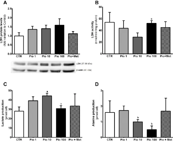
3. A pharmacological concentration of pioglitazone stimulates lactate production by human Sertoli cells ... 32
4. A suprapharmacological concentration of pioglitazone decreases while the combined treatment with metformin increases mitochondrial membrane potential in human Sertoli cells ... 34
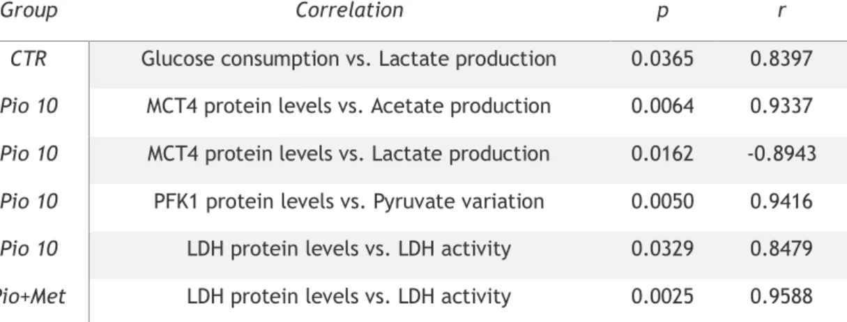
5. The positive correlation between glucose consumption and lactate production detected in non-exposed human Sertoli cells is lost by exposure to pioglitazone alone or in combination with metformin ... 36
V. Discussion ... 37
xiv VII. References ... 43 VIII.Annex I ... 62
List of publications resultant from the work developed during the M.Sc. in Biomedical Sciences ... 62 List of oral communications resultant from the work developed during the M.Sc. in Biomedical Sciences ... 63
xvi
List of Figures
Figure 1: Schematic representation of the human testis and spermatogenesis. ... 5 Figure 2: Representative diagram of the metabolic cooperation established between Sertoli cells and developing germ cells in the testis.. ... 11 Figure 3: Possible mechanism of metformin action in cells. ... 20 Figure 4: Effect of pioglitazone (1 µM, 10 µM, and 100 µM) and the combined action of pioglitazone 1.5 µM and metformin 10 µM (Pio+Met) in glucose uptake by human Sertoli cells (hSCs).. ... 31 Figure 5: Effect of pioglitazone (1 µM, 10 µM, and 100 µM) and the combined action of pioglitazone 1.5 µM and metformin 10 µM (Pio+Met) in pyruvate metabolism in human Sertoli cells (hSCs). ... 32 Figure 6: Effect of pioglitazone (1 µM, 10 µM, and 100 µM) and the combined action of pioglitazone 1.5 µM and metformin 10 µM (Pio+Met) in lactate and alanine metabolism by human Sertoli cells (hSCs). ... 33 Figure 7: Effect of pioglitazone (1 µM, 10 µM, and 100 µM) and the combined action of pioglitazone 1.5 µM and metformin 10 µM (Pio+Met) in mitochondria of human Sertoli cells (hSCs). ... 35
xviii
List of Tables
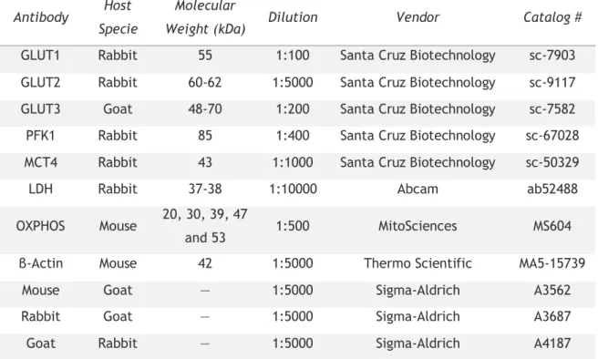
Table 1: List of the primary and secondary antibodies used in this study... 28 Table 2: Pearson correlation coefficients between glycolysis related-steps in hSCs cultured in control conditions as well as in those cultured with pioglitazone alone or in combination with metformin ... 36
xx
List of Abbreviations
1H-NMR Proton nuclear magnetic ressonance
ACAC Acetyl-Coenzyme A carboxylase ACAD Acyl-Coenzyme A dehydrogenase ALT Alanine aminotransferase
AMP Adenosine monophosphate
AMPK Adenosine monophosphate-activated protein kinase ATP Adenosine triphosphate
BSA Bovine serum albumin BTB Blood/testis barrier
CI NADH dehydrogenase (ubiquinone) 1 beta subcomplex subunit 8 CII Succinate dehydrogenase complex, subunit B, iron sulfur CIII Ubiquinol-cytochrome c reductase core protein II
CIV Mitochondrially encoded cytochrome c oxidase I
CoA Coenzyme A
CV ATP synthase alpha-subunit
DM Diabetes mellitus
DMEM Dulbecco’s Modified Eagle Medium DNA Deoxyribonucleic acid
EDTA Ethylene Diamine Tetra Acetic acid
FBS Fetal bovine serum
FSH Follicle-stimulating hormone
GH Growth hormone
GLUT Glucose transporter
GnRH Gonadotropin releasing hormone HBSS Hank’s Balanced Salts Solution hSC Human Sertoli cell
IGF-1 Insulin-like growth factor I IGT Impaired glucose tolerance ITS Insulin–transferrin–sodium selenite
JC-1 5,5′,6,6′-tetrachloro-1,1′,3,3′-tetraethylbenzimidazolylcarbocyanine iodide
LDH Lactate dehydrogenase
xxi LDHC4 Lactate dehydrogenase type C isoform 4
LH Luteinizing hormone
MCT Monocarboxylate transporter
Met Metformin
M-PER Mammalian Protein Extraction Reagent
mRNA Messenger RNA
mtDNA Mitochondrial DNA
MTP Mitochondrial trifunctional protein
NADH Reduced nicotinamide adenine dinucleotide OXPHOS Oxidative phosphorylation
PBS Phosphate buffered saline PCOS Polycystic ovary syndrome PDH Pyruvate dehydrogenase PFK1 Phosphofructokinase 1 PI3K Phosphatidylinositol 3-kinase
Pio Pioglitazone
PPARα Peroxisome proliferator-activated receptor α PPARγ Peroxisome proliferator-activated receptor γ
SC Sertoli cell
SEM Standard error of the mean
SGLT Sodium dependent glucose transporter
SRB Sulforhodamine B
T1DM Type 1 diabetes mellitus T2DM Type 2 diabetes mellitus
T3 Tri-iodothyronine
T4 Thyroxine
TBS Tris-buffered saline solution
1
I. Introduction
1. Testicular physiology
The establishment of male fertility is a complex process that requires specific and controlled interactions between different tissues and cells of the male reproductive tract and accessory glands. The male reproductive tract is formed of heterogeneous tissues, including the testis, efferent ducts, epididymis and vas deferens. The mammalian testis is a complex organ, coated for a capsule constituted by complex fibrous structure in various distinct tissue layers. The major component of the capsule, the tunica albuginea, is distinguished by the existence of fibroblasts interspersed in collagen fibers (Setchell, Maddocks et al. 1994), from which septations extend toward the testicular mediastinum, separating the human testis into 200– 300 lobules (Niederberger 2011) (Figure 1). Tunica albuginea has several physiological functions in male reproduction, including preservation of the interstitial pressure within the testis, boost the transport of sperm out of the testis into the epididymis and control of the blood flow through the testis (Setchell, Maddocks et al. 1994).
Each testicular lobule encloses coiled seminiferous tubules (Figure 1). Within the testis two compartments are formed, the interstitial and tubular, that present different cell types and fluid flow (Weinbauer, Luetjens et al. 2010). The interstitial compartment is the space outside the seminiferous tubules. Here, the most important cells are the Leydig cells that actively produce and secrete testosterone, the main male sexual hormone (Haider 2004, Ge and Hardy 2007) (Figure 1). In humans, the interstitial compartment represents about 12–15% of the total testicular volume, 10–20% of which is composed of Leydig cells. Human testes have approximately 200×106 Leydig cells (Weinbauer, Luetjens et al. 2010). The proliferation rate of these cells in the adult testis is low and influenced by luteinizing hormone (LH) (see below). In addition to Leydig cells, the interstitial compartment contains macrophages, lymphocytes, mesenchymal fibroblasts, extracellular matrix, and small blood capillaries (Haider 2004). The tubular compartment represents about 60-80% of the total testicular volume. The seminiferous tubules are avascular and with no nerves penetrating through their walls (Setchell 1986). Overall, the human testis contains about 600 seminiferous tubules. The lobules filled by seminiferous tubules are separated by extensions of the tunica albuginea that open on both ends into the rete testis (Figure 1). Of note, all the processes involved in the production of male gametes (spermatogenesis), occur within the seminiferous tubules (Weinbauer, Luetjens et al. 2010). These tubules end at the rete testis, which is a network of tubules that empties into the efferent ductules. Spermatozoa are transported through the efferent ductules into the epididymis, and then enter the vas deferens, which through peristalsis propels them to the ejaculatory duct (Niederberger 2011).
2 The tubular compartment contains the germ cells and two different types of somatic cells, the Sertoli and peritubular cells (Niederberger 2011). The seminiferous tubules are sheltered by a lamina, which consists of a basal membrane, a layer of collagen and the peritubular cells (myofibroblastic cells). These cells are stratified and form concentric layers that are separated by collagen layers (Schell, Albrecht et al. 2010). The peritubular cells have an essential role in the transport of sperm (Romano, Tripiciano et al. 2005, Welsh, Saunders et al. 2009), and contribute to several testicular functions by secreting factors involved in cellular contractility (panactin, desmin, gelsolin, smooth muscle myosin and actin), components of the extracellular matrix (collagen, laminin, vimentin, fibronectin, growth factors, fibroblast protein) and adhesion molecules (Albrecht, Rämsch et al. 2006, Schell, Albrecht et al. 2008, Schell, Albrecht et al. 2010, Mayerhofer 2013, Flenkenthaler, Windschüttl et al. 2014).
The Sertoli cells (SCs) extend from the base to the apex of the epithelium in a direct interaction with the developing germ cells (Mruk and Cheng 2004) (Figure 1). About 35–40% of the volume of the germinal epithelium is comprised by these cells. The human testis of reproductive age contains about 800–1200×106 SCs (Zhengwei, Wreford et al. 1998). These somatic cells are commonly referred as “nurse cells”, because they are responsible for providing nutritional and energetic support to developing germ cells, and for creating an immunologically protected space for the development of germ cells (Rato, Alves et al. 2012). Adjacent SCs form tight junctions with each other in such a way that nothing larger than 1 kDa can pass from the outside to the inside of the tubule. The tight junctions formed between adjacent SCs create the Sertoli/blood–testis barrier (BTB) that physically divides the seminiferous epithelium into basal and apical compartments (Figure 1). This barrier regulates the movement of substances, such as nutrients and wastes, in and out of the seminiferous epithelium (Madara 1998, Alves, Rato et al. 2013). The cytoskeleton of SCs is responsible for the organization of the seminiferous epithelium and plays a fundamental role in facilitating germ cells movement. In addition to the tight junctions, other types of associations occur between the SCs to strengthen the BTB, such as adherens junctions (eg: ectoplasmic specialization, a testis-specific adherens junction type) and intermediate filament-based desmosome-like junctions (Pelletier 2001, Mruk and Cheng 2004, Rato, Alves et al. 2012). The relevance of these junctions goes far beyond the physical and nutritional support, as they must undergo extensive restructuring during germ cell migration on the course of spermatogenesis.
Several proteins, products and factors are known to be secreted by SCs, including: proteases, protease inhibitors, hormones, energy substrates, growth factors, paracrine factors, inhibin, transferrin, androgen-binding protein, plasminogen activator, glycoproteins, sulpho-proteins, myo-inositol and other extracellular matrix components (Fritz, Rommerts et al. 1976, Robinson and Fritz 1979, Elkington and Fritz 1980, Skinner and Griswold 1980, Marzowski,
3 Sylvester et al. 1985, O'Brien, Gabel et al. 1993, Griswold 1998). Notably, the number of SCs is also an important determinant of testis size, and this number is directly related to the number of germ cells (Griswold 1998). Germ cells are dependent on SCs not only for structural support, but also for nutritional and energetic support. The SCs convert glucose to lactate that is known to be used as substrate and to influence the survival of germ cells. Although glucose is one of the most used substrates by SCs, they can also metabolize other substrates such as ketone bodies and fatty acids (Griswold 1998, Rato, Alves et al. 2012). These processes are vital for the production and export of lactate that can be conditioned by several factors. For instance, it was recently reported that insulin-deprived SCs altered the expression of metabolism-associated genes implicated in the export and production of lactate, as well as the consumption of glucose and secretion of lactate (Oliveira, Alves et al. 2012). The SCs also control the composition of the seminiferous tubular fluid and the physicochemical milieu where spermatogenesis occurs. The seminiferous tubular fluid serves as a mean of transport of sperm, and also helps to maintain a proper microenvironment required for a normal spermatogenesis (Rato, Socorro et al. 2010, Rato, Alves et al. 2011).
2. Spermatogenesis
Spermatogenesis is a complex biological process that produces spermatozoa in the seminiferous tubules of the mammalian testis. It involves an important balance between self-renewal and differentiation of spermatogonial stem cells to ensure an endless production and release of mature spermatozoa (Sharpe 1994). A fertile man produces more than 40 million spermatozoa per day, beginning at puberty and spanning his entire reproductive life (Cheng and Mruk 2013). The duration of the complete spermatogenic process varies according to species and in human it spans 74–76 days (Sharpe 1994). The SCs are responsible for the movement of germ cells from the base of the tubule toward the lumen and for the release of mature spermatozoa into the lumen. Moreover, as discussed above, the BTB, composed by SCs, divides the epithelium into two compartments: the basal compartment, in which spermatogonia, preleptotene, and leptotene spermatocytes exist; and the adluminal compartment, in which meiotic spermatocytes and spermatids in different stages reside (Hess and de Franca 2008, Cheng, Wong et al. 2010) (Figure 1). Spermatogenesis is controlled by several factors, (endocrine, paracrine and autocrine) and can be divided into four different phases that include: mitosis, meiosis, spermiogenesis and spermiation. In the early steps of spermatogenesis, diploid spermatogonia (2n) proliferate and generate two different populations of cells: one subpopulation of stem cells identical to their progenitors; other subpopulation, the majority, starts a differentiation process and differentiate into spermatozoa. In this process there is a subpopulation of spermatozoa and germ cells in different stages that undergo apoptosis (Dym 1994, Hofmann 2008, Sá, Neves et al. 2008, Sá,
4 Miranda et al. 2013). Spermatogenesis is of great complexity and requires a time-specific period for completion depending of the species. For instance, it requires 6–9 weeks for completion in human. Spermatogonia are the most primitive diploid germ cells (2n) that divide by mitosis and reside on the basement membrane of seminiferous epithelium (Figure 1). In the mitotic phase, spermatogonia are self-renewed or undergo differentiation, with both cases involving several mitotic divisions. Little is known about the division of spermatogonia in humans, but three different types of spermatogonia have been identified in human: type A Dark, type A Pale, and type B (Rowley, Berlin et al. 1971, Dym 1994, Sá, Neves et al. 2008, Sá, Miranda et al. 2013). The main morphological features used to distinguishing between these four types of spermatogonia were the shape and staining characteristics of the nucleus, the position of the nucleolus, the structure of the mitochondrial cristae, the association of the endoplasmatic reticulum with mitochondria, the presence or absence of glycogen in the cytoplasm of the cells and the presence of the filamentous structures of cytoplasm are evaluated to distinguish (Rowley, Berlin et al. 1971). The Type A Dark spermatogonia are suggested to be the least morphologically differentiated and are considered the subpopulation of stem cells. Each spermatogonium is in touch with the basal membrane in the seminiferous tubule. Notably, this proximity progressively decreases with the degree of spermatogonia differentiation. The type A Dark spermatogonia are the flat cells lying parallel to the basal membrane, through the type A pale and type B (Rowley, Berlin et al. 1971, Hess and de Franca 2008). Type B are the most differentiated type of spermatogonia since this differentiation only occurs in two thirds of the type A pale spermatogonia. Type B spermatogonia enter meiosis and originate preleptotene spermatocytes, which in turn differentiate into leptotene, zygotene, pachytene and diplotene spermatocytes.
During these phases the chromosomes condense, form pairs of homologous chromosomes, synapses are completed and then substituted by crossing-over and homologous recombination. In diplotene stage, chromosomes are unsynapsed and the cell divides. The BTB separates these two states of spermatocytes, the preleptotene spermatocytes located in the basal compartment and the pachytene spermatocytes in adluminal compartment (Mruk and Cheng 2004, Cheng and Mruk 2013). Meiosis is characterized by the separation of chromosomes that occurs during the metaphase, anaphase and telophase of the first meiotic division, after which secondary spermatocytes are originated (Phillips, Gassei et al. 2010) (Figure 1). The prophase of the first meiosis lasts 1–3 weeks, whereas the other phases of the first meiosis and the entire second meiosis are completed within 1–2 days in man (Holstein 1994, Weinbauer, Luetjens et al. 2010). Secondary spermatocytes contain haploid chromosomes in duplicate. During the second meiotic division secondary spermatocytes originate four haploid spermatids (n) (Grootegoed, Siep et al. 2000, Weinbauer, Luetjens et al. 2010) (Figure 1). Spermiogenesis and spermiation are the last phases of spermatogenesis in which the spermatids undergo several alterations. During spermatid development, the nucleus elongates and condenses. The nucleus of spermatids contains compacted DNA following the
5 replacement of nucleosomal histones by transition proteins and subsequently by protamines (Meistrich, Trostle‐Weige et al. 1992). Finally, differentiated elongated spermatids and spermatozoa (Figure 1) are produced. For human, originally 12 spermatid maturation steps were described. These steps include nucleus condensation, the formation of a flagellum and the extrusion of a large part of cytoplasm (Weinbauer, Luetjens et al. 2010). The differentiation into the extremely specialized sperm cells is one of the most significant cell developmental processes that occur in biological systems. It involves phases of acrosome development, nuclear elongation and condensation, the formation of middle piece and tail, and the reduction of cytoplasmatic volume (Grootegoed, Siep et al. 2000). The SCs play an essential role in reducing the cytoplasmatic volume of the elongated spermatids. The adhesive contacts and ectoplasmic junctional specializations between SCs and spermatids are shattered and elongated spermatids are thereafter released into the lumen of seminiferous tubule occurs, in a process named spermiation (Griswold 1998, Grootegoed, Siep et al. 2000).
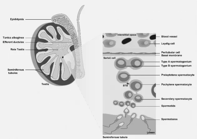
Figure 1: Schematic representation of the human testis and spermatogenesis. The testis is coated by
tunica albuginea and divided in lobules. The seminiferous tubules, where spermatogenesis occurs, are coiled in the testis lobules. The Leydig cells and blood vessels are found in the interstitial space. The seminiferous epithelium is composed of Sertoli cells, which form the Sertoli/blood-testis barrier (BTB), and developing germ cells at different stages. The Sertoli cells adhere to the basal membrane where spermatogonia are also adherent. Type A Spermatogonia divide and develop into type B spermatogonia. The primary spermatocytes (preleptotene and pachytene) start meiosis and divide in secondary spermatocytes, which in turn divide into haploid spermatids that migrate towards the lumen where the fully formed spermatozoa are finally released.
6 2.1 Hormonal regulation of spermatogenesis
Spermatogenesis is an intricate process, controlled by several interacting mechanisms that differ according to the consecutive developmental stages: fetal, infantile, pubertal and adult. It is tightly regulated by the hypothalamus–pituitary–testis axis. Within this axis, neurons of the hypothalamus produce gonadotropin releasing hormone (GnRH). Regulation of gonadotropin secretion involves a complex interaction between stimulation by GnRH from the hypothalamus and feedback control by sex steroids and inhibin from the testes, besides autocrine/paracrine modulation by other factors within the pituitary (Walker and Cheng 2005, Niederberger 2011). Pulsatile GnRH signals stimulate gonadotropic cells in the anterior pituitary to release follicle-stimulating hormone (FSH) and LH that then act on the testis to regulate the spermatogenic potential (Walker and Cheng 2005, Rato, Alves et al. 2012, Alves, Rato et al. 2013). LH is of upmost importance for the stages of immature and mature adult Leydig cells, being the main inducer of these adult cells differentiation and the responsible for the maintenance of its high rates of proliferation (Habert, Lejeune et al. 2001). LH acts on Leydig cells through LH receptors present in the surface of these somatic cells of the testis and stimulates the production of testosterone, a steroid hormone that diffuses into the seminiferous tubules. Testosterone is the main secreted product of the testis, with daily production rate being 5–7 mg in men (Niederberger 2011). Interestingly, testosterone plasma levels are strictly correlated with LH levels and it was described that pulsatile LH concentrations stimulate testosterone secretion in a dose-dependent manner (Walker and Cheng 2005, Cheng and Mruk 2013). SCs are the only testicular cells that express receptors for testosterone and FSH. FSH is a member of the glycoprotein hormone family which binds to its receptor in SCs and has typically been described to be involved in the beginning of pubertal spermatogenesis (Sharpe 1994). This hormone is crucial to stimulate spermatogenesis, as it maintains testicular size, seminiferous tubular diameter and a suitable number of sperm with normal motility (Walker and Cheng 2005, Niederberger 2011). FSH also regulates DNA synthesis in spermatogonia, spermatogonial proliferation and differentiation (Rossi, Albanesi et al. 1991, Dym 1994).
The hypothalamus–pituitary–testis axis is regulated by feedback mechanisms. The production of inhibin by SCs, as well as testosterone and 17β-estradiol by Leydig cells lead to a negative feedback that decreases the secretion of GnRH in the hypothalamus and of LH in the pituitary (Tilbrook and Clarke 2001). Sex steroids seem to play a slight role in the feedback control of FSH secretion while inhibin is the key factor implicated in the testicular regulation of FSH secretion. Notably, there is an inverse relationship between inhibin’s circulating level and FSH ones (Niederberger 2011). In the last years, there has been an increasingly comprehension that sex steroid hormones are involved in several biological mechanisms, including the homeostasis of energy balance and energy metabolism in male reproductive activity (Wade, Schneider et al. 1996, Hill, Elmquist et al. 2008). The GnRH pulse is extremely susceptible to energetic deficits, environmental contaminants and intense exercise (Nindl,
7 Kraemer et al. 2001, Saradha and Mathur 2006, Trumble, Brindle et al. 2010). For instance, Trumble and collaborators (2010) have described that fasting causes suppression of GnRH pulses, which consequently causes a decrease in the levels of LH and testosterone production levels by Leydig cells disturbing male reproductive function.
Androgens are considered the main sex male hormones, namely testosterone, while estrogens are usually referred as the sex female hormones. Nevertheless, androgens and estrogens are present in both sexes. Therefore, sexual distinctions are not qualitative differences, but result from quantitative divergence in hormone concentrations and differential expressions of steroid hormone receptors (Carreau and Hess 2010). In men, most serum estradiol is produced by peripheral aromatization of androgens secreted by Leydig cells. The cytochrome P450 enzyme aromatase is responsible for catalyzing this reaction and is primarily present in adipose tissue but also functions in skin and liver. In the testis, aromatase activity is primarily localized in Sertoli and Leydig cells (Inkster, Yue et al. 1995). It has been shown that estrogens have an essential role in regulating the hypothalamus–pituitary–testis axis and thus indirectly regulate LH and testosterone equilibrium through a feedback loop (O’Donnell, Robertson et al. 2001). Besides from gonadotrophins and steroid hormones, thyroid hormone has also been shown to play an important role in testicular physiology (Cooke, Holsberger et al. 2004, Mendis-Handagama and Ariyaratne 2005). Thyroid gland produces thyroxine (T4) and tri-iodothyronine (T3) (Mendis-Handagama and Ariyaratne 2005). It is known that T3 regulates the maturation and growth of the testis, in rats and other mammalian species, by inhibiting immature SCs proliferation and by stimulating their functional differentiation (Cooke, Hess et al. 1991, Hess, Cooke et al. 1993). Likewise, thyroid hormone has been shown to play a critical role in the onset of Leydig cell differentiation and stimulation of steroidogenesis in postnatal rat testis (Mendis-Handagama and Ariyaratne 2005). Though the mechanisms implicated in the regulatory actions of thyroid hormone in testicular cells are still undefined, the presence of thyroid hormone receptors in human and rat testis throughout development and in adulthood implies that T3 may act via the classical genomic pathway in testis (Benbrook and Pfahl 1987, Jannini, Olivieri et al. 1990).
Insulin-like growth factor I (IGF-1) is also biosynthesized in the testis by SCs (Itoh, Nanbu et al. 1994). Its receptors are identified in Leydig cells, peritubular cells, and spermatocytes (Niederberger 2011). It is known that IGF-1 stimulates the proliferation of Leydig cell precursors, spermatogenesis and spermatid maturation (Itoh, Nanbu et al. 1994). LH, FSH and testosterone may cooperate in spermatogenesis through stimulation of IGF-1 secretion by SCs (Itoh, Nanbu et al. 1994, Niederberger 2011). Growth hormone (GH) is not classically considered as a reproductive hormone, although it has important roles in reproductive function. It plays a role in steroidogenesis and spermatogenesis exerting an endocrine action either directly at gonadal sites or indirectly via IGF-1. GH influences Leydig cell steroidogenesis by regulating the secretion of IGF-1 and by increasing the expression of
8 several genes that code for steroidogenic enzymes, including 3β-hydroxysteroid dehydrogenase, responsible for the conversion of pregnenalone to progesterone (Spiteri-Grech and Nieschlag 1992, Gomez, Weil et al. 1999, Hull and Harvey 2000, Niederberger 2011). Progesterone is another hormone very relevant for male reproduction. The maturation of progesterone to androstenedione is catalyzed by 17α-hydroxylase/ C17–20 lyase (CYP17), while further conversion of androstenedione to testosterone depends on 17β-hydroxysteroid dehydrogenase activity, in Leydig cell smooth endoplasmic reticulum (Niederberger 2011). Therefore, it is essential to unravel the complex hormonal network and signalling involved in the control of spermatogenesis to understand all the mechanisms relevant to male fertility, and unveil its flaws in cases of infertility.
3. Metabolic cooperation in testis
Metabolic cooperation is defined as the interchange of metabolism-associated molecules and metabolic substrates between cells (Hooper and Subak-Sharpe 1981). Although metabolic cooperation between neuronal and other cell types (Pellerin 2003) has been studied for decades, the study of these processes in the testis are relatively recent. In fact, latest advances provided compelling evidence that the metabolic cooperation between SCs and developing germ cells is crucial for the normal development of spermatogenesis (Rato, Alves et al. 2012). Moreover, it has been proposed that alterations in these processes result in serious consequences to male fertility (Alves, Martins et al. 2013). One of the major intervenient in the metabolic cooperation in the testis and to the occurrence of a normal spermatogenesis are SCs (Oliveira, Martins et al. 2015). As discussed, junctions between adjacent SCs form the BTB, which physically divides the seminiferous epithelium (Rato, Alves et al. 2011). Thus, the SCs are responsible for the selective passage of substances from blood plasma and testicular lymph to the rete testis fluid (Setchell 1980). Due to this function, these cells are responsible for providing nutritional support for the developing germ cells (Griswold 1995). Although this latter function of SCs has been underestimated for many years, it is now accepted that there is a close metabolic relationship between these cells and developing germ cells, a condition that is affected by many diseases and drugs (Alves, Dias et al. 2014). It has already been shown that there is a transference of carbohydrates, vitamins, lipids, amino acids and metal ions between SCs and germ cells (Mruk and Cheng 2004). Notably, SCs metabolism presents some distinctive characteristics. Cultured SCs convert a great percentage of glucose to lactate, the major energy substrate to germ cells (Robinson and Fritz 1981). This is a very interesting metabolic behaviour since SCs prefer the pathway of lactate production instead of Krebs cycle, using a less effective pathway concerning to adenosine triphosphate (ATP) production, in a clear evidence of a Warburg-like metabolic effect. Moreover, SCs present a high glycolytic flux, equally to what occurs in cancer cells
9 (Oliveira, Martins et al. 2015). This is a consequence of the metabolic cooperation needed between SCs and developing germ cells for the normal occurrence of spermatogenesis, illustrating the relevance for this process.
Glucose is essential for the occurrence of spermatogenesis. However, it is present in very low levels in the seminiferous tubules due to its rapid metabolism (Voglmayr, Waites et al. 1966, Robinson and Fritz 1979). Moreover, glucose is very hydrophilic and thus, it can pass through the lipid bilayer by simple diffusion in a very inefficient way. Therefore, there are two families of glucose transporters: the glucose transporters (GLUTs) and the sodium dependent glucose transporters (SGLTs) (Thorens 2001, Scheepers, Joost et al. 2004) that are responsible for the transport of this metabolite to cells. From these transporters, GLUTs are present in most tissues and are reported to play a key role in mediating passive glucose transport through membranes (Thorens 2001) (Figure 2). Different isoforms were already identified in SCs, particularly GLUT1 (Carosa, Radico et al. 2005, Galardo, Riera et al. 2008), GLUT2 (Kokk, Veräjänkorva et al. 2003), GLUT3 (Galardo, Riera et al. 2008) and GLUT8 (Carosa, Radico et al. 2005). However, the latter isoform is not reported to be responsible for glucose transport from the extracellular milieu since it has not been localized in the plasma membrane (Piroli, Grillo et al. 2002). Moreover, GLUT8 has been reported to be related with lysosomes and membranes of the endoplasmic reticulum (Schmidt, Joost et al. 2009). Once a molecule of glucose has passed from the blood into the cell, it is gradually metabolized in a controlled sequence of biochemical steps catalyzed by various enzymes. Firstly, it is oxidized into two molecules of pyruvate (Martins, Alves et al. 2013), through a process known as glycolysis (Figure 2). The first rate-limiting step of glycolysis, is the irreversible conversion of fructose-6-phosphate to fructose-1,6-bisphosphate (Chehtane and Khaled 2010). This reaction is catalyzed by phosphofructokinase (PFK) whose activity is also known to be linked with the energy status of the cell (Morgante, Tosti et al. 2011). The pyruvate produced during glycolysis can then follow three main pathways. One of those is the Krebs cycle where pyruvate is transported to the mitochondria and oxidized, as well as acetyl CoA, to produce NADH and FADH2 for ATP synthesis in the respiratory chain (Freedman and Graff 1958, Oliveira, Martins et al. 2015). Another pathway is the conversion of pyruvate into alanine by alanine aminotransferase (ALT). The last pathway that pyruvate can follow is the conversion of this end product of glycolysis to lactate, by lactate dehydrogenase (LDH) (Rato, Alves et al. 2012) (Figure 2). For many years, lactate was reported as a waste product of glycolysis (Gladden 2004). However, this has been challenged and nowadays it is known that lactate is the main energy source for several cells, including the developing germ cells (Boussouar and Benahmed 2004).
In the last years, several authors have debated the role of the lactate produced by SCs in the survival and nutritional support of developing germ cells (Erkkilä, Aito et al. 2002, Boussouar and Benahmed 2004, Gladden 2004). Some studies have shown that lactate production is
10 stimulated by FSH (Mita, Price et al. 1982), as well as epidermal growth factor (Mallea, Machado et al. 1986). It has been shown that both stimulate lactate production by enhancing the glycolytic metabolism of rat SCs, since they increase the glucose uptake into the cells (Hall and Mita 1984, Mallea, Machado et al. 1986). These mechanisms are thought to be highly regulated through a complex signalling network. The greater glucose uptake may possibly result from the interaction between FSH and phosphatidylinositol 3-kinase (PI3K). Deprivation of insulin has also been reported to alter glucose consumption and lactate production by SCs (Oliveira, Alves et al. 2012). Insulin deprivation altered the expression of several genes involved in the production and export of lactate, such as LDH but also induced a similar adaptation in the expression of GLUTs in conditions of glucose deprivation (Riera, Galardo et al. 2009, Oliveira, Alves et al. 2012). Moreover, 5α‐dihydrotestosterone (100 nM) decreased lactate production in human SCs (Oliveira, Alves et al. 2011). This decrease was reported to be due to a decrease in mRNA levels of LDHA, since glucose consumption was increased. Thus, it seems that 5α‐dihydrotestosterone redirects glucose metabolism to Krebs cycle (Oliveira, Alves et al. 2011).
After being produced in SCs through the action of LDH, lactate is exported to the intratubular fluid through monocarboxylate transporters (MCTs) (Figure 2). From the 14 members of this family, only MCT1 to MCT4 are thought to be involved in the transport of lactate (Boussouar and Benahmed 2004). However, not all are present in testicular cells. While MCT1 and MCT4 can be found in SCs, MCT2 can be found in elongated spermatids. Moreover, MCT1 was found in developing germ cells and is reported to play a major role in the import of lactate from the intratubular fluid. Lactate is converted by LDHC4 (Shi, Wang et al. 2005), the specific isoform of LDH in testis, into pyruvate. The latter is then converted by pyruvate dehydrogenase into acetyl-CoA, which enters the mitochondria to be used in the Krebs cycle. Besides lactate, acetate is also produced by SCs and may be used by developing germ cells for lipid synthesis and membrane remodelling (Alves, Socorro et al. 2012) (Figure 2). Acetate has been described as an intermediate metabolite and is the most common intermediate for the synthesis of fatty acids and cholesterol (Alves, Socorro et al. 2012). It results from the conversion of acetyl-CoA by acetyl-CoA hydrolase.
While developing germ cells rely on the lactate produced at high rates by SCs, the resulting mature sperm cells present specific metabolic needs, using external hexoses, such as glucose, as their main substrate (Frenkel, Peterson et al. 1973). Following the uptake, the hexose metabolism is crucial to preserve spermatozoa function and motility (Williams and Ford 2001). Thus, either oxidative phosphorylation and/or glycolysis can be used to provide the energetic needs to spermatogenic cells (Storey 2008).
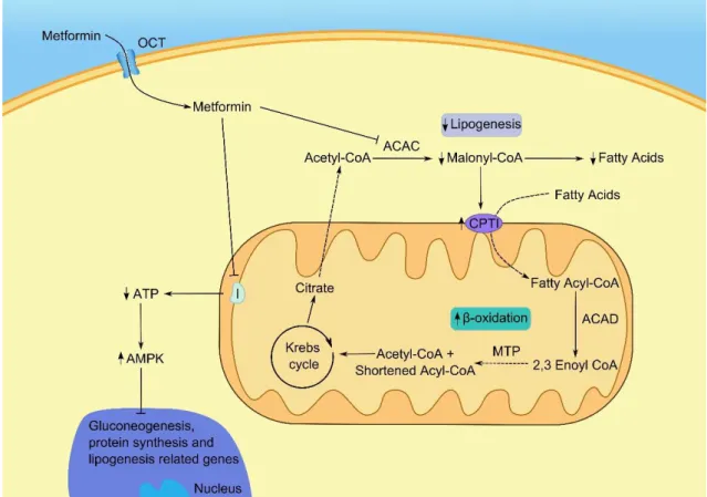
11 Figure 2: Representative diagram of the metabolic cooperation established between Sertoli cells and developing germ cells in the testis. The glucose from the extracellular space enters the Sertoli
cells through glucose transporters (GLUT), and is converted into glucose-6-phosphate (Glucose-6-P). Phosphofructokinase (PFK) catalyzes the rate limiting step of glycolysis, which results in the formation of pyruvate. The pyruvate can then be: converted into alanine by alanine aminotransferase (ALT); converted into lactate by lactate dehydrogenase (LDH); or transported into the mitochondrial matrix to form acetyl-CoA. Pyruvate dehydrogenase (PDH) catalyzes the rate limiting step of the latter conversion of pyruvate. The lactate produced by Sertoli cells is exported to the intratubular fluid by monocarboxylate transporters (MCTs), mainly MCT4. The germ cells uptake the lactate produced by Sertoli cells through MCT2 that is used for ATP production by the isoform LDHC. Moreover, acetyl-CoA may be converted into acetate, which is then exported to the intratubular fluid and may be used by germ cells for membrane remodelling.
12
4. (Pre)-diabetes in brief
DM is one of the most widespread chronic diseases. It is a leading cause of morbidity and mortality, in both, developed and in developing countries. The World Health Organization estimates that 347 million of people have DM (Alberti and Zimmet 1998). According to a recent report, the number of diabetic patients may reach 592 million until the year 2035 (Guariguata, Whiting et al. 2014). These alarming statistics clearly illustrate that is urgent to study the impact of this pathology on cells, organs and whole body. DM is a chronic, metabolic disease of multiple aetiologies, characterized by hyperglycemia that can result from defects in insulin secretion, insulin action or both (American Diabetes Association 2014). Moreover, it causes impaired carbohydrate, fat, and protein metabolism (Hamden, Jaouadi et al. 2011). All body metabolism is affected and thus, DM can lead to a variety of co-morbidities, including cardiovascular diseases, retinopathy, obesity, sexual disorders, nephropathy and diabetic foot (Struijs, Baan et al. 2006, Meneses, Sousa et al. 2015). The most prevalent types of DM are type 1 diabetes mellitus (T1DM) and type 2 diabetes mellitus (T2DM). T1DM, also known as insulin-dependent DM, is characterized by an autoimmune reaction of T lymphocytes on insulin-producing beta cells in genetically susceptible individuals (Killestein 2002, Lehuen, Diana et al. 2010). The destruction of pancreatic beta cells is progressive and results in a dramatic decrease or even elimination of insulin production. Consequently, these patients are dependent on exogenous insulin treatment to survive (Van Belle, Coppieters et al. 2011). The first symptoms of T1DM generally appear before the age of 30 for what it is known as juvenile-onset DM. Accompanying the trends of DM, T1DM has also been drastically increasing in recent years. Of note, it is expected that the prevalence of cases in individuals younger than 5 years will double between 2005 and 2020 (Patterson, Dahlquist et al. 2009).
T2DM is responsible for the vast majority of DM cases (American Diabetes Association 2014). It comprises individuals who present insulin resistance and relative insulin deficiency (Henriksen, Diamond-Stanic et al. 2011). Unlike T1DM, the main triggering factor for T2DM seems to be the new lifestyle of modern societies rather than genetic factors, although the latter can also play a crucial role in the establishment of the disease (Hu, Manson et al. 2001). Insulin, which has a preponderant role in DM, is the main hormone responsible for glucose homeostasis. In the early stages of the disease, the cells have decreased sensitivity to insulin, failing to respond to normal levels in circulation. This characteristic of T2DM is known as insulin resistance (Hu, Manson et al. 2001). Thus, pancreatic beta cells augment the rate of insulin secretion to compensate the resistance of the tissues to this hormone (Kahn 1998). However, with the progression of the disease, beta cells loose the capacity to release adequate amounts of insulin to compensate the insulin resistance, leading to the development of impaired glucose tolerance (IGT) (DeFronzo, Bonadonna et al. 1992). IGT is the main characteristic of pre-diabetes, which is an intermediate condition between normal
13 glucose tolerance and T2DM (Tabák, Herder et al. 2012). In fact, this condition may be identified through an oral glucose tolerance test, once pre-diabetic individuals present glycemic variables that are higher than normal, but lower than T2DM thresholds (Tabák, Herder et al. 2012, Alves, Martins et al. 2013). Like T2DM, pre-diabetes may be triggered by sedentary lifestyle and inadequate diets, such as the ingestion of high caloric and saturated fat rich diets (Rato, Alves et al. 2013). Consequently, these patients present metabolic modifications in diverse organs, leading to an increased probability of developing several co-morbidities, like cardiovascular disease (American Diabetes Association 2014, Buysschaert, Medina et al. 2015). In fact, pre-diabetic patients present a higher risk of stroke (Lee, Saver et al. 2012) and a 2- to 3-fold increase in the risk of developing macroangiopathy (Huxley, Barzi et al. 2006). Moreover, the prevalence of chronic kidney disease is also high among pre-diabetic patients (Plantinga, Crews et al. 2010, Buysschaert, Medina et al. 2015). Despite the similarity between T2DM and pre-diabetes, the latter does not imply the progression to T2DM. Lifestyle and/or drug-based interventions can be done to revert the condition, illustrating the relevance of the early diagnosis of this prodromal stage of T2DM.
5. Modifications in testicular metabolic cooperation promoted
by (pre)-diabetes
The metabolic cooperation between testicular cells is crucial for germ cells development and any alteration in those processes may have drastic consequences for male fertility potential (Alves, Dias et al. 2014, Reis, Moreira et al. 2015). However, the precise molecular mechanisms of testicular glucose metabolism in diabetic patients are far from being unveiled. However, a study performed during the 80s reported that rat SCs and peritubular cells were very sensitive to glucose concentrations. In fact, SCs produced more lactate when they were in contact with a higher glucose concentration (Hutson 1984).
There is increasing information about the precise molecular mechanisms of testicular glucose metabolism in diabetic patients through in vitro and morphological studies of human biopsies from those individuals. Studies using testicular biopsies from diabetic men have shown an extensive vacuolization and a high degree of degeneration in SCs (Cameron, Murray et al. 1985). In addition, and despite germ cells presented normal morphology, the seminiferous tubules were depleted and Leydig cells presented lipid droplets and variable number of vacuoles (Cameron, Murray et al. 1985). In fact, all these alterations have drastic consequences to testicular cells’ metabolism, particularly to the metabolic cooperation discussed above. Moreover, a study described that DM caused a decrease in lactate
14 production and an increase of endogenous oxygen uptake by testicular cells (Sharaf, Kheir El Din et al. 1978).
A recent study described that insulin depletion and hyperglycemia in streptozotocin-induced DM did not regulate the expression of GLUT8 in testes, once its expression was not modified in these conditions (Gómez, Ballester et al. 2009). However, another study explored the effect of insulin deprivation on several markers of apoptotic signalling in cultured rat SCs. The latter showed a significant decrease on mRNA levels of p53, Bax, caspase-9 and caspase-3 followed by a significant increase of Bax and a decrease of caspase-9 protein levels relatively to the control. These results provided clear evidence that insulin deprivation decreases caspase-dependent apoptotic signalling in cultured rat SCs, evidencing a possible mechanism by which the lack of insulin can affect spermatogenesis and fertility (Dias, Rato et al. 2013).
Although the exact mechanisms by which DM modulates glucose metabolism in testicular cells are not easy to follow in vivo, in vitro tests showed some interesting mechanisms. Some studies demonstrated that both rat and human SCs metabolism is influenced by sex steroid hormones, which are known for being altered in DM (Kanter, Aktas et al. 2012). Importantly, treatment with 5α-dihydrotestosterone resulted in decreased glucose consumption, leading to reduced lactate production and, consequently, less energy for developing germ cells (Oliveira, Alves et al. 2011). Besides, the decrease in lactate production may lead to increased apoptosis of developing germ cells once lactate as an important anti-apoptotic effect (Dias, Martins et al. 2013). 17β-estradiol and 5α-dihydrotestosterone also modified gene transcript levels of GLUTs, LDH and MCT4. These proteins are all related with the glycolytic pathway and lactate production or transport, thus these modifications will affect germ cells (Oliveira, Alves et al. 2011, Rato, Alves et al. 2012). Moreover, impaired levels of steroid sex hormones may lead to apoptosis and necrosis of SCs, since they regulate apoptotic signalling pathways (Royer, Lucas et al. 2012, Simoes, Alves et al. 2013). Nevertheless, when analysing the effects of DM in the glycolytic pathway, one cannot neglect the possible involvement of glycogen. Although some reports from the 80s report that SCs possess glycogen and glycogen phosphorylase activity, these reports were not consolidated (Leiderman and Mancini 1969, Slaughter and Means 1983). However, a recent study has shown that T2DM enhances testicular glycogen accumulation in rats, by modulating the availability of the precursors for its synthesis.(Rato, Alves et al. 2015) Thus, the influence of glycogen metabolism to SCs is most likely underestimated, mainly under abnormal physiological conditions since these cells possess the necessary machinery to metabolize glycogen. In fact, when glucose is not available, glycogen is a mobilizable fuel storage that can be readily metabolized (Villarroel-Espíndola, Maldonado et al. 2013). Thus, glycogen should deserve special attention in the future to comprehend its real relevance on these processes, particularly under pathological conditions.
15 Spermatogenesis and sperm maturation are also affected by pH establishment in the several luminal fluids. In fact, disturbances of acid-base homeostasis in the reproductive tract have been associated with male infertility/subfertility in mammals (Pastor-Soler, Pietrement et al. 2005, Bernardino, Jesus et al. 2013). Moreover, it is also known that pre-diabetes cause an alteration in bicarbonate homeodynamics in the lumen of the epididymis (Bernardino, Martins et al. 2013). This disturbance may affect the establishment of a proper environment for sperm storage and viability and, hence, male reproductive potential since the regulation of pH and ionic properties of the seminiferous tubular fluid is essential for spermatogenesis (Rato, Socorro et al. 2010, Martins, Bernardino et al. 2014).
6. Effects of diabetes mellitus in sperm physiology and
metabolism
As previously discussed, the prevalence of DM has been dramatically increasing (Guariguata, Whiting et al. 2014). Interestingly, on the contrary, the fertility rate has declined in the recent decades. In fact, these two events appear to be interconnected for various reasons. One of the possible explanations is an increased frequency of diabetic men on reproductive age (Delfino, Imbrogno et al. 2007). As discussed in the previous subchapters, the metabolism of testicular cells is pivotal for spermatogenesis. Moreover, glucose and insulin are key players in the control of testicular cells metabolic cooperation. Thus, the deregulation promoted by DM in glucose and insulin might be a key factor to the decline in fertility rate observed in countries with high incidence of metabolic diseases, particularly DM and pre-diabetes.
Testicular function is primarily controlled by pituitary hormones: FSH and LH. While LH controls Leydig cell function, FSH regulates spermatogenesis (Schulz and Miura 2002). Thus, both pre-diabetes and DM may affect male reproductive function due to their effects on the endocrine control of spermatogenesis (Agbaje, Rogers et al. 2007). Several of the effects promoted by DM in testicular function have been attributed to the lack of insulin (Ballester, Munoz et al. 2004). In men with these diseases, the absence of the stimulatory effect of insulin and an insulin-dependent decrease in FSH causes a decrease in testosterone production by Leydig cells. As FSH acts on SCs and is crucial to stimulate spermatogenesis, the decrease in FSH also causes a decrease in the sperm output and fertility (Ballester, Munoz et al. 2004). Moreover, diabetic patients present an abnormal feedback of the hypothalamus pituitary axis by gonadal steroids, either due to inefficient steroid transport into effector cells or reduced pituitary sensitivity (Dong, Lazarus et al. 1991, Baccetti, la Marca et al. 2002, Dias, Alves et al. 2014). These modifications have a direct effect on germ cell
16 development, namely during spermatogenesis, spermiogenesis and on sperm metabolism (Chiodini, Di Lembo et al. 2006) leading to abnormalities such as abnormal ultrastructure of ejaculated sperm (Baccetti, la Marca et al. 2002).
DM can cause other sexual disorders including erectile dysfunction (Dey and Shepherd 2002), retrograde ejaculation (Ellenberg and Weber 1966), impotence (Ellenberg 1971) or decreased libido (Nakanishi, Yamane et al. 2004). Some studies have reported anomalous sperm parameters and quality markers in diabetic men. However, the literature shows several contradictory results (Alves and Oliveira 2013). While the majority of the studies report one or more anomalies in sperm parameters of diabetic men, such as lower sperm counts (Ranganathan, Mahran et al. 2002), significant differences in sperm motility (Bartak 1978, Ali, Shaikh et al. 1993, Delfino, Imbrogno et al. 2007) and morphology (Bartak 1978, Delfino, Imbrogno et al. 2007, Rato, Alves et al. 2013), others did not find any significant differences (Padrón, Dambay et al. 1984). However, it is less controversial that diabetic patients present higher levels of glucose and fructose in sperm (Padrón, Dambay et al. 1984, Delfino, Imbrogno et al. 2007). This fact, along with sperm ineffective metabolic control, led to the establishment that impairment of sperm parameters in diabetic men may be related with hexose metabolism in these cells (Padrón, Dambay et al. 1984). In fact, diabetic patients are known to have a defective glucose transport due to a depletion of GLUTs (Handberg, Vaag et al. 1990). Moreover, it was found that DM is associated with increased sperm nuclear and mitochondrial (mtDNA) damage (Agbaje, Rogers et al. 2007), probably due to oxidative damage, which may impair male fertility and reproductive health. Interestingly, a study has shown that the ejaculate of diabetic men contains higher concentrations of spermatozoa with disrupted mitochondrial transmembrane potential, activated caspase-3, reactive oxygen species and fragmented DNA when compared with nondiabetic donors (Roessner, Paasch et al. 2012). Moreover, these results were more pronounced in men with T2DM (Roessner, Paasch et al. 2012). Despite some contradictory studies, it seems clear that diabetic patients have fertility problems. Thus, the discovery of the molecular mechanisms by which DM affects male fertility is essential.
7. Antidiabetic Drugs
In the last decades, the number of diabetic patients has alarmingly increased, particularly due to the increased rates of T2DM. Besides the health problems, this is associated with severe economic and sociologic problems. The treatment of diabetic individuals involves high costs and the amount of money spent every year in their treatment is increasing. There are several antidiabetic drugs in the market that can be used either in monotherapy or in combination. The mechanisms of action for each compound are different and may vary depending of several conditions, including the doses. The antidiabetic drugs aim to control
17 glucose metabolism and, in a non-specialized approach, we can affirm that their gold objective is to lower blood glucose levels. Therefore, most of their mechanisms of action are intimately linked with glucose metabolism. Unfortunately, most of these compounds compensate loss of insulin sensitivity, of insulin action or of insulin secretion, as well as other mechanisms responsible for the disease, but are unable to avoid or treat some of the deleterious effects. In addition, this is a field of research in constant change with the development of new products and intense research.
Glycemic control in diabetic patients is sometimes quite difficult do attain. Therefore, it is urgent that the scientific community provides new and better options to improve the use of the current available drugs and to highlight the most suitable therapies. Of particular relevance is the study of the pathophysiological alterations that diabetic individuals suffer and how the available drugs may affect those processes. There are several options for the treatment of diabetic patients, with distinct modes of action. Notably, some of those compounds were developed or are in development to treat other diseases, particularly obesity, which is a major cause for the establishment of DM. Thus, the complexity of compounds available, their modes of action and their biological activities are hot topics of debate for multiple areas of the human health, particularly for cardiovascular, renal, neurologic and even cancer pathologies.
DM is a multifactorial disease, which indicates that a complex analysis is needed when searching for new targets to treat this disease or the mode of action of compounds with potential antidiabetic activity. Moreover, several of these compounds, if not all, have the ability to alter cellular metabolism in a way that may be of benefit in some organs, but cause damage in others. This is a tricky problem that hampers the development of new drugs and obliges to a constant monitoring of how the patients take each compound alone or how combined therapies may evolve.
7.1 Biguanides
Several glucose-lowering guanidine derivatives were introduced in the 1920s, due to the finding that Galega officinalis, a traditional herb historically used as treatment for DM, was rich in guanidine (Oubre, Carlson et al. 1997). These agents were almost forgotten as insulin became widely available and used (Bailey 1992). It was not until the 1950s that biguanides were re-investigated for the treatment of DM. In the late 1950s, three biguanides with antidiabetic action were reported: phenformin (McKendry, Kuwayti et al. 1959), buformin (Bailey 1992) and metformin (Met) (Gottlieb and Auld 1962). The use of phenformin and buformin has been discontinued in many countries due to a high incidence of lactic acidosis (Misbin 1977), leaving Met as the main biguanide used worldwide (Williams and Palmer 1975,