Faculdade de Farmácia
ANTIBACTERIAL ACTIVITY OF THE CHEMICAL CONSTITUENTS OF THE AFRICAN MEDICINAL PLANT GREWIA HEXAMITA
AGAINST RESISTANT BACTERIA
Sara Filipa Santos do Jogo
Dissertation supervised by Professora Doutora Maria José Umbelino Ferreira and co-supervised by Professora Doutora Noélia Maria da Silva Dias Duarte
Pharmaceutical and Medicinal Chemistry
“Valeu a pena? Tudo vale a pena Se a alma não é pequena. Quem quer passar além do Bojador Tem que passar além da dor. Deus ao mar o perigo e o abismo deu, Mas nele é que espelhou o céu.” Fernando Pessoa, in Mensagem.
IV
The main objective of this work was to contribute to the validation of the use of the medicinal plant Grewia hexamita (Malvaceae) in the treatment of infectious diseases, in the traditional medicine of Mozambique.
Bioassay-guided fractionation of the methanol extract of the roots of Grewia
hexamita led to the isolation of four triterpenes, three pentacyclic, namely lupeol (2.1),
betulin (2.2) and betulinic aldehyde (2.3) and a new tetracyclic triterpene named 3β-caffeoyl-cycloartane (2.4). Two steroids, β-sitosterol (2.5) and 7-oxo-β-sitosterol (2.6), two phenolic compounds, p-hydroxybenzaldehyde (2.7) and vanillin (2.8), as well as S-(+)-pantolactone (2.9), a γ-butyrolactone, were also isolated. Acylation of lupeol (2.1) and betulin (2.2), isolated in large amount, using acetic anhydride and benzoyl chloride, gave rise to four derivatives (2.10-2.13). The structures of the compounds were characterized by their spectroscopic data (IR, MS and one- and two-dimensional NMR). The evaluation of the antibacterial activity was performed by the microdilution method in sensitive Staphylococcus aureus (ATCC 6538) and resistant strains (MRSA ATCC 43866 and VISA CIP 106760) and in a vancomycin-resistant Enterococcus
faecalis strain (VRE FFHB H164). Gram-negative strains, namely Salmonella typhymurium (ATCC 13311), Pseudomonas aeruginosa (ATCC 9027) and Escherichia coli were also used.
The best results were found for the pentacyclic triterpenes lupeol (2.1) and betulin (2.2), which showed significant antibacterial activity against both sensitive S. aureus and MRSA strains (MIC = 30 and 15 μg.mL-1, respectively) and against resistant VISA
strains (MIC 62 μg.mL-1). In turn, betulinic aldehyde (2.3) exhibited MIC = 30 μg.mL-1
and 62 μg.mL-1, against sensitive and MRSA strains, respectively, and no significant
activity against VISA. No inhibitory activities of bacterial growth were observed in Gram-positive E. faecalis VRE FFHB H164 nor in Gram-negative bacteria.
Combination assays, by the checkerboard method, were also performed to evaluate the type of interaction between the compounds and reference antibiotics. It was intended to determine the existence of synergistic effect between them and thus their ability to reverse bacterial resistance. Betulinic aldehyde (2.3) restored synergistically the antibacterial activity of the two β-lactam antibiotics tested, amoxicillin from 62 to 3.8 μg.mL-1, corresponding to a 16-fold reduction (FICI = 0.31) and oxacillin from 62 to 7.5
μg.mL-1 (FICI = 0.37), corresponding to a 8-fold reduction. 7-Oxo-β-sitosterol (2.6) was
also able to interact synergistically with amoxicillin, lowering the antibiotic MIC from 250 μg.mL-1 to 30 μg.mL-1 (FICI = 0.48), corresponding to a 8-fold reduction. Synergistic
effects were also obtained against the VISA CIP 106760 strain, with β-sitosterol (2.5), vanillin (2.8) and pantolactone (2.9).
According to the results obtained, the most active compounds may be promising prototypes for the development of new antibiotics against resistant strains.
Keywords: Grewia hexamita, medicinal plant, antibacterial, Staphylococcus aureus,
VI
Esta dissertação teve como principal objetivo o isolamento e identificação de compostos antibacterianos a partir da planta medicinal africana Grewia hexamita (Malvaceae), de modo a validar cientificamente a sua utilização no tratamento de doenças infeciosas.
Para tal, procedeu-se ao estudo fitoquímico bioguiado do extracto metanólico das raízes, a parte da planta utilizada pelas populações, recorrendo a várias técnicas cromatográficas, nomeadamente cromatografia em coluna e cromatografia em camada fina. Das frações solúveis em acetato de etilo e n-hexano (as que exibiram melhores atividades antibacterianas) foram isolados e caracterizados nove compostos, nomeadamente, três triterpenos pentacíclicos, lupeol (2.1), betulina (2.2) e aldeído betulínico (2.3) e um novo triterpeno tetracíclico designado 3β-cafeoil-cicloartano (2.4), dois esteróides, β-sitosterol (2.5) e 7-Oxo-β-sitosterol (2.6), dois compostos fenólicos, p-hidroxibenzaldeído (2.7) e vanilina (2.8), e uma γ-butirolactona, a S-(+)-pantolactona (2.9). Adicionalmente, foram também preparados dois derivados do lupeol (2.10 e 2.11) e da betulina (2.12 e 2.13) através de reacções de esterificação com o anidrido acético e o cloreto de benzoílo.
As estruturas dos compostos foram estabelecidas com base nos seus dados espetroscópicos (IV, MS e RMN unidimensional - 1H, 13C e DEPT - e bidimensional - 1H-1H-COSY, HSQC e HMBC).
A avaliação da actividade antibacteriana foi efectuada pelo método da microdiluição em meio líquido em estirpes de Staphylococcus aureus sensíveis (ATCC 6538) e resistentes à meticilina e à vancomicina (MRSA ATCC 43866 e VISA CIP 106760, respetivamente) e numa estirpe de Enterococcus faecalis resistente à vancomicina (VRE FFHB H164). A actividade antibacteriana foi também avaliada em bactérias Gram-negativas, nomeadamente Salmonella typhymurium (ATCC 13311),
Pseudomonas aeruginosa (ATCC 9027) e Escherichia coli.
Os triterpenos pentacíclicos lupeol (2.1) e betulina (2.2) foram os mais ativos, apresentando uma actividade antibacteriana significativa contra as estirpes de S. aureus sensíveis (CMI = 30 μg.mL-1) e resistentes à meticilina e à vancomicina (CMI = 15 e 62
μg.mL-1, respetivamente). O aldeído betulínico (2.3) exibiu igualmente actividade
antibacteriana contra as estirpes de S. aureus sensíveis e MRSA resistentes (CMI de 30 μg.mL-1 e 62 μg.mL-1, respectivamente), mas não mostrou actividade significativa contra
as estirpes resistentes à vancomicina. Nenhum dos compostos testados se mostrou ativo contra a estirpe de E. faecalis VRE FFHB H164 nem nas bactérias Gram-negativas.
Com o objectivo de avaliar o tipo de interacção entre os compostos testados e os antibióticos de referência, foram realizados ensaios de combinação recorrendo ao método de checkerboard. O aldeído betulínico (2.3) restaurou sinergicamente a atividade antibacteriana dos dois antibióticos β-lactâmicos testados, a amoxacilina (redução dos valores de CMI de 62 para 3,8 μg.mL-1), correspondendo a uma redução de 16 vezes
(FICI = 0,31) e da oxacilina (variação do valor de CMI de 62 para 7,5 μg. mL-1) (FICI =
0,37), correspondendo a uma redução de 8 vezes. O 7-oxo-β-sitosterol (2.6) também exibiu sinergismo com a amoxicilina alterando o valor de CMI do antibiótico de 250 μg.mL-1 para 30 μg.mL-1 (FICI = 0.48), correspondendo a uma redução de 8 vezes. Foram
também obtidos efeitos sinérgicos dos compostos β-sitosterol (2.5), vanilina (2.8) and pantolactona (2.9) contra a estirpe VISA CIP 106760.
VII resistentes.
Palavras-chave: Grewia hexamita, planta medicinal, antibacteriano, Staphylococcus
VIII
To Professor Maria José Umbelino, my supervisor, for the way she directed and thoroughly revised my work. In addition to your valuable suggestions, criticism and guidance, I am essentially grateful for the availability, perseverance and encouragement you have always conveyed to me.
To Professor Noélia Duarte, an example of competence and dedication, I thank you for your willingness to help me whenever I asked and guidance, which were crucial in never letting me go down, keeping me on the right track.
To Professor Aida Duarte for the availability of her laboratory where the biological tests were performed, always demonstrating availability and attention and to Professor Margarida Madureira who helped me in conducting the antibacterial assays, always being so calm, quick and practical.
To Dr. Silva Mulhovo of the Departamento de Ciências Agropecuárias da Universidade Pedagógica de Maputo for his support in collecting and sending the plant for this study.
To Pedro Russo and Inês Agostinho from the Students Office, you two are fairies in an human body, thanks for answering to my thousand questions and doubts always so patiently and kind.
To the NatProdChem working group, especially father David and mother Shirley, I thank you for all the support, friendship and patience you have provided over the past two years that have contributed to us becoming a small family. To the kids next door, His Royal Highness Jorge Grilo, Eliza little broccoli and Rita Gazela, thank you so much for helping me reach the little clover I became. Actually, Shirley my little piece of chocolate and Rita Gazelicious, this line is all yours because I don't know how to thank you for what you did for me.
To my homies, some from around the world, some living daily with me and others coming from many years ago. Goddamn, I boast your patience and thank you very much for your friendship and emotional support.
Last but certainly not least, to my family my biggest support. To my parents, the most important people in my life who raised me to be the determined, ambitious woman I am today, I will be forever grateful and I will make you very proud of me. To all my cousins, their parents and my grandmother, my forever Ohana thank you. To you Duarte Lopes, love of my life, my husband and best friend, words will never express how grateful I am to have you in my life.
X
Abbreviations and Symbols
BAS β-amyrin synthaseCAS Cycloartenol synthase
CBC Chair-boat-chair
CC Column chromatography
CCC Chair-chair-chair
CEMEC Centro de Estudos Moçambicanos e Etnociências cf. from latin, confer/conferatur
CHCl3 Chloroform
COSY Correlation spectroscopy
CPQ Curcubitiadienol synthase
d doublet
dd doublet of doublets
DCM Dichloromethane
DEPT Distortionless enhancement by polarization transfer
DMAPP Dimethylallyl diphosphate
DNA Deoxyribonucleic acid
DXP Deoxyxylulose-5-phosphate
eq. equivalent
ESI-MS Electrospray ionisation mass spectrometry et al. from latin, et alia
EtOAc Ethyl Acetate
FDP Farnesyl diphosphate
FICI Fractional inhibitory concentration index
FPS Farnesyl pyrophosphate synthase
XI
GGDP Geranyl geranyl diphosphate
HMBC Heteronuclear multiple bond correlation
HSQC Heteronuclear single quantum correlation
Hz Hertz
H2O Water
H2SO4 Sulfuric acid
IPP Isopentenyl diphosphate
IR Infrared
J Coupling constant
m multiplet
MDR Multidrug-resistance
MeOH Methanol
MIC Minimum inhibitory concentration
MRSA Methicillin resistant Staphylococcus aureus
MSSA Methicillin sensitive Staphylococcus aureus
m.p. Melting point
m/z Mass-to-charge ratio
NMR Nuclear magnetic resonance
NPs Natural products
PBPs Penicillin binding proteins
PDR Pandrug resistant
ppm parts per million
QT Triple quadrupole
s singlet
SHC Squalene-hopene cyclase
XII
SQS Squalene synthase
t triplet
TB Tuberculosis
td triplet of doublets
TLC Thin layer chromatography
UV Ultraviolet
VRE Vancomycin resistant Enterococcus
v/v volume per volume
XDR extensively drug resistance
"H Chemical shift in the 1H NMR spectrum
XIV
Table of Contents
ABSTRACT IV
RESUMO VI
ACKNOWLEDGMENTS VIII
ABBREVIATIONS AND SYMBOLS X
FIGURES INDEX XVI
SCHEMES INDEX XVI
TABLES INDEX XVI
1. INTRODUCTION 2
1.1. THE GREWIA GENUS 3
1.2. SECONDARY METABOLITES: DIFFERENTIATION COMPOUNDS CONFERRING ADAPTIVE ROLES 4
1.3. TERPENOID BIOSYNTHESIS 5
1.4. LITERATURE REVIEW 9
1.5. MAJOR FAMILIES OF SECONDARY METABOLITES OF THE GREWIA GENUS 9
1.5.1. TRITERPENES AND STEROIDS 10
1.5.2. ALKALOIDS 12
1.5.3. FLAVONES 13
1.5.4. ANTHOCYANINES 14
1.5.5. LIGNANS:COUMARINOLIGNANS AND NEOLIGNANS 15
1.5.6. OTHER COMPOUNDS 16
1.6. THE THREAT OF ANTIBIOTIC RESISTANCE 18
1.6.1. IMPACT OF RESISTANT BACTERIA ON PUBLIC HEALTH 18 1.6.2. NATURAL PRODUCTS IMPORTANCE IN THE DEVELOPMENT OF NEW DRUGS 20
2. RESULTS AND DISCUSSION 24
2.1. GREWIA HEXAMITA PHYTOCHEMICAL STUDY 24
2.1.1. TRITERPENES 24
2.1.2. STEROIDS 39
XV
2.1.4. LACTONES 44
2.2. ANTIBACTERIAL ACTIVITY 45
2.2.1. PRELIMINARY SCREENING OF THE ANTIBACTERIAL ACTIVITY OF THE CRUDE EXTRACTS 46 2.2.2. ANTIBACTERIAL ACTIVITY OF ISOLATED COMPOUNDS AND DERIVATIVES 46 2.2.3. COMBINATION BETWEEN THE COMPOUNDS AND ANTIBIOTICS 51
3. CONCLUSIONS 56
4. EXPERIMENTAL PROCEDURE 60
4.1. GENERAL INSTRUMENTATIONS AND TECHNIQUES 60
4.2. PHYTOCHEMICAL STUDY OF GREWIA HEXAMITA 60
4.3. BIOASSAY-GUIDED FRACTIONATION: PRELIMINARY SCREENING 61
4.4. EXTRACTION AND ISOLATION 61
4.5. BIOASSAY-GUIDED FRACTIONATION OF THE METHANOL EXTRACT: ETHYL ACETATE SOLUBLE FRACTION
62
4.5.1. STUDY OF FRACTIONS B AND D 65
4.5.2. STUDY OF FRACTION E 66
4.6. BIOASSAY-GUIDED FRACTIONATION OF METHANOL FRACTION: THE N-HEXANE SOLUBLE FRACTION 73
4.6.1. STUDY OF THE FRACTIONS C,E AND G 75
4.6.2. STUDY OF FRACTION D 76
4.6.3. STUDY OF FRACTION HIJ 77
4.7. MOLECULAR DERIVATIZATION OF LUPEOL (2.1) AND BETULIN (2.2) 80
4.7.1. ACETYLATION 80
4.7.2. REACTION WITH BENZOYL CHLORIDE 81
4.8. ANTIBACTERIAL ACTIVITY EVALUATION 83
4.8.1. DETERMINATION OF MINIMUM INHIBITORY CONCENTRATION (MIC) 84
XVI
Figures Index
Figure 1.1 Botanical aspects of the species Grewia hexamita. ____________________ 4 Figure 1.2 Antibiotic resistance. How some bacteria resist antibiotics, remain in the body and transmit their resistance. _____________________________________ 19 Figure 1.3 Ways in which antibiotic-resistant bacteria can proliferate. ____________ 20 Figure 4.1 Phytochemical study of G. hexamita. A and B – fractionation of the EtOAc soluble fraction, C – TLC including the B, D and E fractions. ___________ 63 Figure 4.2 Phytochemical study of G. hexamita. A and B – fractionation of the n-hexane soluble fraction, B – TLC including all the n-hexane fractions. __________ 74
Schemes Index
Scheme 1.1 Biosynthesis of isopentenyl diphosphate (IPP) and dimethylallyl diphosphate (DMAPP) by the mevalonate pathway (A) and the DXP pathway (B) ______ 7 Scheme 1.2 The biosynthetic route to steroids and triterpenes. The enzymes that catalyze the various steps are indicated in boxes. _____________________________ 8 Scheme 2.1 Preparation of lupeol derivatives (2.10 and 2.12). ___________________ 28 Scheme 2.2 Preparation of betulin (2.2) derivatives (2.11 and 2.13). ______________ 31 Scheme 2.3 Ion observed in the mass spectrum of compound 2.4. ________________ 36 Scheme 2.4 Structural fragments established through the COSY experiment and the most relevant correlations observed in the HMBC spectrum of compound 2.4. __ 39 Scheme 2.5 Most relevant correlations observed in the HMBC spectrum of compound
2.9. _________________________________________________________ 45
Scheme 4.1 Study of Grewia hexamita: extraction and fractionation procedures. ____ 62 Scheme 4.2 Fractionation of the EtOAc soluble fraction and isolated compounds. ___ 64 Scheme 4.3 Fractionation of the n-hexane soluble fraction and isolated compounds. _ 75
Tables Index
Table 1.1 Triterpenes isolated and described in literature from Grewia spp. ________ 10 Table 1.2 Steroids isolated and described in literature from Grewia spp. ___________ 12 Table 1.3 Alkaloids isolated and described in literature from Grewia spp. _________ 13 Table 1.4 Flavones isolated and described in literature from Grewia spp. __________ 13 Table 1.5 Anthocyanines isolated and described in literature from Grewia spp. _____ 14
XVII
Table 1.6 Lignans isolated and described in literature from Grewia spp. ___________ 15 Table 1.7 Other compounds isolated and described in literature from Grewia spp. ___ 16 Table 2.1 1H NMR data of lupeol (2.1) and betulin (2.2), (300 MHz, CDCl
3; δ in ppm, J
in Hz). ______________________________________________________ 25 Table 2.2 13C and DEPT NMR data of lupeol (2.1) and betulin (2.2) ______________ 26
Table 2.3 1H NMR data of lupeol-3-acetate (2.10) and lupeol benzoate (2.12) ______ 28
Table 2.4 13C and DEPT NMR data of lupeol-3-acetate (2.10) and lupeol benzoate (2.13)
____________________________________________________________ 29 Table 2.5 1H NMR data of betulin diacetate (2.11) and betulin dibenzoate (2.13) ____ 32
Table 2.6 13C and DEPT NMR data of betulin diacetate (2.11) and betulin dibenzoate
(2.13) _______________________________________________________ 33 Table 2.7 1H NMR data of betulinic aldehyde (2.3) ___________________________ 35
Table 2.8 13C and DEPT NMR data of betulinic aldehyde (2.3) __________________ 35
Table 2.9 1H NMR data of 3β-caffeoyl-cycloartane (2.4) _______________________ 37
Table 2.10 13C and DEPT NMR data of 3β-caffeoyl-cycloartane (2.4) ____________ 38
Table 2.11 1H NMR data of β-sitosterol (2.5) and stigmast-5-en-3β-ol-7-one (2.6) ___ 40
Table 2.12 13C and DEPT NMR data of β-sitosterol (2.5) and stigmast-5-en-3β-ol-7-one
(2.6) ________________________________________________________ 40 Table 2.13 1H, 13C and DEPT NMR data of p-hydroxybenzaldehyde (2.7) _________ 42
Table 2.14 1H, 13C and DEPT NMR data of vanillin (2.8) ______________________ 43
Table 2.15 1H, 13C, DEPT, COSY and HMBC NMR data of pantolactone (2.9) _____ 44
Table 2.16 Preliminary screening of the antibacterial activity of Grewia hexamita crude fractions. ____________________________________________________ 49 Table 2.17 Antibacterial activity (MIC μg.mL-1) of the MeOH extract and the n-hexane
and EtOAc soluble fractions, compounds (2.1-2.13) and antibiotics. ______ 50 Table 2.18 Minimum inhibitory concentration (MIC) of antibiotics alone and combined with isolated compounds. Fractional inhibitory concentration index (FICI) values in the resistant S. aureus strains MRSA ATCC 43866 and VISA CIP 106760. _____________________________________________________ 52 Table 2.19 Minimum inhibitory concentration (MIC) of antibiotics alone and combined with lupeol (2.1) and betulin (2.2) acyl derivatives (2.10-2.13). Fractional inhibitory concentration index (FICI) values in the resistant strains S. aureus ATCC 43866 and CIP 1067 _____________________________________ 54 Table 4.1 Column chromatography of ethyl acetate soluble fraction. ______________ 63 Table 4.2 Column chromatography of fraction E1 ____________________________ 67
XVIII
Table 4.3 Column chromatography of fraction E1.6 ___________________________ 67 Table 4.4 Column chromatography of fraction E1.7 ___________________________ 68 Table 4.5 Column chromatography of fraction E.2.2 __________________________ 68 Table 4.6 Column chromatography of fractions E3.4 and E3.5 __________________ 70 Table 4.7 Column chromatography of fraction E5.2 ___________________________ 70 Table 4.8 Column chromatography of fractions E5.3 and E6.4 __________________ 71 Table 4.9 Column chromatography of n-hexane soluble fraction. ________________ 74 Table 4.10 Column chromatography of fraction D ____________________________ 76 Table 4.11 Column chromatography of fraction HIJ __________________________ 78 Table 4.12 Column chromatography of fraction HIJ1 _________________________ 78 Table 4.13 Column chromatography of fraction HIJ1.3 ________________________ 79
CHAPTER 1
2
1. Introduction
Throughout the history of mankind, humans have turned to Nature to meet their basic needs, including drugs to treat a wide range of diseases. In particular, plants, have being the support of traditional medicine systems (Cragg and Newman 2013). The oldest recorded text for the use of natural products, as therapeutic agents was written in Mesopotamia around 2600 BC (Bernardini et al. 2018). Nowadays, we can enjoy the benefits of herbal medicines thanks to our ancestors who, over thousands of years, discovered plants that had medicinal properties and identified toxic plants (Jamshidi-Kia, Lorigooini and Amini-Khoei 2018).
It is not by chance that natural products are always present in pharmacological research. They are inexhaustible sources of compounds with diverse biological activities, constituting prototypes for the development of several drugs currently used in therapeutics. These compounds, included in secondary metabolites, are molecules that plants produce to control their environment, survive and reproduce (Newman and Cragg 2016). Their contribution to drug development has been extensively documented since its structural diversity and biological activity make them the most valuable sources of drugs and drug leads (Li et al. 2019).
Some species of genus Grewia have been used as folk medicines for a long time in Asian countries and pharmacological studies corroborate these uses revealing that the extracts of this genus are highly bioactive. The knowledge of the extent and mode of inhibition of specific compounds, which are present in the plant extracts, may contribute to the successful application of such natural compounds for treatment of infection disorders like fungal and bacterial diseases (Ullah, Uddin and Siddiqui 2012).
In this dissertation important information is presented in the context of a bioassay-guided phytochemical study of one of the species representing traditional African medicine, Grewia hexamita (Malvaceae family) from Mozambique. The developed work plan had particular incidence in the isolation and identification of the bioactive constituents of this species, together with the evaluation of the antibacterial activity of the isolated compounds.
This dissertation is divided into four distinct parts. The first part, which is the following brief state of the art, seeks an appropriate contextualization to the problem addressed in the laboratory, such as the botanical, chemotaxonomic, phytochemical and
3
subfamily aspects to which the species under study belongs. Some characteristics of the groups of compounds isolated in the study are also mentioned, as well as their relevance in the bacterial resistance to antibiotics. In the second chapter will be presented and discussed the results obtained that allowed to establish the structures of the isolated and derivatized compounds, as well as the results obtained in the evaluation tests of antibacterial activity. In a third chapter all the experimental work involved is related to the isolation and characterization of all compounds, as well as the techniques used in the biological assays. The last part of this dissertation will present the main conclusions regarding this work.
1.1. The Grewia genus
The species studied in this dissertation, Grewia hexamita, corresponds to a species cataloged of the genus Grewia included in the subfamily Grewioideae and belongs to the family of Malvaceae, formely Tiliaceae (Boon and Pooley 2010).
The Malvaceae is a family of flowering plants estimated to contain 243 genera with more than 4225 species. Malvaceae family plant members are distributed worldwide and since ancient times have been used as a folk remedy for the treatment of skin diseases, as an antifertility agent, antiseptic, and carminative (Vadivel, Sriram and Brindha 2016).
The Grewia genus comprises approximately 150 species of small trees and shrubs, distributed in subtropical and tropical regions of the World and is the only genus in the family that yields edible fruits (Zia-Ul-Haq et al., 2013). The name Grewia was given due to Nehemiah Grew, one of the founders of plant physiology science, while the specific name hexamita refers to six threads, but the meaning is obscure. G. hexamita (Glen 2005) is well-known for its nutritional and therapeutic attributes. Despite its diverse use, it has suffered notable disregard, as is evident from the lack of literature on this plant.
Grewia hexamita is a large, multi-stemmed shrub or small tree, 5 m high. It has a
rough, dark grey bark. The branches are reddish brown, with conspicuous lenticels, and are covered in reddish hairs when young. The flowering time is September-December (Burrows et al. 2018).
4
Figure 1.1 Botanical aspects of the species Grewia hexamita. A – Leaves, bark, branch. B – Fruit and flower. C – Seeds.
Many species of this genus are used in folk medicine for the treatment of malaria, diarrhoea, dysentery, typhoid fever, small pox, cough, irritable condition of intestine and bladder, eczema and rheumatism (R.N. Chopra, Nayar, and I.C. Chopra 1956). Anti-bacterial (Grierson and Afolayan 1999), and anti-malarial (Ma et al., 2006) activities have also been reported from this genus.
1.2. Secondary metabolites: differentiation compounds conferring
adaptive roles
The sum of all the biochemical reactions executed by an organism can be defined as metabolism where metabolites are the intermediates and the originated products are usually small molecules. A. Kossel in 1891 introduced the term “secondary” implying that while primary metabolites are present in every living cell capable of dividing, secondary metabolites are present only incidentally and are not primordial purport for organism’s life. Contrary to primary, if secondary metabolites are absent the life of an organism will not curtail although its survival is impaired to a larger extent (Tiwari and Rana 2015, Thirumurugan et al. 2018).
For those reasons, secondary metabolites or natural products can be defined as a varied group of natural metabolic products that are insignificant for vegetative growth of the producing organisms, but they are considered differentiation compounds conferring adaptive roles, for example, by functioning as defense compounds or signaling molecules in ecological interactions, symbiosis, metal transport, competition, and so on (Demain and Fang 2000).
The multitude of secondary metabolite secretions is harvested by human kind to improve their health (antibiotics, immunomodulators, enzyme inhibitors, antitumor
5
agents, and growth promoters of animals and plants), extending the pyramid of healthy nutrition (nutraceuticals and pigments), increasing agricultural productivity (pesticides, insecticides, pheromones and effectors of ecological competition and symbiosis) and hence impacting our society economics in a certain positive way (Thirumurugan et al. 2018).
1.3. Terpenoid biosynthesis
Secondary metabolism has three main starting materials which are shikimic acid, aminoacids and acetate being that the first two are, respectively, the precursors of many aromatic compounds and alkaloids. On the other hand, acetate is either the precursor of prostaglandins, polyacetylenes, and macrocyclic antibiotics via the stepwise addition of C2 units, and isoprenoids (terpenoids) via the mevalonate pathway and the
mevalonate-independent pathway (Mann, 1987; Lange et al, 2000).
Terpenoids (also named isoprenoids) are a large and diverse class of naturally occurring compounds derived from five-carbon isoprene units assembled and modified in multiple ways. Most are polycyclic structures that differ from one another in their basic carbon skeletons as well as in functional groups.
The terpenoid biosynthesis has two major steps, the first one includes the synthesis of the main intermediates: isopentenyl diphosphate (IPP) and dimethylallyl diphosphate (DMAPP) generated by the mevalonate pathway (A) and the deoxyxylulose-5-phosphate pathway (DXP) (B) (Scheme 1.1) (Lange et al 2000). The second step, which is depicted in Scheme 1.2, includes the prenyltransferase-catalyzed condensation of these two C5
units to geranyl diphosphate (GDP) and the subsequent 1’,4-additions of isopentenyl diphosphate to generate farnesyl (FDP) and geranyl geranyl diphosphate (GGDP). These prenyl diphosphates go through a series of cyclizations based on variations of the same mechanistic motif (head-to-tail) to produce the parent skeletons of each class, thereby GDP (C10) origins monoterpenes, FDP (C15) to sesquiterpenes and GGDP (C20) to
diterpenes (Bohlmann et al, 1998). On the other hand, the isoprenoid units may be attached in an irregular way, as in the triterpene squalene (C30), which is a product of two
molecules of farnesyl diphosphate coupled tail-to-tail (Thomas 2004). The terpenoid synthases (cyclases) are responsible for these cyclizations and may be preceded by a
6
range of redox modifications on the present skeletal type to produce other terpenoid metabolites (Bohlmann et al 1998; Duarte 2008), (Scheme 1.2).
Terpenoids can be classified according to the number of isoprene units as hemiterpenes (C5), monoterpenes (C10), sesquiterpenes (C15), diterpenes (C20), sesterterpenes (C25), triterpenes (C30), and tetraterpenes (C40). In particular, triterpenes are one of the largest classes of terpenoids with more than 20,000 different compounds reported to date. Triterpenes and steroids possess the same precursor squalene (Scheme 1.2). However, a distinction is made between steroids and triterpenes based on the way in which these molecules are synthesized. In steroids biosynthesis, 2,3-oxidosqualene is cyclized to lanosterol (in fungi and animals) or cycloartenol (in plants) via the chair-boat-chair (CBC) conformation. In triterpene biosynthesis, in contrast, this substrate is folded into a different conformation – the chair-chair-chair conformation (CCC) – prior to cyclization into a huge array of triterpenes of diverse skeletal types, of which just one (β-amyrin) is shown as an example in Scheme 1.2 (Thimmappa et al. 2014).
7
Scheme 1.1 Biosynthesis of isopentenyl diphosphate (IPP) and dimethylallyl diphosphate (DMAPP) by the mevalonate pathway (A) and the DXP pathway (B) (Lange et al. 2000 and Duarte 2008).
8
Scheme 1.2 The biosynthetic route to steroids and triterpenes. The enzymes that catalyze the various steps are indicated in boxes. Enzyme abbreviations: FPS, farnesyl pyrophosphate synthase; SQS, squalene synthase; SQE, squalene monooxygenase or epoxidase; SHC, squalene-hopene cyclase; CAS, cycloartenol synthase; CPQ, cucurbitadienol synthase; BAS, β-amyrin synthase. Other abbreviations: CBC, chair-boat-chair; CCC, chair-chair-chair (Thimmappa et al. 2014).
9
Thus, steroids are considered to be modified triterpenes containing the tetracyclic ring system of lanosterol and cycloartenol but lacking three methyl groups (Mann, 1987; Dewick 2009). Steroids are important structural components of membranes and also have roles in signaling (as steroidal hormones). On the contrary, triterpenes are not considered as fundamental for development and normal growth, and while they do exist in plants in simple unmodified form, they often build up as conjugates with carbohydrates and other macromolecules, especially as triterpene glycosides. Triterpene glycosides have important ecological and agronomic functions and also have a wide range of commercial applications in the food, pharmaceutical, cosmetics and industrial biotechnology departments (Kemmen et al. 2014).
1.4. Literature review
In this chapter, a bibliographical review of new compounds isolated over the last years is presented for the Grewia genus. Some general characteristics, including biosynthetic aspects, of each of the major classes of secondary metabolites of this genus are also presented. Some of the classes of metabolites recovered were triterpenes, sterols, flavones, lactones and alkaloids.
This bibliographic review was carried out by consulting the "ScienceDirect" database and the "ISI Web of Knowledge" research platform, in the period between January 2010 and July 2019, and were used as the key words Grewia spp., Grewia
hexamita, natural products, triterpenes, antibacterial activity.
1.5. Major families of secondary metabolites of the Grewia genus
To the best of our knowledge, the first phytochemical investigation on Grewia genus can be traced back to 1965. Friedelin was the first pentacyclic triterpenoid reported from G. tiliaefolia (Anjaneyulu et al. 1965) and its presence in G. biloba was also confirmed (Khadeer Ahamed et al. 2010). Since then, plenty of studies regarding chemical and biological aspects of plants within the genus Grewia have been reported.
10
Until now, several alkaloids, including harman, methoxyharman and 6-hydroxyharman (Jaspers et al. 1986), flavone C-glycosides, including vitexin and isovitexin (Jayasinghea et al. 2004), lignans like grewin, nitidanin, bilagrewin (Ma et al. 2006), gulonic acid γ-lactone; 3,21,24 trimethyl-5,7-dihydroxyhentriacontanoic acid δ-lactone (Khadeer Ahamed, Krishna and Dandin. 2010) and triterpenoids (Ahamed, Krishna and Malleshappa 2009; Anjaneyulu et al. 1965; Ma et al. 2006) have been reported.
1.5.1. Triterpenes and steroids
As referred above, friedelin (1.1) was the first pentacyclic triterpenoid reported from G. tiliaefolia (Anjaneyulu et al. 1965). It was also reported from G. biloba along with epi-friedelan-3-ol (1.2) (Ahamed, Krishna, and Malleshappa 2009). Ursene-3,19,28-triol (1.3), α-amyrin (1.4), and ursolic acid (1.5) were isolated from the roots of G. villosa (Bashir, Turner and Rose 1982). Anti-malarial bioassay-directed fractionation of the methanolic extract prepared from a sample of the combined leaves, twigs, and stems of
G. bilamellata led to the isolation of compounds, including two triterpenes 3
α,20-lupandiol (1.6) and 2α,3β-dihydroxy-olean-12-en-28-oic acid (1.7) (Ma et al. 2006). Lupeol (1.8) and betulin (1.9) were reported from G. bicolour, G. tiliaefolia, and G.
damine (Jaspers et al. 1986; Badami et al. 2004). Lupenone (1.10), β-Amyrin (1.11) and
lanost-9(11)-en-12-one (1.12) were isolated from G. asiatica (Abou and Sleem 2005). These compounds are summarized in Table 1.1.
Table 1.1 Triterpenes isolated and described in literature from Grewia spp.
S. No. Compounds Species Reference
1.1 Friedelin G. tiliaefolia, G. biloba G. asiatica Anjaneyulu et al. 1965, Abou and Sleem 2005
1.2 Epi-friedelan-3-ol G. biloba Anjaneyulu et al. 1965
1.3 Ursene-3,19,28-triol G. villosa Bashir, Turner and Rose 1982
1.4 &-Amyrin G. villosa G. asiatica Bashir, Turner and Rose 1982, Abou and Sleem 2005
11 Continuation Table 1.1
1.6 3&-20-Lupandiol G. bilamellata
Ma et al. 2006
1.7 2&,3 -
dihydroxyolean-12-en-28-oic acid G. bilamellata
1.8 Lupeol G. bicolour, G. tiliaefolia, G. damine Badami et al. 2004, Jaspers et al. 1986, Abou and Sleem 2005
1.9 Betulin G. bicolour, G. tiliaefolia, G. damine
1.10 Lupenone G. asiatica Abou and Sleem 2005
O H H 1.1 1.2 H 1.3 H HO HO OH 1.4 H HO H 1.5 H HO OH O H 1.6 HO HO 1.7 HO O OH HO 1.8 HO H H H H 1.9 HO H H H H OH 1.10 O H H H H H 1.11 H HO H H 1.12 H O HO H H H
12
In Table 1.2 are summarized the five steroids (1.13-1.17), including β-sitosterol (1.13) and daucosterol (1.14), reported from different species of this genus (Ma et al. 2006, Jaspers et al. 1986; Ahamed, Krishna, and Malleshappa 2009).
Table 1.2 Steroids isolated and described in literature from Grewia spp.
S. No. Compounds Species Reference
1.13 β-Sitosterol G. bicolour, G. biloba, G. asiatica
Jaspers et al. 1986, Ahamed,
Krishna and Malleshappa 2009,
Abou and Sleem 2005
1.14 Daucosterol G. bilamellata Ma et al. 2006
1.15 Stigmast-7-en-3-ol
G. asiatica Gupta, Sharma and Verma 2012
1.16 Stigmasterol
1.17 Campesterol
1.5.2. Alkaloids
Three alkaloids (Table 1.3), harman (1.18), methoxyharman (1.19) and 6-hydroxyharman (1.20) were isolated from the methanolic extract of G. asiatica, which showed anti-bacterial properties (Jaspers et al. 1986).
1.13 HO H H H 1.14 O H H H H H O H OH HO HO HO 1.15 HO H H H 1.16 HO H H H H 1.17 HO H H H H
13
Table 1.3 Alkaloids isolated and described in literature from Grewia spp.
S. No. Compounds Species Reference
1.18 Harman
G. bicolour Jaspers et al. 1986
1.19 6-Methoxyharman
1.20 6-Hydroxyharman
x
1.5.3. Flavones
Two flavone C-glycosides, vitexin and isovitexin, have been isolated from n-butanol fraction of the methanolic extract of leaves of G. damine (Jayasinghe et al. 2004).
Fruits and flowers of G. asiatica contain narigenin (1.23), quercetin (1.24) and catechin (1.25) (Table 1.4) (Chattopadhyay and Pakrashi 1975).
Table 1.4 Flavones isolated and described in literature from Grewia spp.
S. No. Compounds Species Reference
1.21 Vitexin
G. damine Jayasinghe et al. 2004
1.22 Isovitexin
1.23 Narigenin
G. asiatica Chattopadhyay and Pakrashi 1975
1.24 Quercetin 1.25 Catechin HN N 1.18 HN N 1.19 O HN N 1.20 HO
14
1.5.4. Anthocyanines
Fruits of G. asiatica contain three anthocyanines such as pelargonidin 3,5-diglucoside (1.26), cyanidin-3-glucoside (1.27) (Chattopadhyay and Pakrashi 1975) and cyanidin 3-galactoside (1.28) (Table 1.5) (Nair et al. 2005).
Table 1.5 Anthocyanines isolated and described in literature from Grewia spp.
S. No. Compounds Species Reference
1.26 Pelagornidin 3,5- diglucoside
G. asiatica
Chattopadhyay and Pakrashi 1975
1.27 Cyanidin 3-glucoside
1.28 Cyanidin 3-galactoside Nair et al. 2005
O+ O OH HO OH O O OH HO HO OH O OH OH OH OH 1.26 Cl -O+ OH HO OH O 1.27 Cl -OH HO O OH OH O+ OH HO OH O 1.28 Cl -OH O OH HO OH O HO OH HO O OH HO OH O OH 1.21 O OH O OH 1.22 HO OH HO HO HO O OH O OH 1.23 HO O OH O OH 1.24 HO OH OH O OH OH 1.25 HO OH OH
15
1.5.5. Lignans: Coumarinolignans and Neolignans
Lignans isolated from the plants of the genus Grewia (Table 1.6) covered coumarinolignans and neolignans. Three coumarinolignans, grewin (1.29), nitidanin (1.30), and cleomiscosin D (1.31), were isolated from G. bilamellata and three neolignans have also been reported. They were isolated and characterized from chloroform fraction of G. bilamellata as 8-O-40’ neolignans, guaiacylglycerol- β-coniferyl ether isomers (threo (1.32) and erythron (1.33)) and bilagrewin (1.34) (Ma et al. 2006)..
Table 1.6 Lignans isolated and described in literature from Grewia spp.
S. No. Compounds Species Reference
1.29 Grewin
G.
bilamellata Ma et al. 2006
1.30 Nitidanin
1.31 Cleomiscosin D
1.32 Guaiacylglycerol- β-coniferyl ether (threo) 1.33 Guaiacylglycerol- β-coniferyl ether (erythron) 1.34 Bilagrewin O O O O OH HO HO 1.29 O O OH O HO 1.30 O OH O O OH O HO 1.31 O O O 1.32 O O O OH O O HO H H OH O OH H H 1.33 OH O O H OH H OH O OH H H O O OH O HO 1.34 O O O H H H
16
1.5.6. Other compounds
Three lactones, gulonic acid γ-lactone (1.35), 3,21,24-trimethyl-5,7-dihydroxyhentriacontanoic acid δ-lactone (1.36) and D-erythro-2-hexenoic acid γ-lactone (1.37) are reported from G. tiliaefolia and G. asiatica (Khadeer Ahamed, Krishna and Dandin. 2010).
The presence of vitamins A (1.38) and C (1.39) has also been reported from G.
asiatica (Yadav 1999). Heneicosanoic acid (1.40), an organic acid, was reported from G. biloba (Ahamed, Khrisna and Malleshappa 2009).
Propyl palmitate (1.41), grewinol (1.42) and 2,6-dimethoxy-1-acetonylquinol (1.43) are reported from G. biloba, G. bilamellata, and G. asiatica, respectively (Lakshmi and Chauhan 1976; Ma et al. 2006; Ahamed, Khrisna and Malleshappa 2009).
Finally, 9,12-octadecadienoic acid methyl ester (1.44), α-methyl-l-sorboside (1.45), citric acid trimethyl ester (1.46), nonacosanol (1.47) and docosanol (1.48) were the main compounds identified in G. asiatica pomace extract (Gupta, Sharma and Verma 2012; Zia-Ul-Haq et al. 2013).
All of the previous compounds are summarized in Table 1.7. Table 1.7 Other compounds isolated and described in literature from Grewia spp.
S. No. Compounds Species Reference
1.35 Gulconic acid γ-lactone
G.tiliafolia
Ahamed, Krishna and Dandin et al. 2010
1.36
3,21,24-trimethyl-5,7-dihydroxyhentriacontanoic acid δ-lactone
1.37 D-erythro-2-hexenoic acid γ-lactone
1.38 Vitamin A
G. asiatica Yadav 1999
1.39 Vitamin C
1.40 Heneicosanoic acid
G. biloba Ahamed, Khrisna and Malleshappa 2009
17 Continuation Table 1.7 1.42 Grewinol G. asiatica Lakshmi and Chauhan 1976 1.43 2,6-dimethoxy-1-acetonylquinol Ma et al. 2006
1.44 9,12-octadecadienoic acid methyl ester
Gupta, Sharma and Verma 2012
1.45 α-methyl-1-sorboside
1.46 Citric acid trimethyl ester
1.47 Nonacosanol Zia-Ul-Haq et al. 2013 1.48 Docosanol O O HO OH HO OH H OH 1.38 1.39 1.40 HO O O O 1.41 O O O O 1.43 OH O 1.42 O O 1.44
18
1.6. The threat of antibiotic resistance
1.6.1. Impact of resistant bacteria on public health
In 1928, casually spores from a filamentous fungus contaminated a petri dish with bacterial cultures in Alexander Fleming's laboratory at St Mary's Hospital in London, leading to the discovery of a bactericidal substance later designated and identified as penicillin. The introduction of this compound into therapy took about 12 years, curing patients with bacterial infections that would otherwise have succumbed (Tan and Tatsumura 2015). New antibiotics were discovered and revolutionized healthcare, becoming the foundation of many of the greatest medical advances of the twentieth century. Common but often fatal diseases such as pneumonia and tuberculosis (TB) have been effectively treated (Fair and Tor 2014)
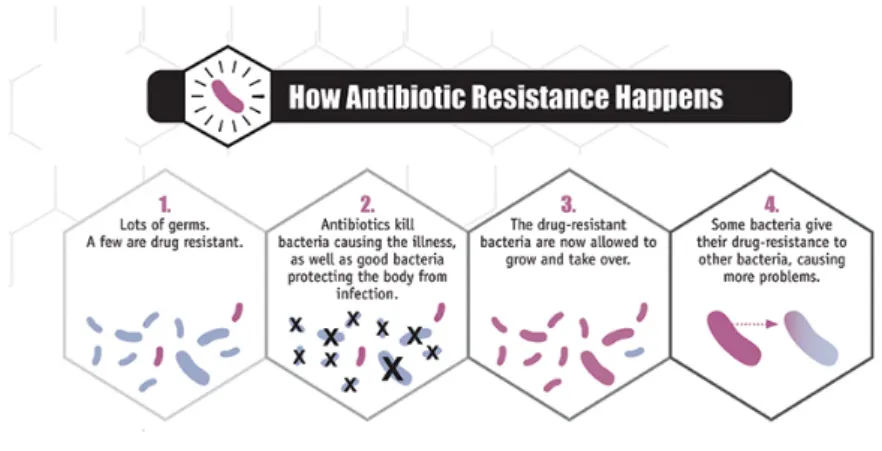
However, bacteria and other pathogens have evolved to resist the new drugs used to combat them (Figure 1.2). This resistance has become a growing public health problem in recent years due to the misuse and overuse of antibiotics in both human and veterinary use (Figure 1.3). Ultimately, resistant microorganisms could evolve to multidrug-resistant forms (MDR) that are defined as the acquired nonsusceptibility to at least one agent in three or more antibiotic categories (Magiorakos et al. 2012). Moreover, extensively drug
O OH O HO HO OH 1.45 O O O O OH O O 1.46 OH 1.47 1.48
19
resistant (XDR) and pandrug resistant (PDR) microorganisms have also became major concerns in clinical because these organisms can be resistant to all currently available antibiotics or remain susceptible only to potentially more toxic agents, leading to limited options for treatment (Magiorakos et al. 2012).
Figure 1.2 Antibiotic resistance. How some bacteria resist antibiotics, remain in the body and transmit their resistance. Adapted from CDC, 2013.
Examples of MDR bacteria are methicillin resistant Staphylococcus aureus (MRSA) and vancomycin resistant Enterococcus (VRE), fluoroquinolone resistant
Pseudomonas aeruginosa, ceftazidime resistant Klebsiella pneumoniae and many other
bacteria. The options for treating these bacteria are increasingly limited resulting in the resurgence of pathologies considered classically treatable. (Hansra and Shinkai 2011; Barnes and Sampson 2011).
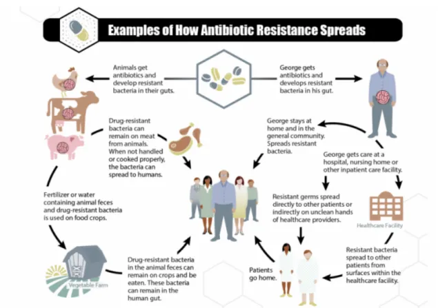
Results from various surveillance programs indicate that there is a high percentage of nosocomial infections caused by MDR bacterial strains such as MRSA and VRE (Fig. 1.3). Joint analysis of data from 15 European countries revealed that over 10% of blood infections are caused by MRSA strains, and several of these countries have MDR strain prevalence rates close to 50% (European Center for Disease Prevention and Control, 2018). The threat stems not only from the high prevalence of resistance among bacteria but also from the rapidly increasing levels of resistance.
20
Figure 1.3 Ways in which antibiotic-resistant bacteria can proliferate. Adapted from CDC, 2013. Antibiotic resistance is a worldwide problem. New forms of antibiotic resistance can cross international borders and spread across continents with ease. Many forms of resistance have spread with remarkable speed and world health leaders have described antibiotic resistant microorganisms as "nightmare bacteria" that pose a catastrophic threat to people in every country of the world (CDC, 2013). At the same time the pace of discovery of new antibiotics has slowed dramatically, (O’Neill 2014). One of the reasons for this decline is the challenges in identifying new chemicals that are both effective and non-toxic (Kalan e Wright, 2011).
1.6.2. Natural Products importance in the development of new drugs
Nowadays the economics of antibacterial research and development is considered “broken” and is commonly quoted as the principal cause for the lack of new therapies but the truth is that discover new antibacterial drugs is exceedingly difficult and science is not yet well advanced to allow the discovery of effective, efficient and non-toxic drugs. This has led to fears of a ‘post-antibiotic era’ (Jackson Czaplewski and Piddock, 2018). Therefore, there is an urgent need to find new approaches to antibiotic discovery.21
A new approach is the concept of the ‘magic bullet’ – that is, a small-molecule drug which is both selectively lethal to bacteria and able to be administered to humans and animals (Czaplewski et al. 2016).
Although assuming the small-molecule approach, how will it be possible to go forward to identify new drugs? The answer is in Natural Products (NPs). Over the past century NPs have supplied a crucial start-point in drug discovery and antibacterial therapies (Brown, Lister and May-Dracka 2014), and as they appear to have a number of undiscovered chemical properties, this strategy offers great chances for exploitation in drug development (Mugumbate and Overington 2015). This realization has led to a renaissance of interest in natural products with antibiotic activity for the identification of new molecules, and their application in antibacterial drug discovery (Johnston and Magarvey 2015).
Currently, existing antibiotics are mostly from natural products whose purpose is to target the bacterial cell wall, DNA or ribosomes. With rare exceptions, these compounds generally have more than one molecular target and exert complex effects on the bacterial cell (Brown and Wright 2016). Taking the example of penicillin, a type of β-lactam antibiotic, covalently alter various target enzymes known as penicillin binding proteins (PBPs) which, in turn, are responsible for the synthesis and remodeling of the bacterial cell wall for growth and division. Some antibiotics target the ribosome by inhibiting protein synthesis, others target some topoisomerase enzymes to block DNA synthesis. It is important to emphasize that, this effect on manifold cellular targets creates a limitation in the frequency of natural resistance that can emerge from mutation in the target gene. Inhibition of the molecular targets of antibiotics usually outcome in complex downstream effects that exceed those of simple enzyme inhibition. Evidence-based, β-lactam antibiotics disarray the bacterial cell-wall synthesis machinery activity in a way far more complex than simple inhibition (Cho, Uehara and Bernhardt 2014).
Systems-biology approaches proposed that reactive oxygen species have been neglected as contributors to cell death (Dwyer, Collins and Walker 2015). Even though this hypothesis remains controversial, there is an enhanced appreciation that bacterial cell death is complex and likely demands the involvement of several cellular pathways. Numerous natural-product antibiotics are the product of selection for these complex traits over millions of years of evolution. For that reason, it is possibly expected that modern methods of drug discovery have yet to hand compounds with efficacy comparable to that
22
of the first generation of natural antibiotics and their semisynthetic derivatives (Brown and Wright 2016).
This experimental work was intended to contribute to the scientific validation of the therapeutic application of G. hexamita in traditional African medicine as an antibacterial agent by isolating some of its bioactive constituents.
CHAPTER 2
24
2. Results and Discussion
2.1. Grewia hexamita phytochemical study
Bioassay-guided fractionation of Grewia hexamita allowed the isolation and identification of four triterpenes, three with a pentacyclic scaffold, lupeol (2.1), betulin (2.2), and betulinic aldehyde (2.3) and a new tetracyclic triterpene named 3β-caffeoyl-cycloartane (2.4). In addition to these compounds, two steroids, β-sitosterol (2.5) and 7-oxo-β-sitosterol (2.6), two phenolic compounds, (-hydroxybenzaldehyde (2.7) and vanillin (2.8), as well as pantolactone (2.9), an γ-butyrolactone, have also been isolated (Figure 2.1). Their structures were characterized by spectroscopic methods mainly 1D-(1H, 13C) and 2D-NMR (DEPT, COSY, HMBC and HMQC) experiments and by
comparisons with literature data. Furthermore, acylation of lupeol (2.1) and betulin (2.2), isolated in large amount, yielded four derivatives (2.10-2.13).
2.1.1. Triterpenes
2.1.1.1. Lupeol (2.1) and betulin (2.2)
Compound 2.1 was isolated as white crystals of m.p. 214-216 ºC and identified as lupeol based on the comparison of its physical and spectroscopic data to those described on the literature (Sai Prakash and Prakash 2012).
The ESI-MS with a protonated molecular ion at m/z 427 and the 13C NMR
spectrum were consistent with the molecular formula C30H50O. The six degrees of
unsaturation are in agreement with the presence of five rings and a double bond.
H HO H H H 1 2 3 4 5 6 7 8 9 10 11 12 26 13 14 27 15 16 17 18 19 20 21 22 23 24 25 28 30 29 2.1
25
The presence of a hydroxyl group was supported by the 1H NMR spectrum by the
existence of an oxymethine signal at δH 3.19 ppm, which was displayed as a double
doublet (J = 11.0 and 5.3 Hz) indicative of its axial orientation (Table 2.1). The existence of an exocyclic double bond was suggested by a doublet of doublets at δH 4.57 (J = 2.4
and 1.3 Hz) and a doublet at δH 4.69 ppm (J = 2.4 Hz) that were assigned to the methylene
protons. The presence of an isopropenyl group was indicated by the vinylic methyl signal at δH 1.68 ppm. Moreover, the 1H NMR spectrum also displayed signals for six tertiary
methyl groups at δH 0.76, 0.79, 0.83, 0.95, 0.97 and δH 1.03 ppm.
The 13C and DEPT NMR spectra (Table 2.2) corroborated the data described
above, showing thirty carbon resonances: six methyls, eleven methylenes (one sp2 at δC
109.5 ppm), seven methines (one oxygenated at δC 79.2 ppm) and six quaternary carbons
(one olefinic at δC 151.1 ppm).
All these data are in agreement with those reported in literature for lupeol [20(29)-lupen-3β-ol] and allowed the identification of compound 2.1 (Sai Prakash and Prakash 2012).
Table 2.1 1H NMR data of lupeol (2.1) and betulin (2.2), (300 MHz, CDCl
3; δ in ppm, J in Hz). 2.1 2.2 Position 1H 1H 3 3.19 dd (11.0; 5.3) 3.19 dd (10.8; 5.3) 19 2.38 td (11.0; 5.6) 2.38 td (11.0; 5.6) 23 0.95 s 0.97 s 24 0.76 s 0.76 s 25 0.83 s 0.83 s 26 1.03 s 1.02 s 27 0.97 s 0.98 s 28 0.79 s 3.80 dd (10.8; 1.5) 3.33 d (10.8) 29 4.57 dd (2.4; 1.3) 4.69 d (2.4) 4.58 dd (2.3; 1.4) 4.68 d (2.2) 30 1.68 s 1.68 s
26
Table 2.2 13C and DEPT NMR data of lupeol (2.1) and betulin (2.2), (300 MHz, CDCl3; δ in ppm, J
in Hz).
2.1 2.2 2.1 2.2
Position 13C DEPT 13C DEPT Position 13C DEPT 13C DEPT
1 38.9 CH2 39.0 CH2 16 35.8 CH2 29.3 CH2 2 27.6 CH2 27.6 CH2 17 43.12 C 47.9 CH 3 79.2 CH 79.1 CH 18 48.5 CH 47.9 CH 4 39.0 C 38.9 C 19 48.1 CH 48.9 CH 5 55.5 CH 55.6 CH 20 151.1 C 150.6 C 6 18.5 CH2 18.5 CH2 21 30.0 CH2 29.9 CH2 7 34.5 CH2 34.4 CH2 22 40.2 CH2 34.1 CH2 8 41.0 C 41.1 C 23 28.2 CH 28.1 CH 9 50.6 CH 50.6 CH 24 15.5 CH3 15.5 CH3 10 37.3 C 37.5 C 25 16.3 CH3 16.3 CH3 11 21.1 CH2 20.9 CH2 26 16.1 CH3 16.1 CH3 12 25.3 CH2 25.4 CH2 27 14.7 CH3 14.9 CH3 13 38.2 CH 37.3 CH 28 18.2 CH3 60.7 CH2 14 42.8 C 42.9 C 29 109.5 CH2 109.8 CH2 15 27.6 CH2 27.2 CH2 30 19.5 CH3 19.2 CH3
Compound 2.2 was isolated as white crystals of m.p. 248-251 ºC and identified as betulin based on the comparison of its physical and spectroscopic data to those described on the literature (Kwaji et al. 2018).
The ESI-MS, with a protonated molecular ion at m/z 443, and 13C NMR spectrum
were consistent with the molecular formula C30H50O2. The six degrees of unsaturation are
in agreement with the presence of five rings and a double bond.
H HO H H H 1 2 3 4 5 6 7 8 9 10 111226 13 14 27 15 16 17 18 19 20 21 22 23 24 25 28 30 29 2.2 OH
27
In the 1H NMR spectrum (Table 2.1), a doublet of doublets at δH 3.19 ppm (J =
10.8 and 5.3 Hz) indicated the presence of α-oriented hydrogen at C-3. Furthermore, two signals at δH 3.80 and 3.33 ppm, corresponding the diastereotopic methylene protons at
C-28, and resonances at δH 4.58 and 4.68 ppm, assigned to the olefinic protons at C-29,
together with a vinylic methyl group at δH 1.68 ppm could also be observed. The structure
of compound 2.2 was further substantiated by the 13C NMR and DEPT spectra that
revealed thirty signals due to five quaternary carbons (one olefinic at δC 150.6 ppm), eight
methines (two oxygenated, at δC 79.1 and 60.7 ppm), twelve methylenes (one sp2 at δC
109.8 ppm) and five methyls (Table 2.2). The olefinic signals at δC 150.6 and 109.8 ppm
corroborated the presence of the characteristic double bond at C-29 of lupane-type triterpenes.
Consequently, based on the comparison of the NMR data with literature, compound 2.2 was found to be lup-20(29)-ene-3,28-diol, commonly known as betulin (Kwaji et al. 2018), which differs from lupeol at C-28, having an hydroxyl group at this position.
2.1.1.1.1. Preparation of lupeol and betulin derivatives
Lupeol (2.1) and betulin (2.2), isolated in large amount, were acylated for increasing the number of compounds and further study their antibacterial activity.
2.1.1.1.2. Lupeol acetate (2.10) and lupeol benzoate (2.12)
Acetylation of lupeol (Scheme 2.1) afforded compound 2.10. This compound was obtained as colorless crystals of m.p. 220 ºC and identified as lupeol acetate based on the comparison of its physical and spectroscopic data with those described on the literature (Barla et al. 2006; da Silva et al. 2018).
H O H H H 1 2 3 4 5 6 7 8 9 10 11 12 26 13 14 27 15 16 17 18 19 20 21 22 23 24 25 28 30 29 2.10 O 1’ 2’
28 Scheme 2.1 Preparation of lupeol derivatives (2.10 and 2.12). a a Reagents and conditions: (i) Pyridine (80 eq.) and Ac
2O (120 eq.), RT, O/N; ii) Pyridine (60 eq.)
and benzoyl chloride (130 eq.), RT, O.N
The molecular formula, C32H52O2, was substantiated by the 13C NMR spectrum
and ESI-MS, which exhibited a protonated molecule ion at m/z 469 [M+H]+. The IR
spectrum displayed an absorption band at 1735 cm-1 for ester carbonyl group.
The NMR data of compound 2.10 (Table 2.3) resembled those obtained for lupeol (2.1). As expected, in the 1H NMR spectrum, the most remarkable differences were the
presence of a new acetyl singlet at δH 2.05 ppm, and the downfield chemical shift of H-3
that appeared in compound 2.10 at δH 4.47 (m).
Table 2.3 1H NMR data of lupeol-3-acetate (2.10) and lupeol benzoate (2.12), (300 MHz, CDCl 3; δ in ppm, J in Hz). Position 2.10 2.12 3 4.47 m 4.72 m 19 2.38 td (11.0; 5.6) 2.39 td (11.0; 5.6) 23 0.83 s 0.92 s 24 0.78 s 0.79 s 25 0.83 s 0.83 s 26 1.02 s 1.05 s 27 0.93 s 1.00 s 28 0.84 s 0.84 s 29 4.56 dd (2.8; 1.4) 4.69 d (2.5) 4.57 dd (2.6; 1.4) 4.69 d (2.6) 30 1.66 s 1.69 s 2’ 2.05 s − 3’ − 8.04a m 4’ − 7.44b m 5’ – 7.54 m 6’ – 7.44b m 7’ – 8.04a m a, b Overlapped signals H HO H H H i) ii) H O H H H 1 2 3 4 5 6 7 8 9 10 111226 13 14 27 15 16 17 18 19 20 21 22 23 24 25 28 30 29 2.10 O 1’ 2’ 2.1 H O H H H 1 2 3 4 5 6 7 8 9 10 111226 13 14 27 15 16 17 18 19 20 21 22 23 24 25 28 30 29 2.12 O 1’ 2’ 3’ 4’ 5’ 6’ 7’
29
In the 13C NMR, the presence of the new signals at δC 171.2 and 21.5 ppm,
together with the paramagnetic effect at C-3 (δC 81.8 ppm) were consistent with the
introduction of a new acetyl group at this position (Table 2.4).
Table 2.4 13C and DEPT NMR data of lupeol-3-acetate (2.10) and lupeol benzoate (2.13), (300 MHz,
CDCl3; δ in ppm, J in Hz).
2.10 2.12 2.10 2.12
Position 13C DEPT 13C DEPT Position 13C DEPT 13C DEPT
1 38.1 CH2 38.3 CH2 20 151.1 C 151.1 C 2 23.8 CH2 23.9 CH2 21 29.9 CH2 29.9 CH2 3 81.8 CH 79.2 CH 22 40.1 CH2 40.1 CH2 4 38.5 C 38.5 CH2 23 28.1 CH 28.3 CH 5 55.5 CH 55.5 CH 24 15.5 CH3 16.1 CH3 6 18.5 CH2 18.4 CH2 25 16.3 CH3 16.9 CH3 7 34.3 CH2 34.3 CH2 26 16.1 CH3 16.4 CH3 8 40.9 C 40.9 C 27 14.6 CH3 14.6 CH3 9 50.4 CH 50.5 CH 28 18.2 CH3 18.2 CH2 10 37.2 C 37.2 CH 29 109.5 CH2 109.5 CH2 11 21.1 CH2 21.1 CH2 30 19.4 CH3 19.4 CH3 12 25.2 CH2 25.2 CH2 1’ 171.2 C 166.4 C 13 37.9 CH 38.1 C 2’ 21.5 CH3 131.1 C 14 42.9 C 42.9 C 3’ − − 129.7 CH 15 27.5 CH2 27.6 CH2 4’ − − 128.4 CH 16 35.7 CH2 35.7 CH2 5’ − − 132.8 CH 17 43.1 C 43.1 CH 6’ − − 128.4 CH 18 48.4 CH 47.9 CH 7’ − − 129.7 CH 19 48.1 CH 48.4 CH
Benzoylation of lupeol afforded compound 2.12 that was obtained as colorless crystals with m.p. 261 ºC. Its IR displayed an absorption band for the ester carbonyl group at 1718 cm-1.
30 .
When comparing with lupeol (2.1) (Tables 2.3 and 2.4), the main differences in the 1H NMR spectrum of compound 2.12, were the presence of downfield signals
corresponding to the aromatic protons at δH 8.04, 7.44 and 7.54 ppm. In turn, in the 13C
NMR, besides the extra signals for the benzene the ring and the carbonyl carbon (δC 166.4
ppm), the most significant changes were in the carbon signals of ring A (Table 2.4), namely at C-3 that was shifted downfield (δC 79.2 ppm).
All the physical and spectroscopic data were in agreement with those described in the literature for lupeol benzoate (Adotey et al. 2012)).
2.1.1.1.3. Betulin diacetate (2.11) and betulin dibenzoate (2.13) Acetylation of betulin afforded compound 2.11 (Scheme 2.2.) isolated as colorless crystals of m.p. 219-221 ºC. H O H H H 1 2 3 4 5 6 7 8 9 10 111226 13 14 27 15 16 17 18 19 20 21 22 23 24 25 28 30 29 2.12 O 1’ 2’ 3’ 4’ 5’ 6’ 7’ H O H H H 1 2 3 4 5 6 7 8 9 10 111226 13 14 27 15 16 17 18 19 20 21 22 23 24 25 28 30 29 2.11 O 1’ 2’ O O 1’’ 2’’
31
Scheme 2.2 Preparation of betulin (2.2) derivatives (2.11 and 2.13).a
a Reagents and conditions: i) Pyridine (110 eq.) and Ac
2O (90 eq.), RT, O/N; ii) Pyridine (110 eq.)
and benzoyl chloride (40 eq.), RT, O.N.
The ESI-MS with a protonated molecular ion at m/z 527 [M+H]+ and the 13C NMR
spectrum were consistent with the molecular formula C34H54O4. The IR spectrum showed
a strong absorption band at 1712 cm–1 corresponding to stretching vibrations of the
carbonyl ester groups.
The NMR data of compound 2.11 (Table 2.5 and 2.6) resembled those found for betulin (2.2). Therefore, in the 1H NMR spectrum, the most notable differences were the
presence of two new acetyl singlets at δH 2.03 and 2.06 ppm, and the signals of H-3 and
H-28 that were shifted downfield, appearing in compound 2.11 at δH 4.45 as a multiplet
and two doublets of doublets at δH 3.83 (J = 11.0 and 1.2 Hz) and δH 4.23 (J = 11.0 and
1.9 Hz), respectively (Table 2.5). In the 13C NMR, the resonances of two methyl groups
at δC 21.2 and δC 21.5 ppm with the corresponding signals for each carbonyl at δC 171.2
and δ 171.8 ppm (Table 2.6), together with the downfield shifts of C-3 (δC 81.8 ppm) and
C-28 (δC 62.9 ppm), were consistent with the introduction of the two new acetyl groups
at this position (Table 2.4).
Compound 2.11 was identified as betulin diacetate based on the comparison of its physical and spectroscopic data to those described on the literature (Salah and Bakibaev 2017). H HO H H H 2.2 OH ii) i) H O H H H 1 2 3 4 5 6 7 8 9 10 111226 13 14 27 1516 17 18 19 20 21 22 23 24 25 28 30 29 2.11 O 1’ 2’ O O 1’’ 2’’ H O H H H 1 2 3 4 5 6 7 8 9 10 111226 13 14 27 1516 17 18 19 20 21 22 23 24 25 28 30 29 2.13 O O 1’’ 2’’ 3’’ 4’’ 5’’ 6’’ 7’’ 1’ 2’ 3’ 4’ 5’ 6’ 7’ O
32
Table 2.5 1H NMR data of betulin diacetate (2.11) and betulin dibenzoate (2.13), (300 MHz, CDCl 3; δ in ppm, J in Hz). 2.11 2.13 Position 1H 1H 3 4.45 m 4.70 m 19 2.43 td(10.9; 5.7) 2.54 td (10.8; 5.7) 23 0.83 s 1.00 s 24 0.83 s 0.92 s 25 0.82 s 0.91 s 26 1.01 s 1.09 s 27 0.95 s 1.02 s 28 3.83 dd (11.0; 1.2) 4.23 dd (11.0; 1.9) 4.10 m 4.54 dd (11.2; 1.8) 29 4.57 dt (2.7; 1.4) 4.67 d (2.3) 4.62 dd (2.3; 1.4) 4.73 d (2.3) 30 1.67 s 1.72 s 2’ 2.06 s − 3’ − 8.05a m 4’ − 7.44b m 5’ − 7.55c m 6’ − 7.44b m 7’ − 8.05a m 2’’ 2.03 s − 3’’ − 8.05a m 4’’ − 7.44b m 5’’ − 7.55c m 6’’ − 7.44b m 7’’ − 8.05a m a, b, c Overlapped signals
Benzoylation of betulin afforded compound 2.13 (Scheme 2.2). This compound was obtained as colorless crystals with m.p. 142 ºC. Its IR displayed absorption bands for the ester carbonyl groups at 1716 cm-1.
H O H H H 1 2 3 4 5 6 7 8 9 10 111226 13 14 27 15 16 17 18 19 20 21 22 23 24 25 28 30 29 2.13 O O 1’’ 2’’ 3’’ 4’’ 5’’ 6’’ 7’’ 1’ 2’ 3’ 4’ 5’ 6’ 7’ O