CASE REPORT
Hydropic leiomyoma of the uterus presenting
as a giant abdominal mass
Mariana Horta,
1Teresa Margarida Cunha,
2Rita Oliveira,
3Paula Magro
41Department of Radiology, Centro Hospitalar Lisboa Ocidental, Lisbon, Portugal 2Department of Radiology, Instituto Português de Oncologia de Lisboa Francisco Gentil, Lisbon, Portugal 3Department of Pathology, Centro Hospitalar Lisboa Ocidental, Lisbon, Portugal 4Department of Surgery, Centro Hospitalar Lisboa Ocidental, Lisbon, Portugal
Correspondence to
Dr Mariana Horta, [email protected]
Accepted 23 August 2015
To cite:Horta M, Cunha TM, Oliveira R,et al.
BMJ Case RepPublished online: [please includeDay Month Year] doi:10.1136/ bcr-2015-211929
SUMMARY
We describe a case of a 35-year-old woman with a pedunculated uterine leiomyoma with diffuse hydropic degeneration presenting as a giant abdominal mass. The patient was admitted in the emergency department because of diffuse abdominal bloating and discomfort. Ultrasonography (US) showed a heterogeneous abdominopelvic mass. Magnetic resonance imaging (MRI) was performed to further characterise and revealed a myometrial pedunculated tumour. Despite its marked T2-signal heterogeneity and volume, there were no other suspiciousfindings to suggest a malignant nature; therefore, fertility-sparing myomectomy was performed. Leiomyomas frequently undergo degenerative changes altering their imaging appearances. Leiomyomas with uncommon degenerative changes may be difficult to differentiate from malignant myometrial tumours, based solely on imaging. To the best of our knowledge, a diffuse hydropic degeneration imaging appearance has only been described twice in the literature. We describe the imaging appearance of this rare form of leiomyoma drawing attention to its differential diagnosis.
BACKGROUND
We describe a case of a woman of childbearing age with a giant abdominal mass diagnosed as a pedun-culated uterine leiomyoma with diffuse hydropic change.
Giant pedunculated leiomyomas with degenera-tive changes may be misinterpreted as ovarian malignant tumours on ultrasound (US) imaging, since they may present as masses with heteroge-neous echogenicity. Moreover, they can make it
dif-ficult to visualise the adnexa and vascular pedicle between the uterus and juxta-uterine mass. Therefore, MRI is crucial in the preoperative diag-nosis of such lesions in order to determine the origin and nature of the tumour.
Typical degenerative forms of leiomyomas have characteristic MRI appearances. However, a leio-myoma with uncommon degenerative changes, such as the tumour presented, may be a diagnostic challenge and sometimes difficult to differentiate from its malignant counterparts, based solely on imaging. This may lead to a more aggressive surgi-cal approach (non-fertility sparing surgery) in a woman of childbearing age.
Leiomyomas with diffuse hydropic changes are rare, with only two cases reported in the English literature. We describe the imaging and histopatho-logical appearance of this rare form of degener-ation, drawing attention to its differential diagnosis characteristics.
CASE PRESENTATION
A 35-year-old nulliparous woman with diffuse abdominal bloating and discomfort was admitted to our hospital emergency department. Her per-sonal medical history was unremarkable.
INVESTIGATIONS
On physical examination, a 20 cm mobile and pain-less pelvic mass was detected. Other physical assess-ments were negative. Routine laboratory data were in the normal range, apart from a slightly raised cancer antigen 125 (CA-125) serum level of 123 U/mL (reference range 0–35 U/mL).
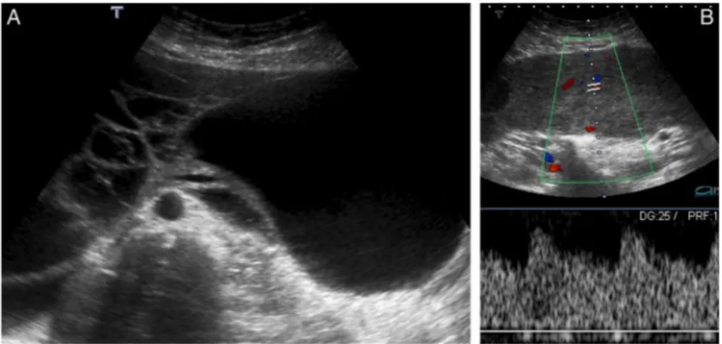
Pelvic transabdominal and transvaginal US were performed, revealing a large, relatively well-defined, multilobulated abdominopelvic mass, measuring approximately 20×30×8 cm (figure 1). The mass had heterogeneous echogenicity, with prominent vascularised solid as well as cystic com-ponents (figure 1). No apparent relation to the uterus was seen and the ovaries could not be depicted; a small amount of pelvic ascites was also present.
Since the ovaries could not be seen and the serum levels of CA-125 were slightly elevated, the possibility of an ovarian neoplasm was raised; therefore, a MRI was performed to identify the origin of the tumour and to further characterise the lesion.
MRI showed a giant pedunculated abdominopelvic mass arising from the posterior uterine fundus, which was clearly demonstrated by the presence of multiple vessels between the uterus and the juxta-uterine mass (‘bridging vessels’sign) (figure 2A; arrow); thesefindings were consistent with the pres-ence of a tumour of myometrial origin, such as a leio-myoma on the benign side or a leiomyosarcoma on the malignant side.
The mass was lobulated, very well-defined and encapsulated, with a small protruding anterior segment herniating through an umbilical defect (figure 2A; dashed arrow). There were no signs of invasion of the adjacent structures.
areas showing low signal on T1-weighted images and high T2 signal (figures 2B, C; arrows). No necrosis or areas of increased T1 signal that could suggest haemorrhage were seen.
A small amount of ascites was detected only in the pelvis (figure 2A; asterisks).
No inguinal, pelvic or para-aortic lymphadenopathies, and no peritoneal implants or hepatic metastases were detected.
Both ovaries were within their normal appearance and a sub-mucosal myometrial leiomyoma, displaying low T2 signal was seen in the anterior uterine corpus, measuring 2.4×1.5 cm (figures 2A(cross), B).
DIFFERENTIAL DIAGNOSIS
The differential diagnosis of a giant heterogeneous myometrial pedunculated subserosal tumour should include leiomyomas
with degenerative changes on the benign side and leiomyosarco-mas on the malignant side.
The definite diagnosis of a leiomyosarcoma is histopatho-logical (based on the assessment of nuclear atypia, mitotic activ-ity and tumour cell necrosis). However, apart from the presence of a large intermediate to high signal T2 mass, there were no imagingfindings to suggest this diagnosis (irregular or infiltrative margins; inhomogeneity with necrosis and/or haemorrhage; lymphadenopathies and/or distant metastasis).
The four main degenerative forms of leiomyomas are: hyaline; myxoid; haemorrhagic and cystic. Rare forms include: lipomatous variants and diffuse hydropic leiomyomas.
However, there were also enhancing cord-like solid components with moderate T2 hypo-intense signal and iso-signal to the muscle in T1-weighted images, which are not characteristic of any of the commonest degenerative forms of leiomyomas. Histopathologically, these were revealed to be strand-like areas of tumour cells within a vast oedematous stroma that was some-times combined with hyalinisation, which is characteristic of hydropic degeneration. Therefore, the tumour was diagnosed as a leiomyoma with diffuse hydropic degeneration. The US and MRI characteristics of this type of leiomyoma have only been previously described twice in the literature.
TREATMENT
Since the tumour had no MRI features to suggest a malignant myometrial tumour and seemed resectable, fertility-sparing myomectomy was performed. The pelvic and abdominal cavities were explored during surgery, and neither peritoneal implants nor lymphadenopathies were found.
OUTCOME AND FOLLOW-UP
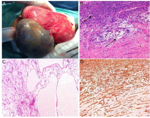
The tumour, weighing 2801 g and measuring 24×30×8 cm, exhibited a smooth, shiny surface. On cut surface, it was a solid, multicystic whitish-grey mass with smooth and thin walls. Wateryfluid was seen extruding from it.
The histopathological examination revealed a tumour com-posed of hypercellular areas of intersecting fascicles of spindle cells with eosinophilic fibrillary cytoplasm and cigar-shaped nuclei (figure 3). These cells were within a vast oedematous stroma where some vessels and collagen deposition could be seen (figure 3). Moreover, hypocellular and pseudomulticystic areas, without epithelial covering, were seen (figure 3). These areas werefilled with pale eosinophilicfluid.
No cytological atypia, mitosis or necrosis was identified. The diagnosis of a leiomyoma with extensive hydropic degener-ation was made.
To date, the patient has undergone 7 months of follow-up, without major complications.
DISCUSSION
Uterine leiomyomas are the most common uterine neoplasms. They are more likely to occur as asymptomatic masses in women in their fourth andfifth decades of life.1The presence of symptoms is related to the tumoural size, with up to one-third of women presenting with uterine bleeding, pelvic pain and pressure.1 Infertility is also associated with the
pres-ence of leiomyomas, especially those of a submucosal type. Transvaginal US in conjunction with transabdominal US is the
first imaging modality to assess a pelvic mass. The US diagnosis of typical leiomyomas is usually straightforward, as they usually present as well-defined, solid hypoechoic masses in a submuco-sal, intramural or subserosal localisation. However, peduncu-lated leiomyomas may pose diagnostic problems on US, as occurred in our case.
Pedunculated leiomyomas are tumours that are connected to the uterus via a vascular pedicle. They can be submucosal or subserosal. We show the latter type, which was confused with an ovarian mass on transabdominal and transvaginal B mode, as well as on Doppler US imaging.
This misinterpretation especially occurs when large tumours make it difficult to visualise the adnexa, and to depict the vascu-lar pedicle between the uterus and the juxta-uterine mass on Doppler imaging. Moreover, some of these tumours may detach from their stalk, forming independent masses (the so-called parasitic leiomyomas).
When the adnexal or uterine origin of the tumour cannot be determined by US imaging, MRI should be performed. In these cases, oblique sequences (ie, the axial plan of the ovary, which corresponds to the parallel plan of the endometrial cavity) are a problem-solving tool.
Furthermore, MRI in this particular case was also useful to further characterise the lesion, since it showed marked US heteroge-neous echogenicity, displaying both cystic and vascular solid areas.
It is well known that 10% of leiomyomas are variant forms represented by different types of histological degeneration or cellular histological subtypes.1Unlike typical leiomyomas, which
characteristically display low signal on T2-weighted images, the different degenerating forms of leiomyoma have a variable appearance on MRI. These forms tend to occur in large tumours that enlarge and outgrow their blood supply.2 3
The most common type of degeneration is hyaline, which can be present in up to 60% of cases.3Hyaline degeneration usually
shows low signal on T2-weighted images, and sometimes a
‘cobblestone appearance’and variable enhancement on postcon-trast imaging.4
Leiomyomas with myxoid degeneration show the presence of soft mucoid areas with hyaluronic acid-rich mucopolysaccharide gelatinous foci, sometimes intermixed with cystic areas. These typically show very high T2 signal and mild enhancement after contrast administration.2
Red degeneration usually occurs in pregnancy and is repre-sented by multiple haemorrhagic infarctions. On MRI, leiomyo-mas with red degeneration tend to present peripheral or diffuse high signal on T1-weighted images and variable signal intensity on T2-weighted images.
Cystic degenerated leiomyomas show non-enhancement and high T2 signal.
Diffuse hydropic change is a very rare form of degeneration characterised by watery oedema that may be combined with hyalinisation, resulting in a cord-like pattern growth of the tumoural cells.1 The presence of a large amount of fluid may result in large tumours.3 The accumulation of watery oedema
can be sometimes be misinterpreted as a myxoid matrix and, therefore, these types of leiomyomas may be misdiagnosed as myxoid leiomyomas or as leiomyosarcomas. Another confound-ing factor is, when the extensive hydropic degeneration presents beyond the limits of a leiomyoma, it mimics an infiltrative border of a myxoid leiomyosarcoma.5
To the best of our knowledge, this is the third case where imaging findings of diffuse hydropic leiomyomas are shown. Both previous reports described a diffuse hydropic leiomyoma in pregnancy.3 6
In one of the cases, the tumour appeared as a unilocular cystic mass with irregular walls on both US and MRI.3 The
other case reported a large and lobulated complex appearing mass on US that on MRI displayed heterogeneous mainly high T2 signal with strands of low signal within.6Thick blood vessels were present but no fatty components were seen. This radio-logical appearance was similar to the case we present here.
Atypical degenerating and cellular forms of leiomyomas with high signal on T2-weighted images can be a diagnostic challenge for radiologists, as they can mimic malignant tumours.7 “Pseudo-Meigs’ syndrome” has been associated with hydropic leiomyomas, and the case we report was also associated with ascites and elevated serum levels of CA-125, which complicated the differential diagnosis.3
Uterine sarcomas account for approximately 1–3% of uterine tumours, of which leiomyosarcoma is the most frequent.1 7The
reported incidence of sarcomatous degeneration of leiomyomas is 0.1–0.8%.8
As atypical forms of leiomyomas, leiomyosarcomas present as intermediate to high signal T2 masses. They tend to be irregular, infiltrative, inhomogeneous enhancing tumours associated with
Although a retrospective study conducted by Thomassin-Naggaraet al7suggested that the combination of T2 signal inten-sity with the analysis of diffusion-weighted imaging would be the best way to differentiate leiomyosarcomas from atypical forms of leiomyomas, both conventional and functional MRI have limited value in the diagnosis of these types of tumours since there is a sig-nificant overlap with their benign counterparts.9 10Therefore, the definite diagnosis is exclusively histopathological and based on the assessment of three major histological features: mitotic activity, nuclear atypia and tumour cell necrosis. None of these character-istics were observed in the case we here present.
Learning points
▸ Pedunculated subserosal leiomyomas may be difficult to
distinguish from adnexal masses by US imaging alone, therefore MRI with oblique sequences should be performed to determine the origin of the tumour.
▸ The diagnosis of a typical leiomyoma is usually
straightforward on US imaging. However, tumours with degenerating changes and those showing rapid growth in postmenopausal women should be further characterised by MRI.
▸ Leiomyomas with atypical degenerating changes such as
diffuse hydropic change, usually present as a diagnostic challenge for the radiologist, since they can mimic malignancy.
▸ Although there are imaging criteria that can suggest the
diagnostic of leiomyosarcomas, there is significant overlap with atypical forms of their benign counterpart in both conventional and functional MRI. Therefore, their diagnosis is exclusively histopathological.
Competing interests None declared.
Patient consent Obtained.
Provenance and peer reviewNot commissioned; externally peer reviewed.
REFERENCES
1 Kurman RJ, Carcangiu ML, Herrington CS,et al. Classification of tumours of the ovary. In:WHO classification of tumours. Vol 6. 4th edn. Lyon: IARC, 2014:135–41.
2 Mazziotti S, Ascenti G, Racchiusa S,et al. Retroperitoneal growth of degenerated myxoid uterine leiomyoma mimicking sarcoma.Clin Radiol 2012;67:616–17.
3 Awad EE, El-agwani AS, Elhabashy AM,et al. A giant uterine myometrium cyst mimicking an ovarian cyst in pregnancy: an uncommon presentation of hydropic degeneration of uterinefibroid.Egypt J Radiol Nucl Med2015;46:529–34. 4 Murase E, Siegelman ES, Outwater EK,et al. Uterine leiomyomas: histopathologic
features, MR imagingfindings, differential diagnosis, and treatment.Radiographics 1999;19:1179–97.
5 Toledo G, Oliva E. Smooth muscle tumors of the uterus: a practical approach.Arch Pathol Lab Med2008;132:595–605.
6 Heffernan E, Kobel M, Spielmann A. Hydropic leiomyoma of the uterus presenting in pregnancy: imaging features.Br J Radiol2009;82:e164–7.
Copyright 2015 BMJ Publishing Group. All rights reserved. For permission to reuse any of this content visit http://group.bmj.com/group/rights-licensing/permissions.
BMJ Case Report Fellows may re-use this article for personal use and teaching without any further permission.
Become a Fellow of BMJ Case Reports today and you can:
▸ Submit as many cases as you like
▸ Enjoy fast sympathetic peer review and rapid publication of accepted articles ▸ Access all the published articles
▸ Re-use any of the published material for personal use and teaching without further permission
For information on Institutional Fellowships contact [email protected]